

Endoscopic transconjunctival optic nerve sheath fenestration for progressive idiopathic visual field deficit: a case report
- PMID: 34693780
- PMCID: PMC8638564
- DOI: 10.1177/03000605211048362
Endoscopic transconjunctival optic nerve sheath fenestration for progressive idiopathic visual field deficit: a case report
Abstract
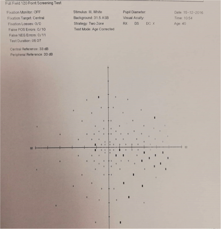
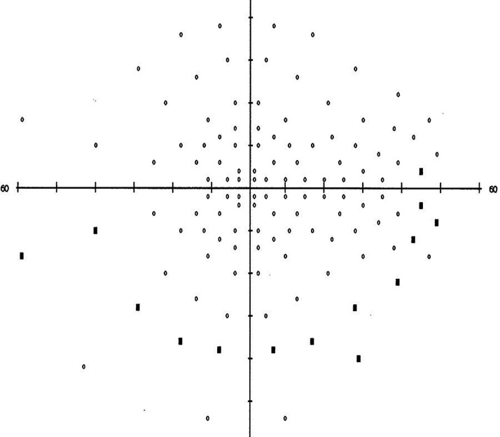
Intra-orbital optic nerve sheath fenestration (ONSF) is an effective option in patients with progressive vision loss due to idiopathic intracranial hypertension. Most proposed techniques involve surgical trauma and require disinsertion of the medial rectus muscle; thus, less invasive surgical procedures are needed. Here, a feasible and effective technique of endoscopic intra-orbital ONSF through a conjunctival incision is presented, in a patient with a progressively compromised visual field, papilloedema, and distended subarachnoid space around the optic nerves. The retrobulbar segment of the optic nerve was exposed for incision, avoiding manipulation of the lateral orbital rim bones and irritation of the ciliary microvessels and nerves. The patient regained the entire visual field. ONSF was safely and effectively performed endoscopically through a narrow corridor gained by brushing away the orbital fat with minimal traction on the medial rectus muscle. The small postoperative wound was associated with faster and easier convalescence, and less tissue trauma versus conventional open approaches.
Keywords: Optic nerve sheath fenestration; endoscopic surgery; endoscopy; intra-orbital; ocular surgery; vision loss.
Conflict of interest statement
Figures


References
-
- Elnahry AG, Elnahry GA. Management of idiopathic intracranial hypertension during the COVID-19 pandemic. Rev Recent Clin Trials 2021; 16: 122–125. - PubMed
-
- Chan JW. Current concepts and strategies in the diagnosis and management of idiopathic intracranial hypertension in adults. J Neurol 2017; 264: 1622–1633. - PubMed
Publication types
MeSH terms
LinkOut - more resources
Full Text Sources
