. 2021 Nov;23(11):1028-1051.
doi: 10.1177/1098612X211020162.

The Feline Cardiomyopathies: 2. Hypertrophic cardiomyopathy

Affiliations

The Feline Cardiomyopathies: 2. Hypertrophic cardiomyopathy

Mark D Kittleson et al. J Feline Med Surg. 2021 Nov.

Abstract

Practical relevance: Hypertrophic cardiomyopathy (HCM) is the most common form of feline cardiomyopathy observed clinically and may affect up to approximately 15% of the domestic cat population, primarily as a subclinical disease. Fortunately, severe HCM, leading to heart failure or arterial thromboembolism (ATE), only occurs in a small proportion of these cats.

Patient group: Domestic cats of any age from 3 months upward, of either sex and of any breed, can be affected. A higher prevalence in male and domestic shorthair cats has been reported.

Diagnostics: Subclinical feline HCM may or may not produce a heart murmur or gallop sound. Substantial left atrial enlargement can often be identified radiographically in cats with severe HCM. Biomarkers should not be relied on solely to diagnose the disease. While severe feline HCM can usually be diagnosed via echocardiography alone, feline HCM with mild to moderate left ventricular (LV) wall thickening is a diagnosis of exclusion, which means there is no definitive test for HCM in these cats and so other disorders that can cause mild to moderate LV wall thickening (eg, hyperthyroidism, systemic hypertension, acromegaly, dehydration) need to be ruled out.

Key findings: While a genetic cause of HCM has been identified in two breeds and is suspected in another, for most cats the cause is unknown. Systolic anterior motion of the mitral valve (SAM) is the most common cause of dynamic left ventricular outflow tract obstruction (DLVOTO) and, in turn, the most common cause of a heart murmur with feline HCM. While severe DLVOTO is probably clinically significant and so should be treated, lesser degrees probably are not. Furthermore, since SAM can likely be induced in most cats with HCM, the distinction between HCM without obstruction and HCM with obstruction (HOCM) is of limited importance in cats. Diastolic dysfunction, and its consequences of abnormally increased atrial pressure leading to signs of heart failure, and sluggish atrial blood flow leading to ATE, is the primary abnormality that causes clinical signs and death in affected cats. Treatment (eg, loop diuretics) is aimed at controlling heart failure. Preventive treatment (eg, antithrombotic drugs) is aimed at reducing the risk of complications (eg, ATE).

Conclusions: Most cats with HCM show no overt clinical signs and live a normal or near-normal life despite this disease. However, a substantial minority of cats develop overt clinical signs referable to heart failure or ATE that require treatment. For most cats with clinical signs caused by HCM, the long-term prognosis is poor to grave despite therapy.

Areas of uncertainty: Genetic mutations (variants) that cause HCM have been identified in a few breeds, but, despite valiant efforts, the cause of HCM in the vast majority of cats remains unknown. No treatment currently exists that reverses or even slows the cardiomyopathic process in HCM, again despite valiant efforts. The search goes on.

Keywords: Cardiomyopathies; echocardiography; gene mutation; hypertrophic cardiomyopathy; mitral valve; myocardial diseases; systolic anterior motion.


Conflict of interest statement

The authors declared no potential conflicts of interest with respect to the research, authorship, and / or publication of this article.

Figures

Figure 1
Figure 1
Gross pathologic specimen of a heart showing markedly thickened left ventricular walls and papillary muscles and an enlarged left atrium. At the upper left there is a thrombus in the left auricle (white asterisk). Another thrombus is present in the body of the left atrium on the right (black asterisk). S = interventricular septum; F = left ventricular free wall; P = base of the papillary muscles; A = body of the left atrium
Figure 2
Figure 2
Cross-sectional view of a gross pathologic specimen of a heart from a cat with lymphoma (pale areas) that invaded the left ventricular myocardium
Figure 3
Figure 3
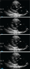
Sequential echocardiographic frames that demonstrate systolic anterior motion of the mitral valve (SAM). (a) Early systole. The mitral valve is closed. The left ventricular outflow tract (LVOT) is to the right of the LV label. A small section of the septal (anterior) leaflet of the mitral valve is being pulled into the LVOT by the papillary muscle at the apex of the ventricle. A chorda tendinea connecting the two is barely visible. (b) Mid-systole. The arrow points at the section of the septal leaflet of the mitral valve that is being simultaneously pulled into the LVOT by the papillary muscle (P) and pushed into the LVOT by blood flow that comes up underneath that section of leaflet tip. (c) Late systole. The tip of the mitral valve leaflet and its associated chorda now have the appearance of a cane. The mitral valve tip is partially occluding the LVOT. (d) End-systole. The SAM is further occluding the LVOT. LV = left ventricular lumen; Ao = aorta; LA = left atrium
Figure 4
Figure 4
M-mode echocardiogram from a cat with systolic anterior motion of the mitral valve (SAM). The SAM label overlies the region of SAM. The MV (mitral valve) label lies within the opening of the MV in diastole. SAM is visible in all cardiac cycles except the one after the MV label
Figure 5
Figure 5
Continuous wave Doppler trace from a cat with systolic anterior motion of the mitral valve (SAM). The peak velocity is elevated due to the dynamic left ventricular outflow tract obstruction caused by the SAM. The trace is shaped like a scimitar (superimposed graphic on the far right side) and is due to the region of obstruction progressively worsening throughout systole, especially in late systole (late peaking)
Figure 6
Figure 6
Histopathologic image of left ventricular myocardium from a Maine Coon cat with HCM showing cardiomyocyte disarray: the longitudinal axes of the cardiomyocytes converge and diverge instead of coursing in parallel
Figure 7
Figure 7
Echocardiographic images from cats with HCM. (a) Right parasternal long-axis view showing severe global thickening of the left ventricle (LV). (b) Right parasternal short-axis view from a different cat with severe global thickening of the LV. (c) Right parasternal short-axis view of the LV from a cat with severe thickening of the left ventricular free wall and a less thick interventricular septum. LA = left atrium
Figure 8
Figure 8
Right parasternal short-axis echocardiographic images from a cat with severe HCM showing hypertrophied and hyperechoic left ventricular papillary muscles in diastole (a) and at end-systole showing end-systolic cavity obliteration (b)
Figure 9
Figure 9
Right parasternal short-axis view of the aorta (Ao), body of the left atrium (LA) and left atrial appendge (LAA). The LA and LAA are severely enlarged
Figure 10
Figure 10
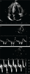
Tissue Doppler imaging (TDI) of a normal cat and a cat with severe HCM. (a) A left apical four-chamber view is used for TDI echocardiography of the lateral mitral annulus. The white bars represent the position of the pulsed wave Doppler gate. Images (b) and (c) show TDI myocardial velocity of the lateral mitral annulus in a cat with severe HCM and a normal cat, depicted with different velocity scales. The normal cat (c) had a higher heart rate (HR; 220 beats per minute [bpm] vs 115 bpm in the cat with HCM) and fusion of the early (E’) and late (A’) diastolic waves to form an EA’ wave. Fusion of the E’ and A’ diastolic waves does not affect the peak velocity in normal cats. Peak diastolic velocity and systolic velocity are greatly reduced in the cat with HCM (b), indicating diastolic dysfunction. S’ = systolic myocardial velocity
None
None
None
None
None
None

References

    1. Cirino AL, Ho C. Hypertrophic cardiomyopathy overview. In: Adam MP, Ardinger HH, Pagon RA, et al. (eds). GeneReviews®. Seattle, WA: University of Washington, Seattle, 1993. Available at: http://www.ncbi.nlm.nih.gov/books/NBK1768/. - PubMed
    1. Ommen SR, Mital S, Burke MA, et al.. 2020 AHA/ACC guideline for the diagnosis and treatment of patients with hyper-trophic cardiomyopathy: a report of the American College of Cardiology/American Heart Association Joint Committee on Clinical Practice Guidelines. ] Am Coll Cardiol 2020; 76: e159–240. - PubMed
    1. Maron BJ. Clinical course and management of hypertrophic cardiomyopathy. N Engl ] Med 2018; 379: 655–668 - PubMed
    1. Ferasin L, Sturgess CP, Cannon MJ, et al.. Feline idiopathic cardiomyopathy: a retrospective study of 106 cats (1994-2001). ] Feline Med Surg 2003; 5: 151–159. - PMC - PubMed
    1. Fox PR, Keene BW, Lamb K, et al.. International collaborative study to assess cardiovascular risk and evaluate long-term health in cats with preclinical hypertrophic cardiomyopathy and apparently healthy cats: The REVEAL Study. J Vet Intern Med 2018; 32: 930–943. - PMC - PubMed