. 2021:234:205-221.
doi: 10.1007/978-3-030-77360-1_10.

Development of the Mouse Placenta

Affiliations

Development of the Mouse Placenta

Sourav Panja et al. Adv Anat Embryol Cell Biol. 2021.

Abstract

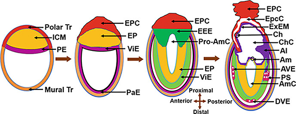
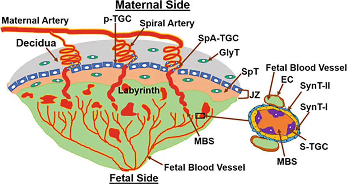
Placenta forms as a momentary organ inside the uterus with a slew of activities only when the woman is pregnant. It is a discoid-shaped hybrid structure consisting of maternal and embryonic components. It develops in the mesometrial side of the uterus following blastocyst implantation to keep the two genetically different entities, the mother and embryo, separated but connected. The beginning and progression of placental formation and development following blastocyst implantation coincides with the chronological developmental stages of the embryo. It gradually acquires the ability to perform the vascular, respiratory, hepatic, renal, endocrine, gastrointestinal, immune, and physical barrier functions synchronously that are vital for fetal development, growth, and safety inside the maternal environment. The uterus ejects the placenta when its embryonic growth and survival supportive roles are finished; that is usually the birth of the baby. Despite its irreplaceable role in fetal development and survival over the post-implantation progression of pregnancy, it still remains unclear how it forms, matures, performs all of its activities, and starts to fail functioning. Thus, a detailed understanding about normal developmental, structural, and functional aspects of the placenta may lead to avoid pregnancy problems that arise with the placenta.

Keywords: Blastocyst; Decidua; Implantation; Labyrinth; Placenta.


Figures

Fig. 1
Fig. 1
Preimplantation stages of the mouse embryo. Blastocoel (BC), Epiblast (EP), Inner cell mass (ICM), Zona pellucida (ZP), Primitive endoderm (PE), Trophectoderm (Tr)
Fig. 2
Fig. 2
The emergence of cell lineages from the blastocyst. Inner cell mass (ICM) and trophectoderm (Tr)
Fig. 3
Fig. 3
Upper panel: Photographs of the preimplantation day 4 receptive uterus and post-implantation uteri with implantation sites from days 5 to 8 of pregnancy. Blastocyst implantation sites (IS) on days 5 and 6 were visualized by intravenous injection of Chicago Blue dye. Lower panel: Photomicrographs (20×) of Hematoxylin- and Eosin-stained sections of the day 4 preimplantation uterus (longitudinal section) and implantation sites (cross sections) of days 5–8 of pregnancy. Antimesometrial (AM) side, Blastocyst (Bl), Circular muscle (CM), Embryo (Em), Longitudinal muscle (LM), Luminal epithelium (LE), Mesometrial side (M); Primary decidual zone (PDZ), Secondary decidual zone (SDZ)
Fig. 4
Fig. 4
Schematic drawings of uterine and embryonic changes during early pregnancy (days 4–6 of pregnancy). Antimesometrial (AM) side, Embryo (Em), Inner cell mass (ICM), Luminal epithelium (LE), Mesometrial (M) side, Primary decidual zone (PDZ), Secondary decidual zone (SDZ), Trophectoderm (Tr)
Fig. 5
Fig. 5
Schematic drawing of the synchronous development of the placenta and the embryo following blastocyst implantation. Allantois (Al), Amnion (Am), Amniotic cavity (AmC), Anterior visceral endoderm (AVE), Chorion (Ch), Chorionic cavity (ChC), Distal visceral endoderm (DVE), Ectoplacental cone (EPC), Ectoplacental cavity (EpcC), Extra-embryonic ectoderm (EEE), Epiblast (EP), Extra-embryonic mesoderm (ExEM), Inner cell mass (ICM), Parietal endoderm (PaE), Primitive endoderm (PE), Primitive streak (PS), Pro-amniotic cavity (Pro-AmC), Visceral endoderm (ViE), and Trophectoderm (Tr)
Fig. 6
Fig. 6
Photomicrographs of hematoxylin- and eosin-stained histological sections of placentae from days 9–19 of pregnancy. Decidua (DC), Junctional zone (JZ), Labyrinth (LB)
Fig. 7
Fig. 7
A schema of a mouse matured placenta. Endothelial cell (EC), Glycogen trophoblast (GlyT), Junctional zone (JZ), Maternal blood sinusoid (MBS), Parietal TGC (p-TGC), Sinusoidal TGC (S-TGC), Spiral artery-associated TGC (SpA-TGC), Spongiotrophoblast (SpT), Syncytotrophoblast I (SynT-I), Syncytotrophoblast II (SynT-II)

References

    1. Abe KI, Funaya S, Tsukioka D, Kawamura M, Suzuki Y, Suzuki MG, Schultz RM, Aoki F (2018) Minor zygotic gene activation is essential for mouse preimplantation development. Proc Natl Acad Sci U S A 115(29):E6780–E6788. Available from: PM:29967139 - PMC - PubMed
    1. Arora R, Papaioannou VE (2012) The murine allantois: a model system for the study of blood vessel formation. Blood 120(13):2562–2572. Available from: PM:22855605 - PMC - PubMed
    1. Azevedo PN, Pelajo-Machado M (2018) Mechanism of hematopoiesis and vasculogenesis in mouse placenta. Placenta 69:140–145. Available from: PM:29680159 - PubMed
    1. Bauer ST, Bonanno C (2009) Abnormal placentation. Semin Perinatol 33(2):88–96. Available from: PM:19324237 - PubMed
    1. Bevilacqua EM, Abrahamsohn PA (1988) Ultrastructure of trophoblast giant cell transformation during the invasive stage of implantation of the mouse embryo. J Morphol 198(3):341–351. Available from: PM:3221406 - PubMed

LinkOut - more resources