. 2021 May;15(3):277-290.
doi: 10.1049/nbt2.12020. Epub 2021 Feb 7.

Polycaprolactone/gelatin electrospun nanofibres containing biologically produced tellurium nanoparticles as a potential wound dressing scaffold: Physicochemical, mechanical, and biological characterisation

Affiliations

Polycaprolactone/gelatin electrospun nanofibres containing biologically produced tellurium nanoparticles as a potential wound dressing scaffold: Physicochemical, mechanical, and biological characterisation

Mohsen Doostmohammadi et al. IET Nanobiotechnol. 2021 May.

Abstract

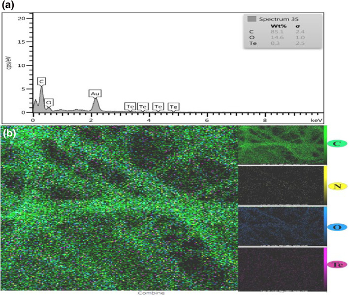
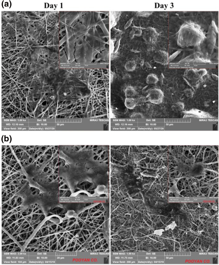
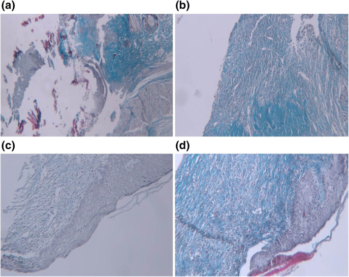
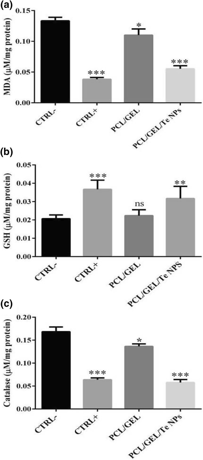
The biologically synthesised tellurium nanoparticles (Te NPs) were applied in the fabrication of Te NP-embedded polycaprolactone/gelatin (PCL/GEL) electrospun nanofibres and their antioxidant and in vivo wound healing properties were determined. The as-synthesised nanofibres were characterised using scanning electron microscopy (SEM), energy-dispersive X-ray (EDX) spectroscopy and elemental mapping, thermogravimetric analysis (TGA), and Fourier-transform infrared (FTIR) spectroscopy. The mechanical properties and surface hydrophobicity of scaffolds were investigated using tensile analysis and contact angle tests, respectively. The biocompatibility of the produced scaffolds on mouse embryonic fibroblast cells (3T3) was evaluated using MTT assay. The highest wound healing activity (score 15/19) was achieved for scaffolds containing Te NPs. The wounds treated with PCL/GEL/Te NPs had inflammation state equal to the positive control. Also, the mentioned scaffold represented positive effects on collagen formation and collagen fibre's horizontalisation in a dose-dependent manner. The antioxidative potency of Te NP-containing scaffolds was demonstrated with lower levels of malondialdehyde (MDA) and catalase (∼3 times) and a higher level of glutathione (GSH) (∼2 times) in PCL/GEL/Te NP-treated samples than the negative control. The obtained results strongly demonstrated the healing activity of the produced nanofibres, and it can be inferred that scaffolds containing Te NPs are suitable for wound dressing.


Conflict of interest statement

The authors declare no conflict of interest.

Figures

FIGURE 1
FIGURE 1
Scanning electron microscopy (SEM) micrographs and fibre size distribution of (a) polycaprolactone/gelatin (PCL/GEL) and (b) PCL/GEL/tellurium nanoparticles (Te NPs)
FIGURE 2
FIGURE 2
(a) Energy‐dispersive X‐ray (EDX) analysis of polycaprolactone (PCL)/gelatin (GEL)/tellurium nanoparticles (Te NPs) and (b) FESEM–EDX elemental mapping of Te NPs encapsulated with PCL/GEL electrospun scaffold
FIGURE 3
FIGURE 3
a) Fourier‐transform infrared (FTIR) spectra of polycaprolactone (PCL), gelatin (GEL), tellurium nanoparticles (Te NPs) nanofibres and (b) Thermo‐gravimetric analysis of PCL/GEL, Te NPs, and PCL/GEL/Te NPs nanofibres
FIGURE 4
FIGURE 4
Tensile stress‐strain curves of polycaprolactone (PCL), PCL/gelatin (GEL), and PCL/GEL/tellurium nanoparticle (Te NP) nanofibres
FIGURE 5
FIGURE 5
Water contact angle of polycaprolactone (PCL) (a1), PCL/gelatin (GEL) (a2), and PCL/GEL/tellurium nanoparticle (Te NP) nanofibres (a3). (b) In vitro degradation of different nanofibres during 4 weeks
FIGURE 6
FIGURE 6
Antibacterial susceptibility disk diffusion test. polycaprolactone (PCL)/gelatin (GEL)/tellurium nanoparticles (Te NPs) against (a) Escherichia coli, (b) Pseudomonas aeruginosa, (c) Bacillus subtilis and PCL/GEL against (d) Eschercia coli, (e) Pseudomonas aeruginosa, and (f) Bacillus subtilis
FIGURE 7
FIGURE 7
Cell viability indicated by MTT assay of 3T3 cells seeded on polycaprolactone (PCL)/gelatin (GEL), PCL/GEL/tellurium nanoparticles (Te NPs), and tissue culture plate after 24, 48, and 72 h
FIGURE 8
FIGURE 8
Scanning electron microscopy (SEM) micrograph of 3T3 cells seeded on (a) polycaprolactone (PCL)/gelatin (GEL) and (b) PCL/GEL/tellurium nanoparticle (Te NP) scaffolds after 24  and 72 h
FIGURE 9
FIGURE 9
Microscopic images of haematoxylin and eosin (H&E) stained wound sections. (a) Negative control (×40): ulcer bed with extensive granulation tissue formation, (b) positive control (×40): ulcer bed with extensive granulation tissue formation and covered by fibrinous exudate (c) polycaprolactone (PCL)/gelatin (GEL) fibres (×40): ulcer bed with extensive granulation tissue formation and covered by fibrinous exudate, and (d) PCL/GEL/tellurium nanoparticle (Te NP) fibres (×40): extensive granulation tissue formation and covered by fibrinous exudate
FIGURE 10
FIGURE 10
Microscopic images of trichrom stained wound sections. (a) Negative control (×40): no mature collagen fibres in ulcer bed (blackish arrow), (b) positive control (×40): minimal horizontal mature collagen fibres, (c) polycaprolactone (PCL)/gelatin (GEL) fibres (×40): moderate amount of early and mature collagen fibres with mixed (mainly horizontal and mild vertical), and (d) PCL/GEL/tellurium nanoparticle (Te NP) fibres (×40): mild amount of early and moderate mature collagen fibres that showing mixed pattern of orientation mainly horizontal
FIGURE 11
FIGURE 11
Oxidative stress assays of skin samples including (a) malondialdehyde (MDA), (b) glutathione (GSH), and (c) catalase. ns denotes not significant as compared to control. *indicates significantly different values as compared to the negative control (p‐value <0.05). ***indicates significantly different values as compared to the negative control (p‐value <0.001)

Similar articles

Cited by

References

    1. Xie, Z. , et al.: Dual growth factor releasing multi‐functional nanofibers for wound healing. Acta Biomater. 9(12), 9351–9359 (2013) - PMC - PubMed
    1. Aduba, D.C. , Yang, H. : Polysaccharide fabrication platforms and biocompatibility assessment as candidate wound dressing materials. Bioengineering (Basel). 4(1), 1 (2017) - PMC - PubMed
    1. Ahmed, R. , et al.: Novel electrospun chitosan/polyvinyl alcohol/zinc oxide nanofibrous mats with antibacterial and antioxidant properties for diabetic wound healing. Int. J. Biol. Macromol. 120, 385–393 (2018) - PubMed
    1. Augustine, R. , et al.: Electrospun polycaprolactone membranes incorporated with Zno nanoparticles as skin substitutes with enhanced fibroblast proliferation and wound healing. Rsc. Adv. 4(47), 24777–24785 (2014)
    1. Akturk, O. , et al.: Wet electrospun silk fibroin/gold nanoparticle 3d matrices for wound healing applications. Rsc. Adv. 6(16), 13234–13250 (2016)

LinkOut - more resources