

Treatment of tumour tissue with radio-frequency hyperthermia (using antibody-carrying nanoparticles)
- PMID: 34694718
- PMCID: PMC8675787
- DOI: 10.1049/nbt2.12061
Treatment of tumour tissue with radio-frequency hyperthermia (using antibody-carrying nanoparticles)
Abstract
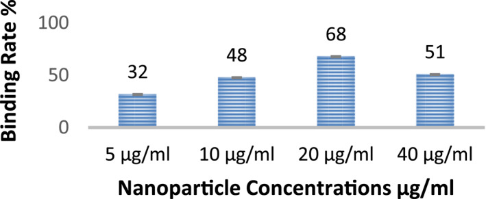
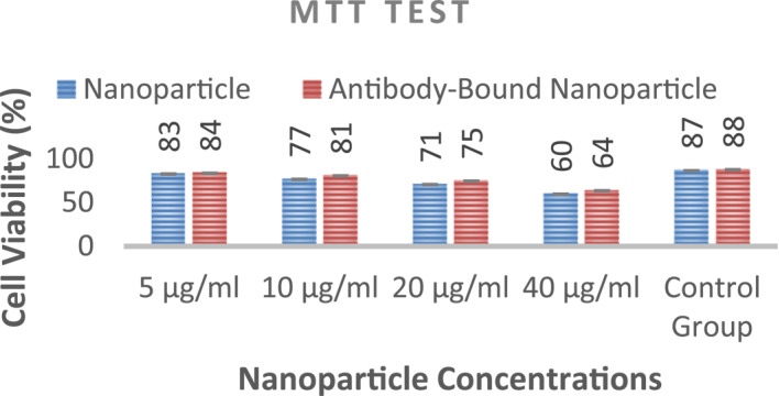
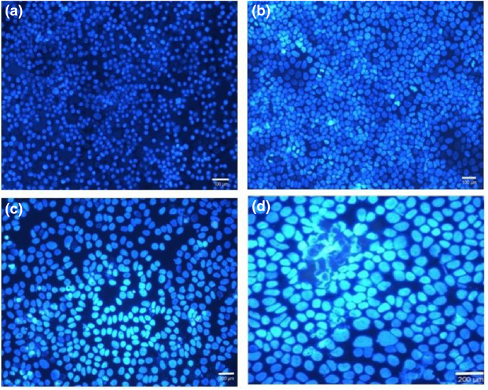
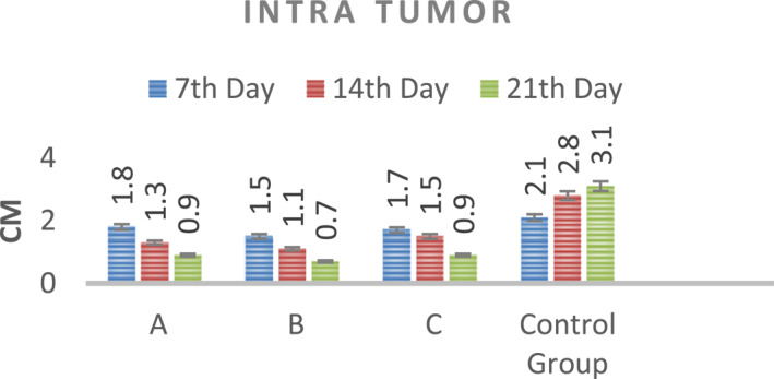
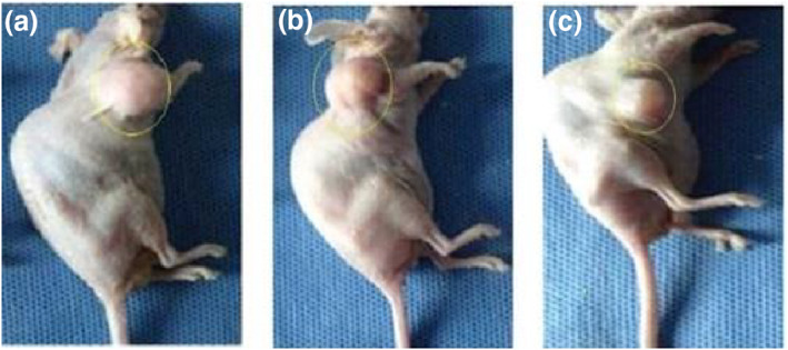
Intelligent inorganic nanoparticles were designed and produced for use in imaging and annihilating tumour cells by radio-frequency (RF) hyperthermia. Nanoparticles synthesised to provide RF hyperthermia must have magnetite properties. For this purpose, magnetite nanoparticles were first synthesised by the coprecipitation method (10-15 NM). These superparamagnetic nanoparticles were then covered with gold ions without losing their magnetic properties. In this step, gold ions are reduced around the magnetite nanoparticles. Surface modification of the gold-coated magnetic nanoparticles was performed in the next step. A self-assembled monolayer was created using cysteamine (2-aminoethanethiol) molecules, which have two different end groups (SH and NH2 ). These molecules react with the gold surface by SH groups. The NH2 groups give a positive charge to the nanoparticles. After that, a monoclonal antibody (Monoclonal Anti-N-CAM Clone NCAM-OB11) was immobilised by the 1-ethyl-3-(3-dimethylaminopropyl)carbodiimide/N-hydroxysuccinimide method. Then, the antenna RF system (144.00015 MHz) was created for RF hyperthermia. The antibody-nanoparticle binding rate and cytotoxicity tests were followed by in vitro and in vivo experiments. As the main result, antibody-bound gold-coated magnetic nanoparticles were successfully connected to tumour cells. After RF hyperthermia, the tumour size decreased owing to apoptosis and necrosis of tumour cells.
© 2021 The Authors. IET Nanobiotechnology published by John Wiley & Sons Ltd on behalf of The Institution of Engineering and Technology.
Figures


Similar articles
-
Nanoparticle-mediated radiofrequency capacitive hyperthermia: A phantom study with magnetic resonance thermometry.Int J Hyperthermia. 2015;31(8):831-9. doi: 10.3109/02656736.2015.1096968. Epub 2015 Nov 10. Int J Hyperthermia. 2015. PMID: 26555005
-
Radio-thermo-sensitivity Induced by Gold Magnetic Nanoparticles in the Monolayer Culture of Human Prostate Carcinoma Cell Line DU145.Anticancer Agents Med Chem. 2020;20(3):315-324. doi: 10.2174/1871520620666191216113052. Anticancer Agents Med Chem. 2020. PMID: 31840615
-
Magnetic Hyperthermia Nanoarchitectonics via Iron Oxide Nanoparticles Stabilised by Oleic Acid: Anti-Tumour Efficiency and Safety Evaluation in Animals with Transplanted Carcinoma.Int J Mol Sci. 2022 Apr 11;23(8):4234. doi: 10.3390/ijms23084234. Int J Mol Sci. 2022. PMID: 35457052 Free PMC article.
-
A review on hyperthermia via nanoparticle-mediated therapy.Bull Cancer. 2017 May;104(5):452-461. doi: 10.1016/j.bulcan.2017.02.003. Epub 2017 Apr 3. Bull Cancer. 2017. PMID: 28385267 Review.
-
Cancer hyperthermia using magnetic nanoparticles.Biotechnol J. 2011 Nov;6(11):1342-7. doi: 10.1002/biot.201100045. Epub 2011 Aug 26. Biotechnol J. 2011. PMID: 22069094 Review.
References
-
- Greenlee, R.T. , et al.: Cancer statistics, 2001, CA A Cancer J Clin, 51, pp. 15–36. (2001) - PubMed
MeSH terms
Substances
LinkOut - more resources
Full Text Sources
Medical
Research Materials
Miscellaneous
