. 2021 Nov 23;15(11):18374-18384.
doi: 10.1021/acsnano.1c07856. Epub 2021 Oct 25.

Immunotargeting of Nanocrystals by SpyCatcher Conjugation of Engineered Antibodies

Affiliations

Immunotargeting of Nanocrystals by SpyCatcher Conjugation of Engineered Antibodies

Cassio C S Pedroso et al. ACS Nano. .

Abstract

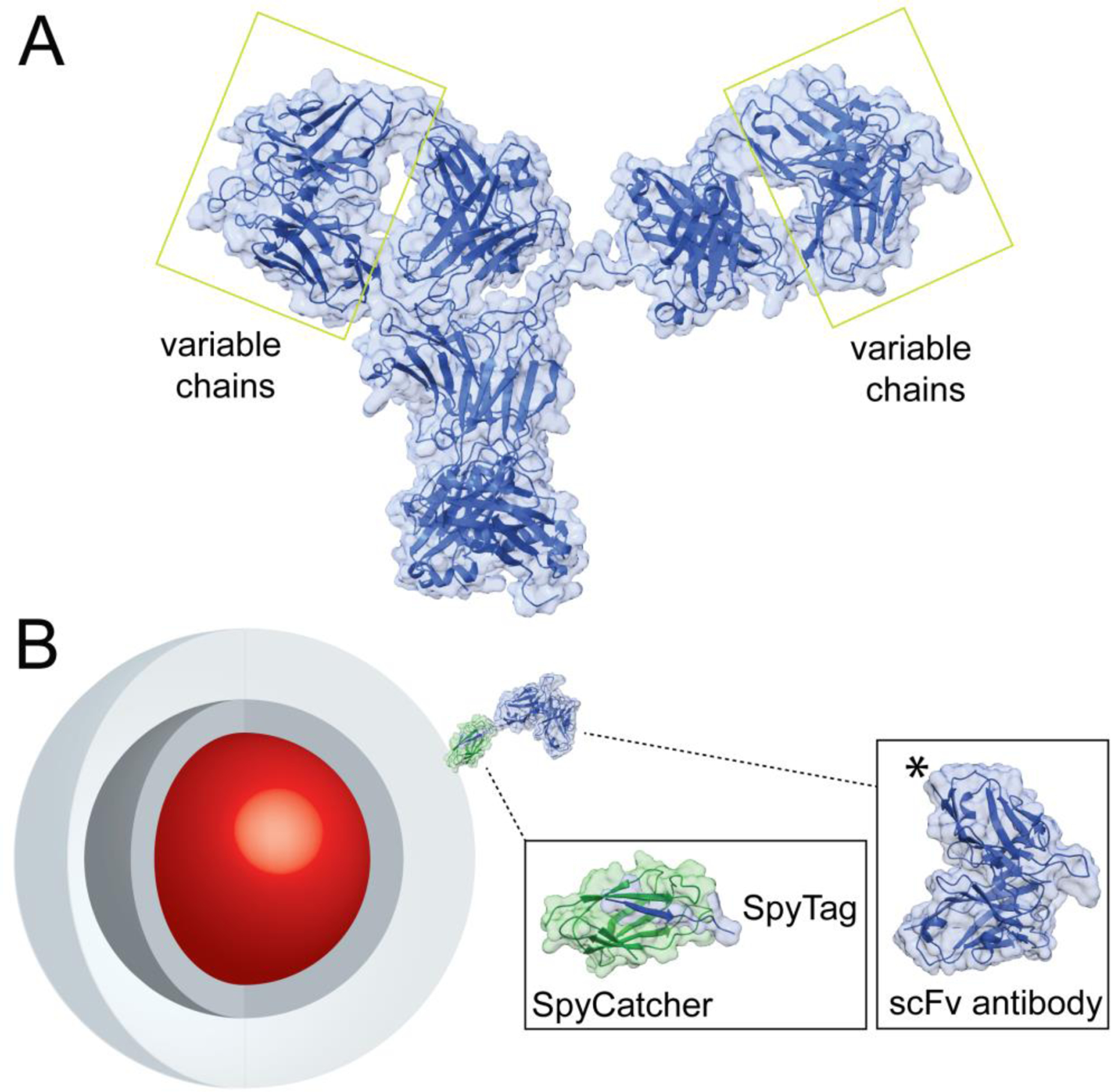
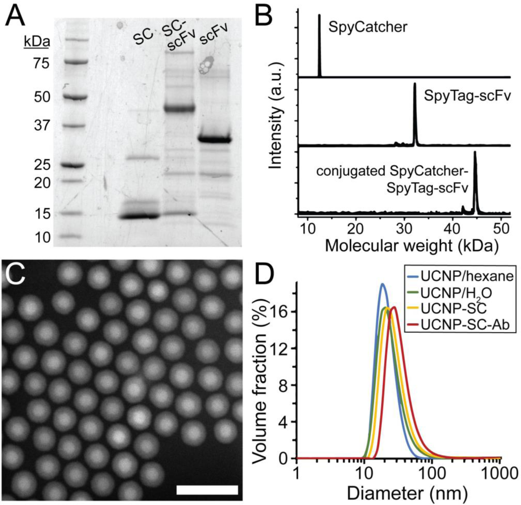
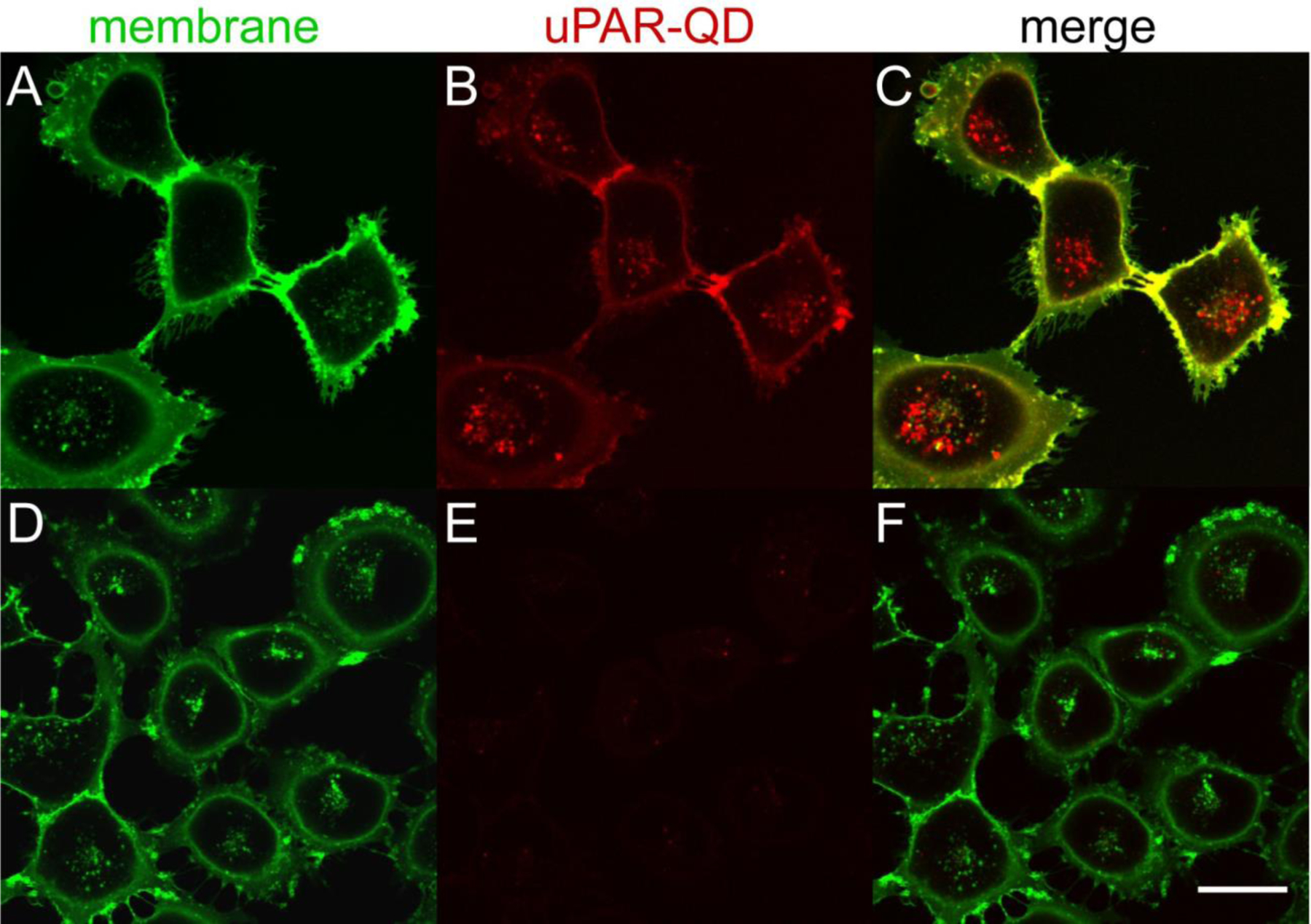
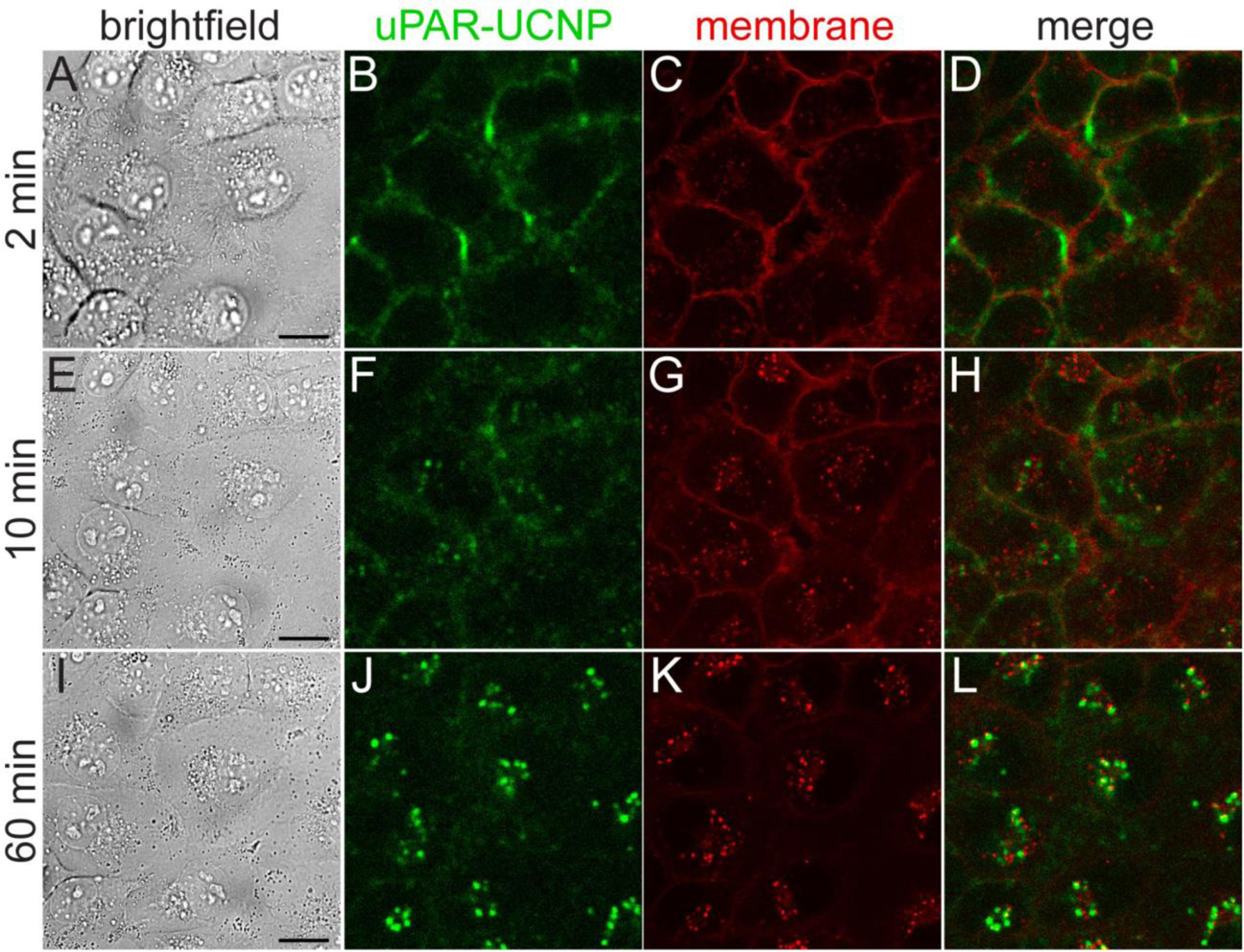
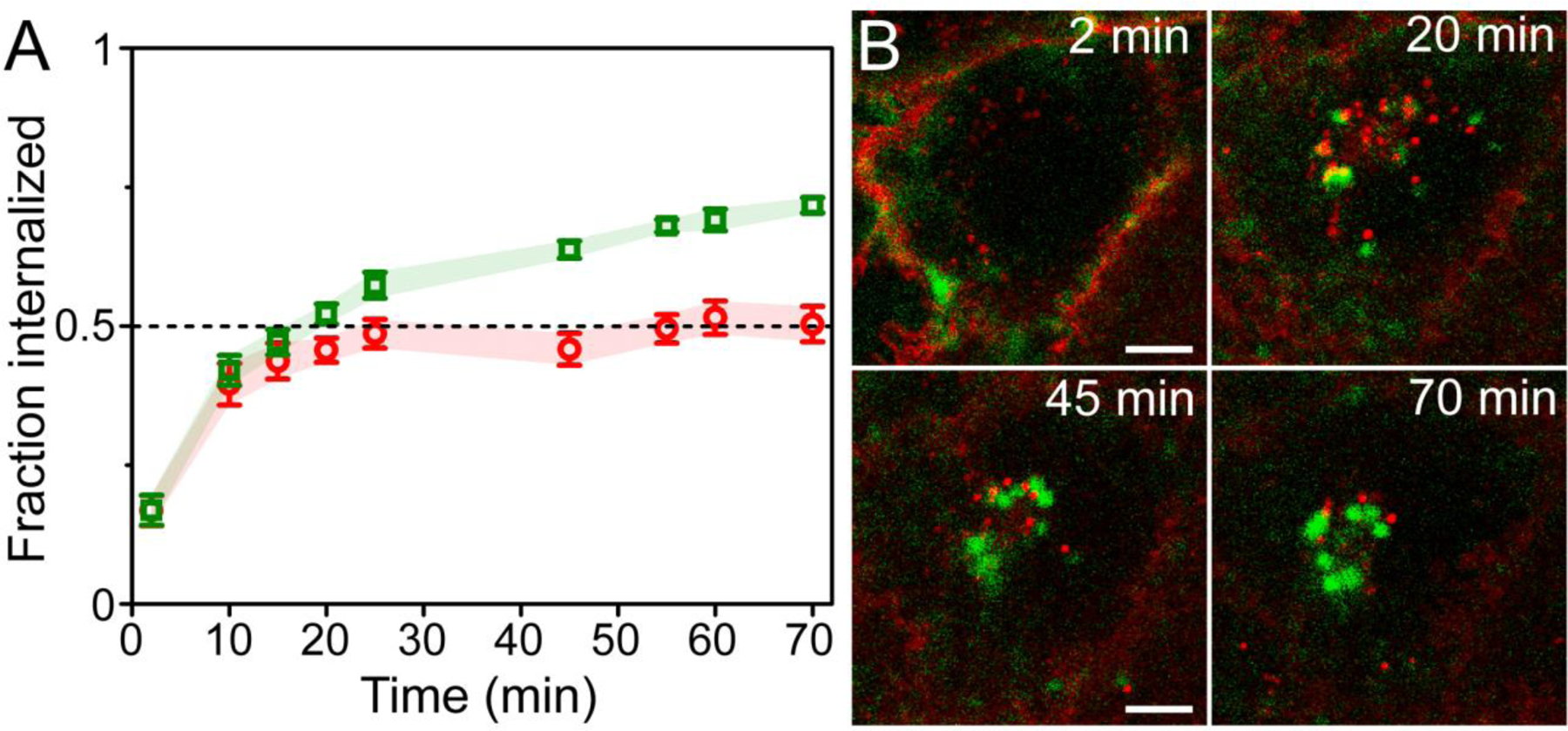
Inorganic nanocrystals such as quantum dots (QDs) and upconverting nanoparticles (UCNPs) are uniquely suited for quantitative live-cell imaging and are typically functionalized with ligands to study specific receptors or cellular targets. Antibodies (Ab) are among the most useful targeting reagents owing to their high affinities and specificities, but common nanocrystal labeling methods may orient Ab incorrectly, be reversible or denaturing, or lead to Ab-NP complexes too large for some applications. Here, we show that SpyCatcher proteins, which bind and spontaneously form covalent isopeptide bonds with cognate SpyTag peptides, can conjugate engineered Ab to nanoparticle surfaces with control over stability, orientation, and stoichiometry. Compact SpyCatcher-functionalized QDs and UCNPs may be labeled with short-chain variable fragment Ab (scFv) engineered to bind urokinase-type plasminogen activator receptors (uPAR) that are overexpressed in many human cancers. Confocal imaging of anti-uPAR scFv-QD conjugates shows the antibody mediates specific binding and internalization by breast cancer cells expressing uPAR. Time-lapse imaging of photostable scFv-UCNP conjugates shows that Ab binding causes uPAR internalization with a ∼20 min half-life on the cell surface, and uPAR is internalized to endolysosomal compartments distinct from general membrane stains and without significant recycling to the cell surface. The controlled and stable conjugation of engineered Ab to NPs enables targeting of diverse receptors for live-cell study of their distribution, trafficking, and physiology.

Keywords: antibodies; bioconjugation; cancer cells; quantum dots; receptor trafficking; uPAR; upconverting nanoparticles.


Conflict of interest statement

Competing Financial Interests

The authors declare no competing financial interests.

Figures

Figure 1.
Figure 1.. Design of nanoparticle-SpyCatcher-scFv antibody conjugates.
A) Structure of full IgG Ab (PDB file 1HZH). Variable chains that bind antigens and are used to engineer miniaturized scFv Ab are highlighted. B) Schematic of NP-SpyCatcher-scFv Ab conjugates. Core/shell nanocrystals (red and dark gray), encapsulated in amphiphilic polymers (light gray), conjugated to a single-Cys SpyCatcher (PDB file 4MLI) and SpyTag-scFv (PDB file 5YD5) chimera. The SpyTag peptide is added at the scFv C terminus, marked (*) in the structure.
Figure 2.
Figure 2.. Synthesis of scFv Ab-conjugated aUCNPs.
(A) Expression and reaction of SpyCatcher (SC) and SpyTag-scFv Ab (ST-scFv), monitored by SDS-PAGE. Partially purified His-tagged scFv were incubated with an excess of SC. (B) MALDI mass spectra of purified SC and ST-scFv before and after conjugation. (C) High-angle annular dark field transmission electron micrograph of core/shell NaEr0.8Yb0.2F4 @ NaY0.8Gd0.2F4 aUCNPs. Scale bar is 50 nm. (D) Diameters of nanoparticles determined by dynamic light scattering (DLS), of hydrophobic (blue), PMAO-encapsulated (teal), SpyCatcher-functionalized (yellow), and scFv-conjugated (red) UCNPs. Mean diameters (n = 5) are 19, 21, 24, and 28 nm, respectively.
Figure 3.
Figure 3.. Specificity of anti-uPAR-QD internalization by uPAR-expressing breast cancer cells.
Confocal images of live MDA-MB-231 cells incubated with (A-C) scFv-QD conjugates (610 nm emission peak) for 60 min at 37 °C or (D-F) SpyCatcher-QD conjugates without scFv Ab. (A,D) AlexaFluor 488-WGA fluorescence of the plasma membrane and endolysosomal pathway; (B,E) QD emission with or without surface anti-uPAR scFv. (C,F) Overlay of WGA and anti-uPAR-QD emission. QDs were excited at 405 nm and emission collected from 605 – 690 nm. AlexaFluor488 was excited at 488 nm and emission collected from 495 – 575 nm. Scale bar is 30 μm.
Figure 4.
Figure 4.. Kinetics of anti-uPAR-UCNP internalization in breast cancer cells.
Live MDA-MB-231 cells treated with 2G10 scFv-UCNP immunoconjugates at 37 °C, imaged by: (A,E,I) brightfield; (B,F,J) upconverted confocal microscopy for anti-uPAR-UCNP imaging; and (C,G,K) standard confocal microscopy for lipophilic fluorophore Cellmask Orange. UCNPs were excited at 980 nm and emission collected from 380 – 750 nm. Cellmask Orange was excited at 561 nm and emission collected from 495 – 575 nm. (D,H,L) Overlay of anti-uPAR-UCNP and membrane emission. Frames 2 min (A-D), 10 min (E-H), and 60 min (I-L) of full 70-min time lapse (Supplementary movie 1) are shown here. Scale bars are 20 μm.
Figure 5.
Figure 5.
Differences of internalization and recycling between anti-uPAR scFv-UCNPs and a lipophilic membrane stain in breast cancer cells. (A) Fraction internalized of 2G10 scFv-UCNP immunoconjugates in live MDA-MB-231 cells during incubation at 37°C (mean ± SEM; n = 6 cells). (B) Confocal images of anti-uPAR-UCNPs (green squares) and lipophilic membrane fluorophore (red circles) in live MDA-MB-231 cells showing differences in intracellular localization between probes. Details from frames at 2, 20, 45 and 70 min of full 70-min time-lapse (Supplementary movie 1) are shown. Scale bars are 10 μm.

References

    1. Freise AC; Wu AM In Vivo Imaging with Antibodies and Engineered Fragments. Mol. Immunol 2015, 67 (2 Pt A), 142–152. 10.1016/j.molimm.2015.04.001. - DOI - PMC - PubMed
    1. Specht EA; Braselmann E; Palmer AE A Critical and Comparative Review of Fluorescent Tools for Live-Cell Imaging. Annu. Rev. Physiol 2017, 79 (1), 93–117. 10.1146/annurev-physiol-022516-034055. - DOI - PubMed
    1. Basu K; Green EM; Cheng Y; Craik CS Why Recombinant Antibodies - Benefits and Applications. Curr. Opin. Biotechnol 2019, 60, 153–158. 10.1016/j.copbio.2019.01.012. - DOI - PMC - PubMed
    1. Wu AM; Senter PD Arming Antibodies: Prospects and Challenges for Immunoconjugates. Nat. Biotechnol 2005, 23 (9), 1137–1146. 10.1038/nbt1141. - DOI - PubMed
    1. Rathore AS; Sarker A; Gupta RD Recent Developments toward Antibody Engineering and Affinity Maturation. Protein Pept. Lett 2018, 25 (10), 886–896. 10.2174/0929866525666180925142757. - DOI - PubMed

Publication types

MeSH terms

Substances