. 2021 Oct 25;17(10):e1009726.
doi: 10.1371/journal.ppat.1009726. eCollection 2021 Oct.

S-farnesylation is essential for antiviral activity of the long ZAP isoform against RNA viruses with diverse replication strategies

Affiliations

S-farnesylation is essential for antiviral activity of the long ZAP isoform against RNA viruses with diverse replication strategies

Dorota Kmiec et al. PLoS Pathog. .

Abstract

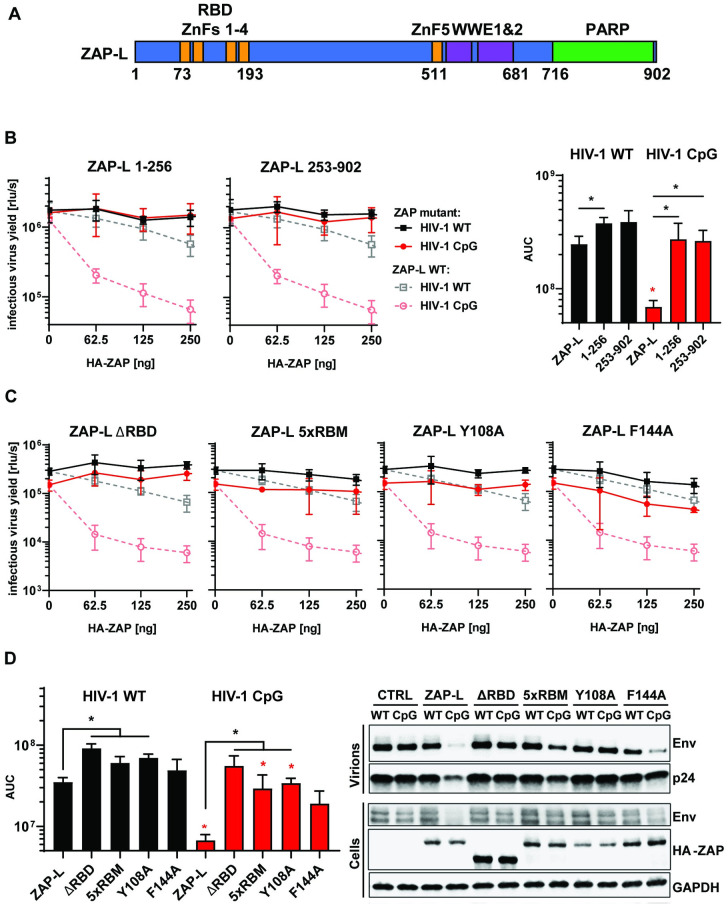
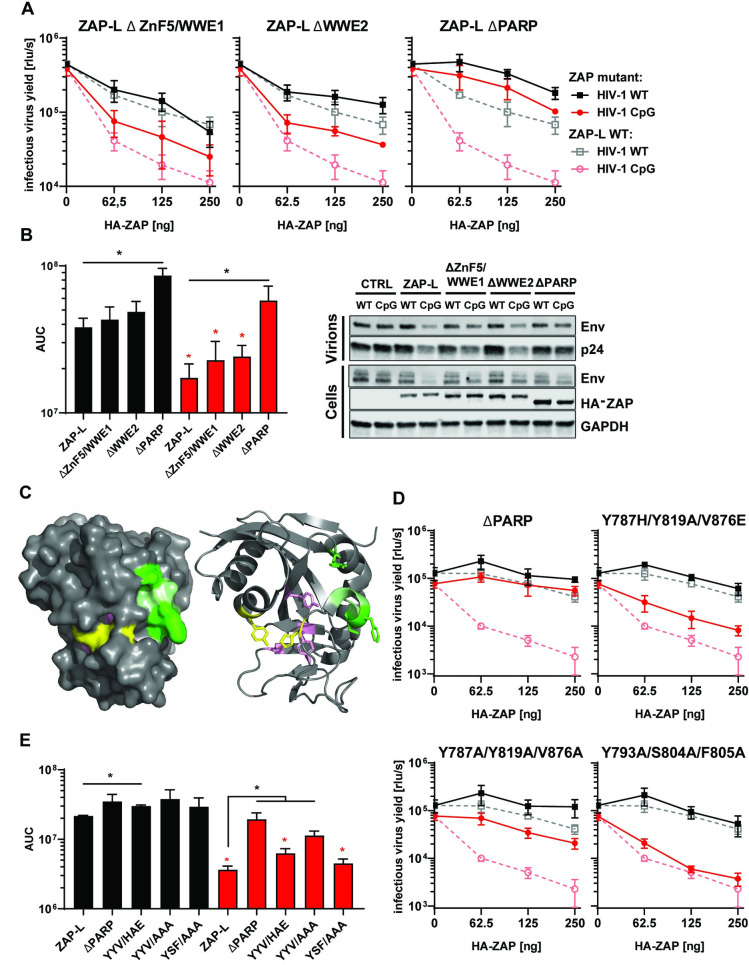
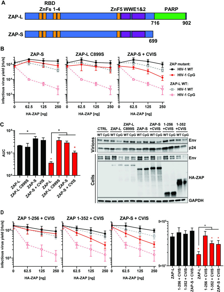
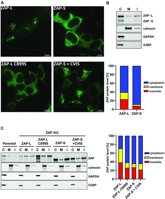
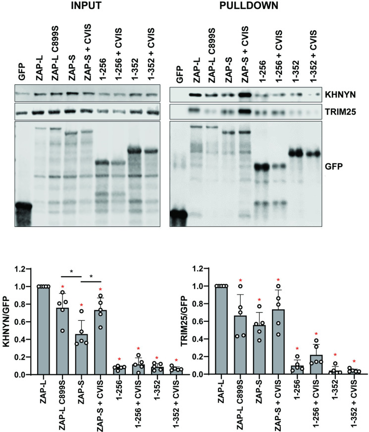
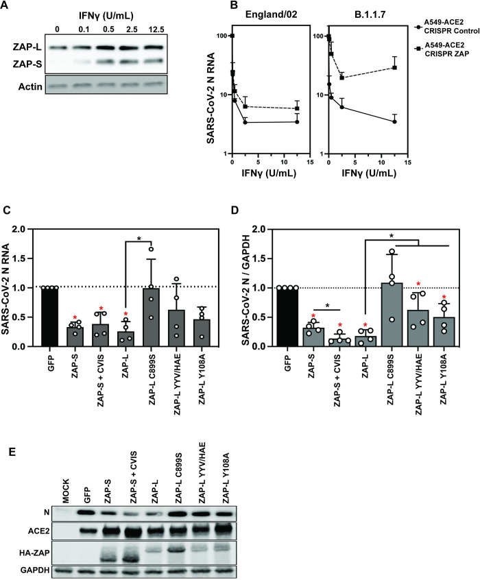
The zinc finger antiviral protein (ZAP) is a broad inhibitor of virus replication. Its best-characterized function is to bind CpG dinucleotides present in viral RNAs and, through the recruitment of TRIM25, KHNYN and other cofactors, target them for degradation or prevent their translation. The long and short isoforms of ZAP (ZAP-L and ZAP-S) have different intracellular localization and it is unclear how this regulates their antiviral activity against viruses with different sites of replication. Using ZAP-sensitive and ZAP-insensitive human immunodeficiency virus type I (HIV-1), which transcribe the viral RNA in the nucleus and assemble virions at the plasma membrane, we show that the catalytically inactive poly-ADP-ribose polymerase (PARP) domain in ZAP-L is essential for CpG-specific viral restriction. Mutation of a crucial cysteine in the C-terminal CaaX box that mediates S-farnesylation and, to a lesser extent, the residues in place of the catalytic site triad within the PARP domain, disrupted the activity of ZAP-L. Addition of the CaaX box to ZAP-S partly restored antiviral activity, explaining why ZAP-S lacks antiviral activity for CpG-enriched HIV-1 despite conservation of the RNA-binding domain. Confocal microscopy confirmed the CaaX motif mediated localization of ZAP-L to vesicular structures and enhanced physical association with intracellular membranes. Importantly, the PARP domain and CaaX box together jointly modulate the interaction between ZAP-L and its cofactors TRIM25 and KHNYN, implying that its proper subcellular localisation is required to establish an antiviral complex. The essential contribution of the PARP domain and CaaX box to ZAP-L antiviral activity was further confirmed by inhibition of severe acute respiratory syndrome coronavirus 2 (SARS-CoV-2) replication, which replicates in double-membrane vesicles derived from the endoplasmic reticulum. Thus, compartmentalization of ZAP-L on intracellular membranes provides an essential effector function in ZAP-L-mediated antiviral activity against divergent viruses with different subcellular replication sites.


Conflict of interest statement

The authors have declared that no competing interests exist.

Figures

Fig 1
Fig 1. RNA binding is crucial for ZAP’s antiviral activity.
(A) Schematic showing domain organisation of long isoform of ZAP (ZAP-L): four N-terminal zinc fingers form RNA binding domain (RBD), fifth zinc finger (ZnF5) and two WWE domains are located in the central part and catalytically inactive Poly(ADP-ribose) polymerase (PARP) domain is at the C-terminal part. (B) Infectious virus yield measured by TZM-bl infectivity assay in relative light units per second [rlu/s] obtained from HEK293T ZAP KO cells co-transfected with wild type (WT; black) HIV-1 and CpG-enriched mutant (CpG-high; red) viruses and increasing doses of pcDNA HA-ZAP constructs encoding the full-length ZAP-L (dashed line), 1-256aa or 253-902aa parts of the protein (solid lines) (left panel). Area Under the Curve (AUC) calculated from the same titration experiments (right panel). (C) Infectious virus yield from HEK293T ZAP KO co-transfected with WT and mutant virus and increasing concentration of pcDNA HA-ZAP with truncated ZAP 253–902 (ΔRBD), ZAP-L mutant unable to bind RNA (V72A/Y108A/F144A/H176A/R189A; 5xRBM) or ZAP-L with substitutions at positions in direct contact with bound RNA CpG (Y108A and F144A) and (D, left panel) derived AUC values. (right panel) representative western blot of produced virions and ZAP transfected (250ng) cells showing viral Env and Gag (p24) proteins as well as HA-tagged ZAP and GAPDH loading control. Mean of n = 3 +/- SD. * p<0.05 for HIV-1 CpG compared to HIV-1 WT for the same ZAP construct. * p < 0.05 for the comparisons demarked by the lines.
Fig 2
Fig 2. Determinants of ZAP’s function located outside RBD.
(A) Infectious virus yield from HEK293T ZAP KO co-transfected with WT (black) and mutant (red) virus and increasing concentration of pcDNA HA-ZAP-L control (dashed line) or mutated pcDNA HA-ZAP with deleted ZnF5 and first WWE domain (Δ511–563; ΔZnF5/WWE1), second WWE (Δ594–681; ΔWWE2) or PARP domain (Δ716–902; ΔPARP). (B) Corresponding AUC values and representative western blot (250ng). (C) Position of studied residues in crystal structure of ZAP’s PARP domain. Residues under positive selection in primates are shown in green, canonical triad positions in pink and residues forming the salt bridge which closes the NAD+ binding grove are shown in yellow. (D) Infectious virus yield from HEK293T ZAP KO co-transfected with WT (black) and mutant (red) virus and increasing concentration of pcDNA HA-ZAP-L control (dashed line), missing PARP domain or carrying amino acid substitutions in alternate triad motif (Y786H/Y818A/V875E, Y786A/Y818/V875A), or residues under positive selection (Y793A/S804A/F805A) (solid lines). (E) Corresponding AUC values. Mean of n = 3 +/- SD. * p<0.05 for HIV-1 CpG compared to HIV-1 WT for the same ZAP construct. * p < 0.05 for the comparisons demarked by the lines.
Fig 3
Fig 3. Contribution of CaaX motif to ZAP’s antiviral activity.
(A) Schematic showing ZAP-L, which contains PARP domain and CaaX motif (amino acids “CVIS”) and short isoform of ZAP (ZAP-S). (B) Infectious virus yield from HEK293T ZAP KO co-transfected with WT (black) and mutant (red) virus and increasing concentration of ZAP-L (dashed line), ZAP-S, ZAP-L with mutated crucial cysteine (C899S) in CaaX or ZAP-S with added CVIS motif (solid lines). (C) Corresponding AUC values and representative western blot (250ng). (D) Infectious virus yield from HEK293T ZAP KO co-transfected with both viruses and pcDNA encoding truncated ZAP (1–256 or 1–352) with added CVIS motif and corresponding AUC values. Mean of n = 3–5 +/- SD. * p<0.05 for HIV-1 CpG compared to HIV-1 WT for the same ZAP construct. * p < 0.05 for the comparisons demarked by the lines.
Fig 4
Fig 4. Cellular distribution of ZAP-S, ZAP-L and their CaaX motif mutants.
(A) Confocal microscopy images of live HEK293T ZAP KO cells 24h after transfection with 250ng of GFP-tagged ZAP isoforms or ZAP-L with inactivated CaaX (C899S) and ZAP-S with added CaaX motif (+CVIS). Size bar 10μm. (B) Representative western blot and quantification of ZAP present in cell fractionation samples of parental HEK293Ts (mean of n = 5) or (C) ZAP KO cells following transfection of 60ng HA-ZAP constructs (mean of n = 3). Cytoplasmic (C), membrane (M) and insoluble (I) fractions are shown. Calnexin serves as a marker for membrane protein and G3BP and GAPDH are cytoplasmic protein controls.
Fig 5
Fig 5. ZAP cofactors KHNYN and TRIM25 bind most efficiently to wild type ZAP-L.
Upper left panel: Representative western blot of GFP-ZAP isoforms and mutant proteins expressed in HEK293T ZAP KO cells. Upper right panel: GFP-ZAP, TRIM25 and KHNYN co-immunoprecipitated using GFP-binding magnetic beads. Input and pulldown samples were stained for GFP as well as endogenous KHNYN and TRIM25. Lower panels: quantification of KHNYN and TRIM25 co-immunoprecipitated with GFP-ZAP normalized to the relative GFP signal. Mean of n = 5 + SD. * p < 0.05 for the sample compared to ZAP-L. * p < 0.05 for the comparisons demarked by the lines.
Fig 6
Fig 6. ZAP-L requires the CaaX box to restrict SARS-CoV-2.
(A) Western blot showing IFNγ-mediated induction of ZAP-L and S in A549-ACE2 cells. (B) IFNγ treatment restricts early-pandemic SARS-CoV-2 strain England02 and B.1.1.7 variant of concern infection less potently in CRISPR ZAP cells than CRISPR Control cells. SARS-CoV-2 RNA in the supernatant was measured by qRT-PCR. Mean of n = 3 + SD. (C) Viral RNA in the supernatant of HEK293T ZAP KO cells transfected with pcDNA encoding human ACE2 and indicated ZAP isoforms/ mutants or GFP control plasmid, 48h after infection with SARS-CoV-2 England 2 strain at 0.01 MOI. Quantification of qRT-PCR detecting viral nucleocapsid (N) RNA in the cell supernatant and (D) SARS-CoV-2 N protein levels in the infected cells, with a representative western blot (E). Mean of n = 4 + SD. * p < 0.05 for the sample compared to GFP. * p < 0.05 for the comparisons demarked by the lines.

References

    1. Kluge SF, Sauter D, Kirchhoff F. SnapShot: antiviral restriction factors. Cell. 2015;163(3):774– e1. doi: 10.1016/j.cell.2015.10.019 - DOI - PubMed
    1. Ryman KD, Meier KC, Nangle EM, Ragsdale SL, Korneeva NL, Rhoads RE, et al. Sindbis virus translation is inhibited by a PKR/RNase L-independent effector induced by alpha/beta interferon priming of dendritic cells. J Virol. 2005;79(3):1487–99. doi: 10.1128/JVI.79.3.1487-1499.2005 - DOI - PMC - PubMed
    1. Gao G, Guo X, Goff SP. Inhibition of retroviral RNA production by ZAP, a CCCH-type zinc finger protein. Science. 2002;297(5587):1703–6. doi: 10.1126/science.1074276 - DOI - PubMed
    1. Kerns JA, Emerman M, Malik HS. Positive selection and increased antiviral activity associated with the PARP-containing isoform of human zinc-finger antiviral protein. PLoS genetics. 2008;4(1):e21. doi: 10.1371/journal.pgen.0040021 - DOI - PMC - PubMed
    1. Nchioua R, Kmiec D, Muller JA, Conzelmann C, Gross R, Swanson CM, et al. SARS-CoV-2 Is Restricted by Zinc Finger Antiviral Protein despite Preadaptation to the Low-CpG Environment in Humans. mBio. 2020;11(5):e01930–20. doi: 10.1128/mBio.01930-20 - DOI - PMC - PubMed

Publication types