Review
. 2021 Sep 22;13(10):1892.
doi: 10.3390/v13101892.

The Not-So-Good, the Bad and the Ugly: HPV E5, E6 and E7 Oncoproteins in the Orchestration of Carcinogenesis

Affiliations
Review

The Not-So-Good, the Bad and the Ugly: HPV E5, E6 and E7 Oncoproteins in the Orchestration of Carcinogenesis

Om Basukala et al. Viruses. .

Abstract

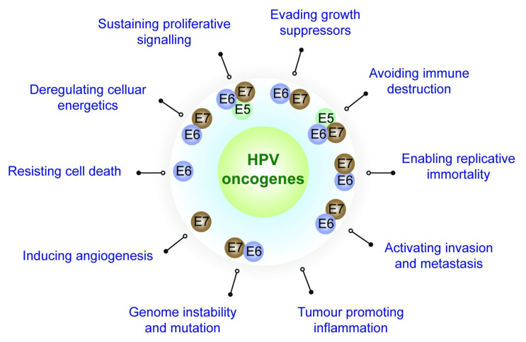
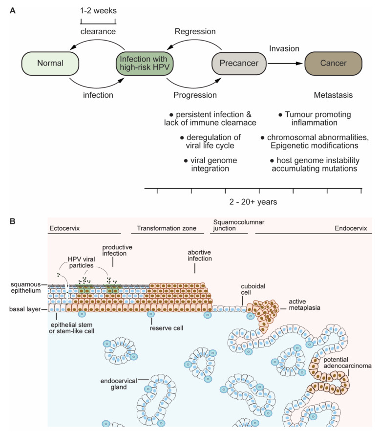
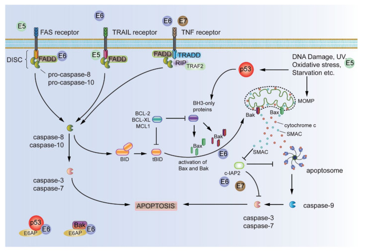
Infection with HPV starts with the access of the viral particles to basal cells in the epidermis, potentially via microtraumas to the skin. The basal cells are able to keep away these pathogens in normal circumstances through a robust immune response from the host, as HPV infections are, in general, cleared within 2 to 3 weeks. However, the rare instances of persistent infection and/or in cases where the host immune system is compromised are major risk factors for the development of lesions potentially leading to malignancy. Evolutionarily, obligatory pathogens such as HPVs would not be expected to risk exposing the host to lethal cancer, as this would entail challenging their own life cycle, but infection with these viruses is highly correlated with cancer and malignancy-as in cancer of the cervix, which is almost always associated with these viruses. Despite this key associative cause and the availability of very effective vaccines against these viruses, therapeutic interventions against HPV-induced cancers are still a challenge, indicating the need for focused translational research. In this review, we will consider the key roles that the viral proteins play in driving the host cells to carcinogenesis, mainly focusing on events orchestrated by early proteins E5, E6 and E7-the not-so-good, the bad and the ugly-and discuss and summarize the major events that lead to these viruses mechanistically corrupting cellular homeostasis, giving rise to cancer and malignancy.

Keywords: E5; E6; E7; HPV; carcinogenesis; viral oncoproteins.


Conflict of interest statement

The authors declare no conflict of interest.

Figures

Figure 1
Figure 1
The hallmarks of cancer adapted from Hanahan and Weinberg 2016 [2]. A schematic illustrating eight distinct functional capabilities and two facilitators that are necessary conditions for the manifestation of malignant disease—cancer—and the expression of the HPV oncoproteins responsible for attaining these functions.
Figure 2
Figure 2
From infection to the development of cancer and malignancy, mediated by HPV (adapted and modified from References [22,23]. (A) Infection with HPV is normally cleared with 1 to 2 weeks; however, in certain individuals, persistent infection and a lack of immune clearance can lead to deregulation of the viral life cycle and viral genome integration, making them major risk factors for tumour development and progression towards cancer and malignancy. The progression towards invasive cancer and metastasis involves several changes, including chromosomal abnormalities, epigenetic modifications, genome instability and accumulating mutations and tumour-promoting inflammation, taking 2 to 20-plus years. (B) A cartoon representing infection by HPVs and progression to cancer. Infection with HPV is thought to occur via microtraumas in the epithelium, allowing access of the virus to the basal cell layer. HPV maintains its genome in the basal cells, and, as these cell divide, there is coordinated expression of early viral proteins, including E6 and E7, that allows the differentiation-determined cells to reinitiate the cell cycle. As these cells reach the upper squamous layers, with a concomitant expression of viral late gene products L1 and L2, new virions are released upon desquamation. Various regions of the cervix composed of stratified epithelium of the ectocervix, the transformation zone and the columnar epithelium of the cervix and endocervix are indicated in the cartoon, suggesting major sites of productive infections leading to the release of viral particles and abortive infection associated with deregulated HPV gene expression, potentially leading to squamous cell carcinoma and adenocarcinoma.
Figure 3
Figure 3
Schematic representation of HPV oncoproteins E5, E6 and E7. HPV16-E5 is an 83-amino acid-long transmembrane protein with three transmembrane domains (TMDI-III). HPV-16 E6 is an 158 amino acid protein with two CXXC (Cys-X-X-Cys,) motifs, which participate in coordinating with zinc ions and are indicated in the schematic. The C-terminus PDZ-binding motif (PBM) sequence ‘ETQV’ is shown, which can be phosphorylated by AKT, Chk2 via PKA and Chk1 kinases. HPV-16 E7 is a cytoplasmic phosphoprotein. The position of the conserved regions (CR1, CR2 and CR3) and CXXC (Cys-X-X-Cys) motifs, which participate in coordinating zinc ions, are also indicated. The LXCXE motif and CKII phosphorylation site in the CR2 region are important for targeting the pRB and related pocket proteins.
Figure 4
Figure 4
Evading growth suppressors. Unlike normal cells, where the growth and proliferation are tightly controlled, cells expressing HPV oncoproteins act to evade the key tumour suppressors pRB and p53. HPV-E7 can associate with pRB and inactivate the repressive function of the pRB/E2F transcription complex, leading to the expression of S-phase genes (cell cycle regulators, including cdc25a, cyclin E and A and replication enzymes and others) in otherwise cell cycle-exited and differentiating keratinocytes, leading to re-entry to the S phase. Further, high-risk E7 can downregulate pRB via proteasomal degradation via the cullin-2 ubiquitin ligase complex. HPV-16 E7 associates with cdk inhibitors p21Cip1 and p27Kip1, abrogates the inhibition of cdk2 activity and enhances the transcriptional activation of cdc25a, leading to the dephosphorylation of inhibitory phosphorylation in cdk-2/cyclin E/A. This leads to activation of the p53 tumour suppressor; however, high-risk E6 can inactivate and degrade p53 via the ubiquitin proteasome pathway involving the E6AP ubiquitin ligase complex. ub—ubiquitin and pp—2-25 residue peptides.
Figure 5
Figure 5
Resisting cell death. One of the major roles played by HPV oncoproteins in resisting cell death is evading apoptosis. Both extrinsic and intrinsic apoptotic pathways are deregulated to attain this function by HPV-E5, -E6 and -E7. The extrinsic apoptotic pathway is activated upon receptor trimerization and the subsequent recruitment of adaptor molecules and procaspase 8 to the DISC. The activation of caspase 8 then leads to the activation of downstream executioner caspases 3 and 7, leading to cell death/apoptosis. The intrinsic apoptotic pathway is activated by external stimuli (UV-radiation, oxidative stress, DNA damage, starvation, etc.), leading to the formation of pores in the mitochondrial membrane and release of mitochondrial inner membrane proteins (cytochrome c, SMAC) into the cytosol. Released cytochrome c and pro-caspase 9 form the apoptosome, leading to activating caspase 9, which, in turn, activates downstream executioner caspases 3 and 7, leading to apoptosis. E5 can downregulate the Fas receptor and perturb the formation of the DISC complex, thus abrogating the extrinsic apoptotic pathway. Further, E5 can perturb ROS-induced Bax activation and inhibit the apoptotic response to UV B radiation. E6 can block extrinsic pathways by binding the death domain, leading to its proteasomal degradation. E6 inactivates p53, Bax and Bak, thus abrogating MOMP and the release of cytochrome c and, thus, inhibiting the intrinsic apoptotic pathway. E6 can also inhibit antiapoptotic c-IAP2, blocking the formation of the apoptosome and activation of the executioner caspases. E7 seems to have a dual function in activating and abrogating apoptosis; however, E7 has been demonstrated to perturb TNF receptor-induced apoptosis by upregulating c-IAP2 and suppressing caspase 8. FADD—Fas-associated protein with death domain, TNF—tumour necrosis factor, TRAIL—FasL and TNF-related apoptosis-inducing ligand, TRAF2—TNF receptor-associated factor 2, DISC—death-induced signalling complex, c-IAP2—cellular inhibitor of apoptosis protein 2, RIP—receptor interacting protein and MOMP—mitochondrial outer membrane permeabilization.

Similar articles

Cited by

References

    1. Hanahan D., Weinberg R.A. Hallmarks of cancer: The next generation. Cell. 2011;144:646–674. doi: 10.1016/j.cell.2011.02.013. - DOI - PubMed
    1. Hanahan D., Weinberg R.A. Holland—Frei Cancer Medicine. Wiley-Blackwell Publishers; Hoboken, NJ, USA: 2016. Biological Hallmarks of Cancer; pp. 1–10.
    1. Plummer M., de Martel C., Vignat J., Ferlay J., Bray F., Franceschi S. Global burden of cancers attributable to infections in 2012: A synthetic analysis. Lancet Glob. Health. 2016;4:e609–e616. doi: 10.1016/S2214-109X(16)30143-7. - DOI - PubMed
    1. De Martel C., Plummer M., Vignat J., Franceschi S. Worldwide burden of cancer attributable to HPV by site, country and HPV type. Int. J. Cancer. 2017;141:664–670. doi: 10.1002/ijc.30716. - DOI - PMC - PubMed
    1. Kombe Kombe A.J., Li B., Zahid A., Mengist H.M., Bounda G.A., Zhou Y., Jin T. Epidemiology and Burden of Human Papillomavirus and Related Diseases, Molecular Pathogenesis, and Vaccine Evaluation. Front. Public Health. 2020;8:552028. doi: 10.3389/fpubh.2020.552028. - DOI - PMC - PubMed

Publication types

Substances