

Identification, Mechanism, and Treatment of Skin Lesions in COVID-19: A Review
- PMID: 34696346
- PMCID: PMC8540564
- DOI: 10.3390/v13101916
Identification, Mechanism, and Treatment of Skin Lesions in COVID-19: A Review
Abstract
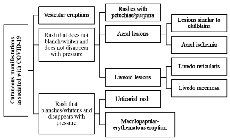
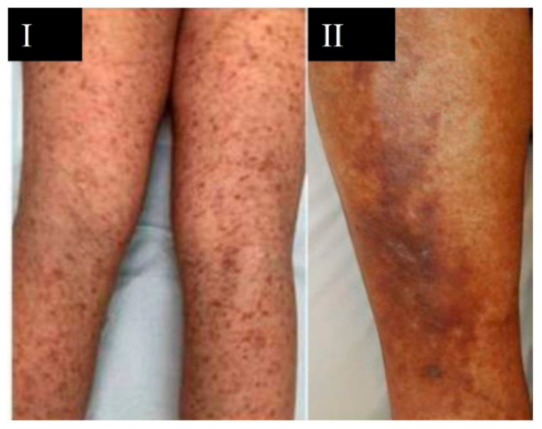
Coronavirus disease 2019 (COVID-19) is a multisystem disease caused by Severe Acute Respiratory Syndrome Coronavirus 2 (SARS-CoV-2), that primarily causes respiratory symptoms. However, an increasing number of cutaneous manifestations associated with this disease have been reported. The aim of this study is to analyze the scientific literature on cutaneous manifestations associated with SARS-CoV-2 by means of a narrative literature review until June 2021. The search was conducted in the following electronic databases: Medline (PubMed), SciELO, and Cochrane Library Plus. The most common cutaneous manifestations in patients with COVID-19 are vesicular eruptions, petechial/purpuric rashes, acral lesions, liveoid lesions, urticarial rash, and maculopapular-erythematous rash. These manifestations may be the first presenting symptoms of SARS-CoV-2 infection, as is the case with acral lesions, vesicular eruptions, and urticaria. In relation to severity, the presence of liveoid lesions may be associated with a more severe course of the disease. Treatment used for dermatological lesions includes therapy with anticoagulants, corticosteroids, and antihistamines. Knowledge of the dermatologic manifestations associated with SARS-CoV-2 contributes to the diagnosis of COVID-19 in patients with skin lesions associated with respiratory symptoms or in asymptomatic patients. In addition, understanding the dermatologic lesions associated with COVID-19 could be useful to establish a personalized care plan.
Keywords: COVID-19; SARS-CoV-2; coronaviruses; cutaneous manifestations; skin lesions.
Conflict of interest statement
The authors declare no conflict of interest.
Figures


Similar articles
-
Severe urticarial rash as the initial symptom of COVID-19 infection.BMJ Case Rep. 2021 Mar 25;14(3):e241793. doi: 10.1136/bcr-2021-241793. BMJ Case Rep. 2021. PMID: 33766974 Free PMC article.
-
Skin Manifestations Associated with COVID-19: Current Knowledge and Future Perspectives.Dermatology. 2021;237(1):1-12. doi: 10.1159/000512932. Epub 2020 Nov 24. Dermatology. 2021. PMID: 33232965 Free PMC article. Review.
-
Pathobiology of Cutaneous Manifestations Associated with COVID-19 and Their Management.Viruses. 2022 Sep 6;14(9):1972. doi: 10.3390/v14091972. Viruses. 2022. PMID: 36146777 Free PMC article. Review.
-
Cutaneous Manifestations in the Context of SARS-CoV-2 Infection (COVID-19).Actas Dermosifiliogr (Engl Ed). 2020 Nov;111(9):734-742. doi: 10.1016/j.ad.2020.08.002. Epub 2020 Aug 31. Actas Dermosifiliogr (Engl Ed). 2020. PMID: 32882184 Free PMC article. Review. English, Spanish.
-
Cutaneous manifestations of SARS-CoV-2 infection: a clinical update.J Eur Acad Dermatol Venereol. 2020 Nov;34(11):2499-2504. doi: 10.1111/jdv.16774. Epub 2020 Jul 20. J Eur Acad Dermatol Venereol. 2020. PMID: 32585074 Free PMC article. Review.
Cited by
-
Comorbidities, Associated Diseases, and Risk Assessment in COVID-19-A Systematic Review.Int J Clin Pract. 2022 Oct 31;2022:1571826. doi: 10.1155/2022/1571826. eCollection 2022. Int J Clin Pract. 2022. PMID: 36406478 Free PMC article.
-
Neutrophil heterogeneity and aging: implications for COVID-19 and wound healing.Front Immunol. 2023 Nov 28;14:1201651. doi: 10.3389/fimmu.2023.1201651. eCollection 2023. Front Immunol. 2023. PMID: 38090596 Free PMC article. Review.
-
Viral Diseases in Dermatology.Viruses. 2023 Feb 13;15(2):513. doi: 10.3390/v15020513. Viruses. 2023. PMID: 36851727 Free PMC article.
-
Defining the short-term and long-term skin manifestations of COVID-19: insights after more than three years of the pandemic.Rom J Morphol Embryol. 2023 Jul-Sep;64(3):291-304. doi: 10.47162/RJME.64.3.01. Rom J Morphol Embryol. 2023. PMID: 37867347 Free PMC article. Review.
-
Efficacy of Therapeutic Exercise in Reversing Decreased Strength, Impaired Respiratory Function, Decreased Physical Fitness, and Decreased Quality of Life Caused by the Post-COVID-19 Syndrome.Viruses. 2022 Dec 15;14(12):2797. doi: 10.3390/v14122797. Viruses. 2022. PMID: 36560801 Free PMC article. Review.
References
-
- World Health Organization Questions and Answers about the COVID-19 Transmission. 2021. [(accessed on 10 August 2021)]. Available online: https://www.who.int/es/news-room/q-a-detail/coronavirus-disease-COVID-19....
-
- Center for Systems Science and Engineering at Johns Hopkins University Interactive Real-Time Web-Based COVID-19 Dashboard. [(accessed on 23 December 2020)]. Available online: https://coronavirus.jhu.edu/map.html.
-
- Coronavirus Incubation Period (COVID-19)—Worldometer. 2021. [(accessed on 10 August 2021)]. Available online: https://www.worldometers.info/coronavirus/coronavirus-incubation-period.
Publication types
MeSH terms
LinkOut - more resources
Full Text Sources
Medical
Miscellaneous
