. 2021 Sep 25;13(10):1929.
doi: 10.3390/v13101929.

Primary HSV-2 Infection in Early Pregnancy Results in Transplacental Viral Transmission and Dose-Dependent Adverse Pregnancy Outcomes in a Novel Mouse Model

Affiliations

Primary HSV-2 Infection in Early Pregnancy Results in Transplacental Viral Transmission and Dose-Dependent Adverse Pregnancy Outcomes in a Novel Mouse Model

Allison M Felker et al. Viruses. .

Abstract

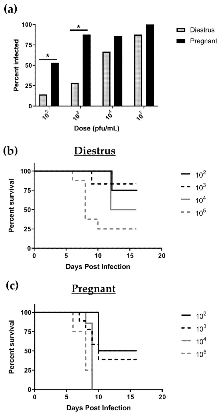
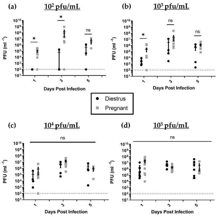
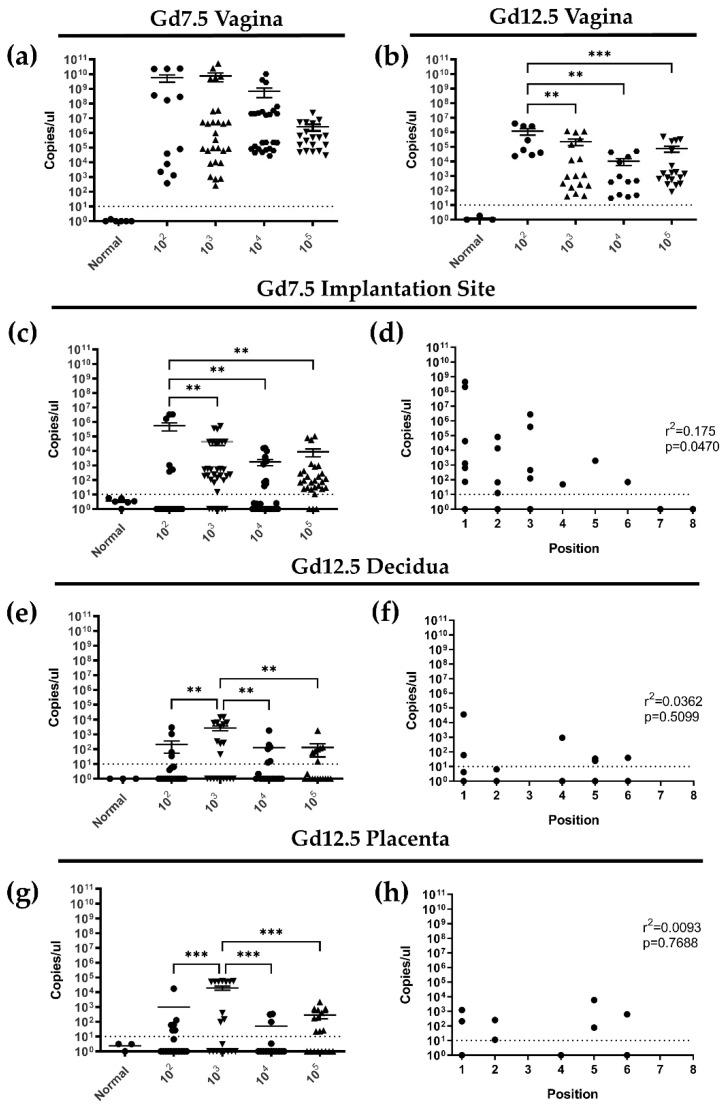
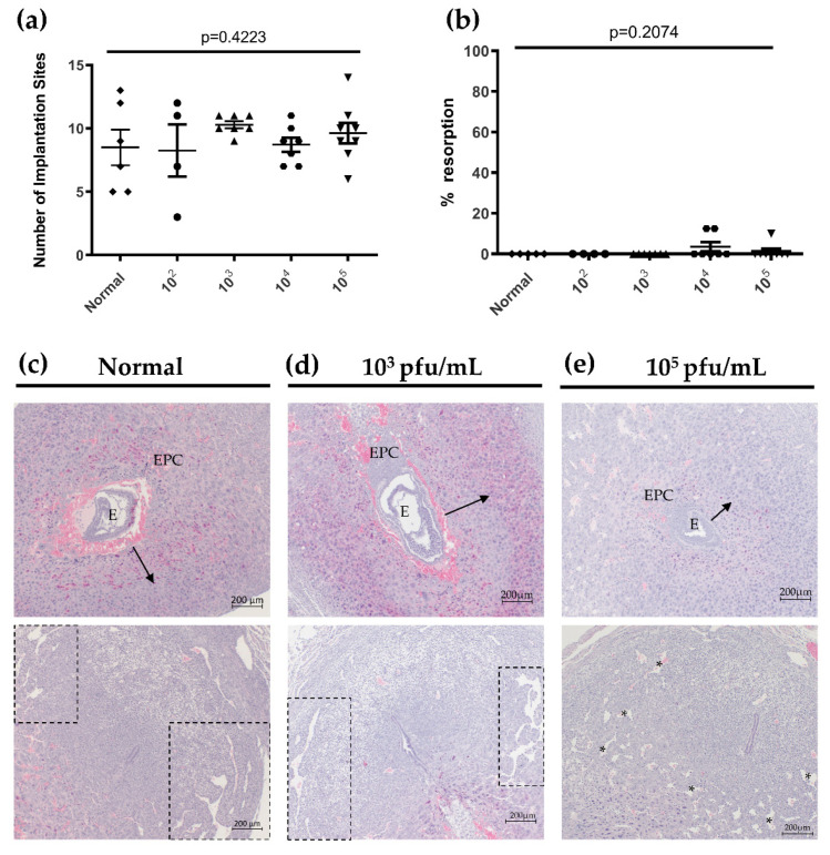
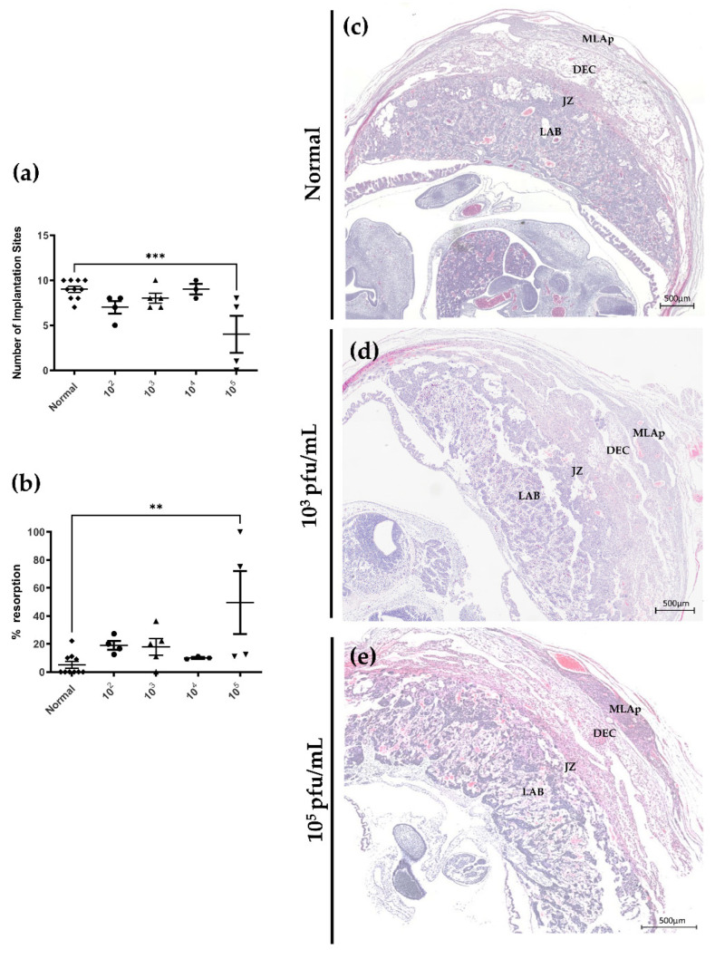
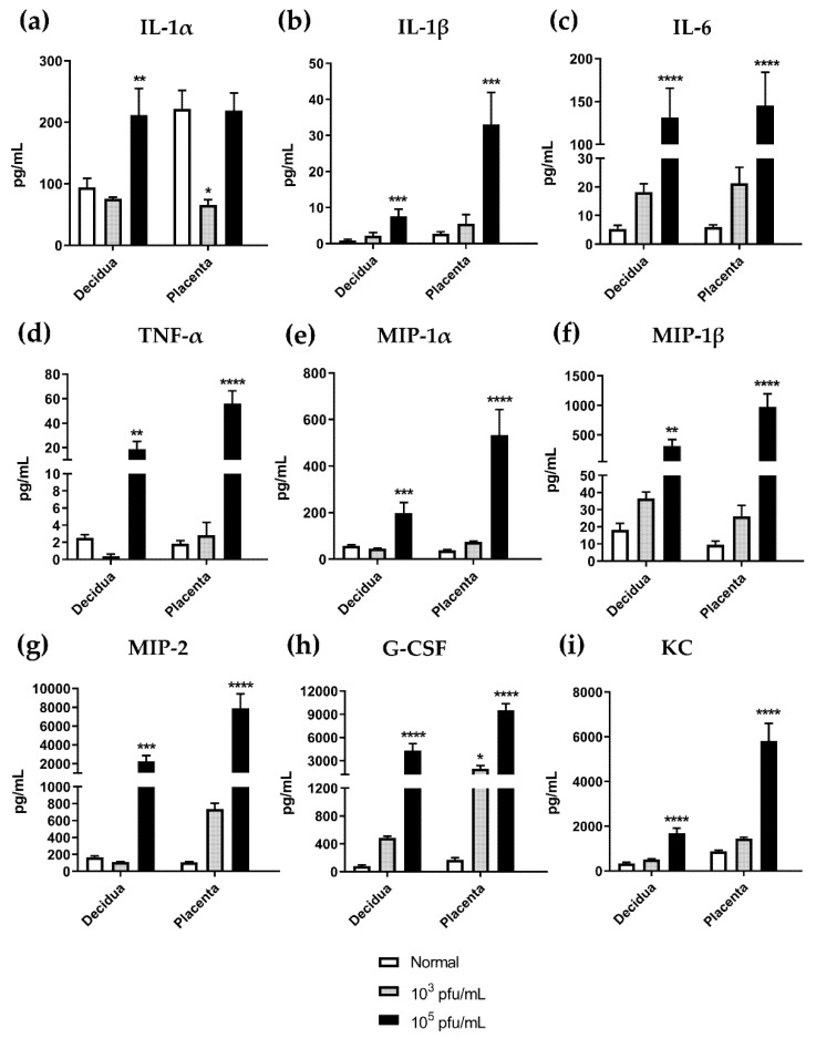
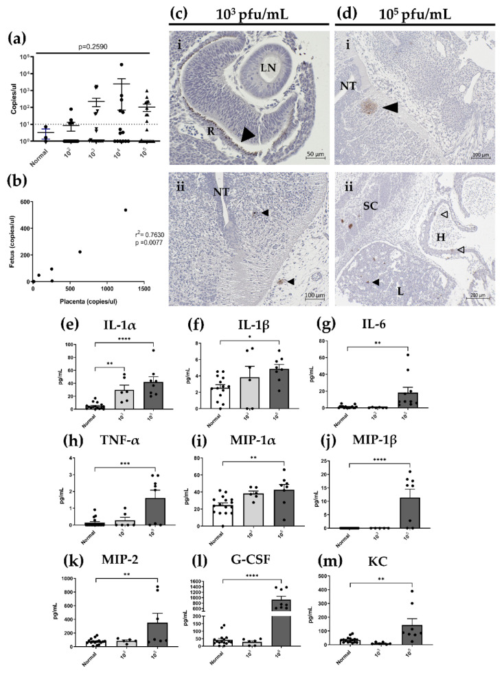
Herpes simplex virus type 2 (HSV-2) infection affects 24 million births annually and is associated with adverse pregnancy outcomes, including neonatal herpes; however, the mechanisms underlying in utero transmission of HSV-2 are largely unknown. We examined the effects of primary HSV-2 infection during early pregnancy on gestational outcomes in a novel, clinically relevant mouse model. Pregnant C57BL/6 mice were infected intravaginally with 102-105 pfu/mL HSV-2 on gestation day (gd) 4.5. Controls were infected, nonpregnant, diestrus-staged mice and pregnant, uninfected mice. Compared to nonpregnant mice, pregnant mice were 100-fold more susceptible to HSV-2 infection. Three days post-inoculation (gd7.5), viral DNA was present in implantation sites, but pregnancy outcomes were largely unaffected by infection. Eight days post-inoculation (gd12.5), HSV-2 DNA persisted in placental tissues, resulting in inflammation and hemorrhage. Fetal and placental weights were reduced and fetal loss was observed with high viral doses. HSV-2 DNA and increased expression of pro-inflammatory mediators were detected in fetal tissues at gd12.5, signifying viral transmission and fetal infection, even with low viral doses. This mouse model shows a dose-dependent effect of primary HSV-2 infection on pregnancy outcomes and suggests that fetal loss may occur due to placental inflammation, thus providing valuable insight into in utero transmission of HSV-2.

Keywords: HSV-2; mouse model; neonatal herpes; placental pathology; pregnancy; viral infection.


Conflict of interest statement

The authors declare no conflict of interest.

Figures

Figure 1
Figure 1
Pregnant mice are more susceptible to intravaginal HSV-2 infection than nonpregnant, diestrus-staged controls. (a) Following intravaginal inoculation with varying doses of HSV-2, productive infection was established in pregnant mice at greater rates than diestrus-staged controls, particularly at low viral doses of 102 and 103 pfu/mL. (b) Nonpregnant, diestrus-staged mice infected with HSV-2 were able to survive up to 16 days post infection (dpi). (c) HSV-2-infected, pregnant mice had decreased survival compared to diestrus-staged controls with 100% mortality observed by 9 dpi with high viral doses (104 and 105 pfu/mL). * p < 0.05.
Figure 2
Figure 2
Viral shedding in vaginal washes collected from nonpregnant, diestrus-staged and pregnant mice at 1, 3 and 5 days post infection (dpi). (a) Following intravaginal (IVAG) infection with 102 pfu/mL HSV-2, pregnant mice had increased viral shedding in vaginal washes collected at 1 and 3 dpi compared to diestrus controls. (b) At a viral dose of 103 pfu/mL, higher rates of viral shedding were observed in pregnant mice compared to diestrus mice 1 dpi, but significant differences were absent at 3 and 5 dpi. In mice infected with high viral doses of (c) 104 and (d) 105 pfu/mL, there were no differences in viral titers in vaginal washes collected on any day. Data expressed as mean ± SEM. ns = not significant, * p < 0.05.
Figure 3
Figure 3
HSV-2 is detected in the vaginal tract by qPCR, ascends directionally into gd7.5 implantation sites, and persists in placental tissues 8 days post infection (dpi). (a) Quantification of HSV-2 DNA by qPCR revealed high viral loads within vaginal tissue at gd7.5 with all viral doses. (b) HSV-2 DNA persisted in vaginal tissue collected at gd12.5 but was detected at lower quantities than at gd7.5. (c) Quantification of HSV-2 DNA in gd7.5 implantation sites by qPCR. (d) HSV-2 viral load in gd7.5 implantation sites is dependent on location along the uterine horn, with higher copy numbers detected in implantation sites close to the cervix (position 1) as compared to those close to the ovary (position 8). (e) Quantification of HSV-2 DNA by qPCR in the decidua at gd12.5. (f) At gd12.5, HSV-2 viral load in the decidua is independent of implantation site position along the uterine horn. (g) Quantification of HSV-2 DNA by qPCR in the placenta at gd12.5. (h) HSV-2 viral load in the placenta is independent of implantation site position along the uterine horn. Data expressed as mean ± SEM. ** p < 0.01, *** p < 0.001.
Figure 4
Figure 4
HSV-2 infection does not result in adverse pregnancy outcomes at gd7.5. (a) The number of healthy implantation sites in mice infected with 102–105 pfu/mL of HSV-2 did not vary significantly from uninfected normal controls at gd7.5 (n = 4–8 mice per group). (b) The percent resorption of pregnancies from infected mice did not differ from uninfected controls. Only mice infected with high viral doses (104 and 105 pfu/mL) had resorptions at gd7.5 (n = 4–8 mice per group). (c) Implantation sites from normal mice showed radial trophoblast invasion (arrow) and lateral decidual sinusoid formation (boxes). (d) In gd7.5 implantation sites from low dose (103 pfu/mL) infected animals, radial trophoblast invasion (arrow) and sinusoid formation (boxes) were similar to controls. (e) In high dose (105 pfu/mL) infected mice, embryos appeared smaller, trophoblast outgrowth was diminished (arrow), and lateral decidual sinusoids (*) were disorganized compared to control implantation sites. EPC: ectoplacental cone; E: embryo. Data expressed as mean ± SEM.
Figure 5
Figure 5
Pregnancy outcomes are affected by HSV-2 infection at gd12.5 resulting in placental pathology. (a) HSV-2 infection resulted in reductions in the number of healthy implantation sites compared to controls at gd12.5 (n = 3–10 mice per group). (b) Percent resorption was increased compared to controls in HSV-2-infected mice (n = 3–10 mice per group). (c) Implantation sites from normal gd12.5 mice consisted of 4 clearly defined and organized placental layers. (d) In mice infected with low dose HSV-2 (103 pfu/mL), placental layers remained distinct but there was evidence of tissue dissociation. (e) In mice infected with high dose HSV-2 (105 pfu/mL), placental layers were highly disorganized and tissue disintegration was evident. MLAp: mesometrial lymphoid aggregate of pregnancy; DEC: decidua; JZ: junctional zone; LAB: labyrinth. Data expressed as mean ± SEM. ** p < 0.01, *** p < 0.001.
Figure 6
Figure 6
HSV-2 infection compromises tissue integrity and vascular remodeling resulting in decreased fetal growth at gd12.5. (a) H&E staining of decidua from normal mice at gd12.5. (b) Decidua from mice infected with low dose HSV-2 (103 pfu/mL) showed signs of hemorrhage and necrosis (*). (c) With high dose HSV-2 (105 pfu/mL) tissue pathology became more apparent with rampant hemorrhage and necrosis in decidual tissue (*). (d) H&E staining of the placenta labyrinth in normal mice consists of intricate vascular branching patterns and thin interhemal membranes. (e) In animals infected with low dose HSV-2 (103 pfu/mL), the labyrinth consisted of long, straight vascular spaces with less vessel branching and thicker interhemal membranes than controls. (f) In the labyrinth of high dose (105 pfu/mL) infected mice, there were regions of extensive hemorrhage (*) and vessel branching was impaired. (g) Compared to controls, high dose (104 and 105 pfu/mL), but not low dose (102 and 103 pfu/mL), infection with HSV-2 significantly increased wall-lumen ratios of spiral arteries compared. (h) Maternal vascular spaces in the labyrinth were increased significantly from controls with all doses of HSV-2. (i) Fetal vascular spaces in the labyrinth were increased significantly for all viral doses greater than 102 pfu/mL compared to normal controls. (j) Gd12.5 fetuses dissected from implantation sites of infected animals weighed less than control fetuses at all viral doses (n = 4–9 fetuses per group). (k) Fetal placentas dissected from implantation sites of animals infected with a high (105 pfu/mL) viral dose had significantly reduced weights compared to controls (n = 4–9 placentas per group). Data expressed as mean ± SEM. * p < 0.05, ** p < 0.01, *** p < 0.001, **** p < 0.0001.
Figure 7
Figure 7
HSV-2 infection alters cytokine/chemokine profiles in the decidua and placenta at gd12.5. Cytokines in tissue homogenates from normal mice and mice infected with low (103 pfu/mL) and high (105 pfu/mL) dose HSV-2 were analyzed by 32-plex array and 9 analytes were selected for further analysis: (a) IL-1α, (b) IL-1β, (c) IL-6, (d) TNF-α, (e) MIP-1α, (f) MIP-1β, (g) MIP-2, (h) G-CSF, and (i) KC. (a) IL-1α was significantly increased in the decidua of high dose infected animals and significantly decreased in the placenta of low dose infected animals. (bi) Compared to normal controls, high dose HSV-2 infection significantly increased cytokine/chemokine levels in decidua and placenta homogenates. (h) Compared to controls, G-CSF was significantly increased in placenta tissue homogenates with both low and high dose HSV-2 infection. Data expressed as mean ± SEM. * p < 0.05, ** p < 0.01, *** p < 0.001, **** p < 0.0001.
Figure 8
Figure 8
In utero HSV-2 transmission into fetuses results in congenital HSV-2 infection and inflammatory responses at gd12.5. (a) HSV-2 DNA was detected in fetal homogenates by qPCR with comparable levels for all viral doses. (b) There was a significant correlation between HSV-2 DNA levels in matched fetal and placental tissues, but HSV-2 DNA was not detected in fetal homogenates in the absence of placental infection. Detection of HSV-2 DNA in placental tissues did not guarantee HSV-2 detection in matched fetal homogenates. (c) In fetuses from mice infected with low dose HSV-2 (103 pfu/mL), HSV-2 was detected in (i) the developing retina and (ii) neuroepithelium surrounding the neural tube (arrowheads). (d) HSV-2 staining in fetuses from mice infected with high dose HSV-2 (105 pfu/mL) showed disseminated expression of HSV-2 in fetal tissues including the (i) neuroepithelium around the neural tube (arrowhead), (ii) heart (open arrowheads), and liver (closed arrowhead). (e) IL1α expression levels were significantly increased compared to controls in fetuses from mice infected with both low (103 pfu/mL) and high (105 pfu/mL) dose HSV-2. (fm) Expression of all other pro-inflammatory cytokines and chemokines in fetal homogenates was significantly increased in fetuses from pregnancies complicated by high, but not low, dose HSV-2 infection. H: heart, L: liver, LN: lens, NT: neural tube, R: retina, SC: spinal column. Data expressed as mean ± SEM. * p < 0.05, ** p < 0.01, *** p < 0.001, **** p < 0.0001.

Similar articles

Cited by

References

    1. James C., Harfouche M., Welton N.J., Turner K.M., Abu-Raddad L.J., Gottlieb S.L., Looker K.J. Herpes simplex virus: Global infection prevalence and incidence estimates, 2016. Bull. World Health Organ. 2020;98:315–329. doi: 10.2471/BLT.19.237149. - DOI - PMC - PubMed
    1. Kaushic C., Ashkar A.A., Reid L.A., Rosenthal K.L. Progesterone increases susceptibility and decreases immune responses to genital herpes infection. J. Virol. 2003;77:4558–4565. doi: 10.1128/JVI.77.8.4558-4565.2003. - DOI - PMC - PubMed
    1. Gillgrass A.E., Fernandez S.A., Rosenthal K.L., Kaushic C. Estradiol regulates susceptibility following primary exposure to genital herpes simplex virus type 2, while progesterone induces inflammation. J. Virol. 2005;79:3107–3116. doi: 10.1128/JVI.79.5.3107-3116.2005. - DOI - PMC - PubMed
    1. Teepe A.G., Allen L.B., Wordinger R.J., Harris E.F. Effect of the estrous cycle on susceptibility of female mice to intravaginal inoculation of herpes simplex virus type 2 (HSV-2) Antiviral Res. 1990;14:227–235. doi: 10.1016/0166-3542(90)90004-Q. - DOI - PubMed
    1. Lee Y., Dizzell S.E., Leung V., Nazli A., Zahoor M.A., Fichorova R.N., Kaushic C. Effects of Female Sex Hormones on Susceptibility to HSV-2 in Vaginal Cells Grown in Air-Liquid Interface. Viruses. 2016;8:241. doi: 10.3390/v8090241. - DOI - PMC - PubMed

Publication types

MeSH terms

Supplementary concepts

Grants and funding