. 2021 Sep 29;13(10):1961.
doi: 10.3390/v13101961.

Norovirus Epidemiology and Genetic Diversity in Leipzig, Germany during 2013-2017

Affiliations

Norovirus Epidemiology and Genetic Diversity in Leipzig, Germany during 2013-2017

Nora Ennuschat et al. Viruses. .

Abstract

Globally and in all age groups, noroviruses are a main cause of gastroenteritis. To assess their local epidemiology and genetic diversity, stool samples of 7509 inpatients with gastrointestinal complaints from all age groups were analyzed. After detection of norovirus genogroup I and II RNA by real-time RT-PCR, viral capsids were genotyped by partial nucleic acid sequencing. In the case of GII.2 strains, polymerase genotypes were also assessed. Between October 2013 and September 2017, presence of norovirus RNA was shown in 611 samples (8.1%), of which 610 (99.8%) were typed successfully. Norovirus positivity rate was higher in patients aged below five years (14.8%) than in older patients (5.7%). Among the 611 norovirus positive samples, GII.4 (56.6%) strains prevailed, followed by GII.6 (11.3%), GII.3 (11.0%) and GII.2 (9.5%). The most common genogroup I (GGI) genotype was GI.3 (3.6%). In addition, rare genotypes such as GII.13, GII.14 and GII.26 were detected. Interestingly, GII.3 infections were most common in children under the age of five years. Assessment of polymerase genotypes in GII.2 viruses showed a shift from P2 to P16, with higher diversity in P2 sequences. The varying distribution of norovirus genotypes depending on season, age and setting of infection highlights the importance of frequent genotyping as a basis for vaccine development and needful adjustments.

Keywords: anti-norovirus vaccines; diarrhea; genotyping; molecular epidemiology; viral diversity; viral gastroenteritis.


Conflict of interest statement

The authors declare no conflict of interest.

Figures

Figure 1
Figure 1
Monthly distribution of detected norovirus GGI and GGII strains, Leipzig University Hospital, October 2013–September 2017.
Figure 2
Figure 2
Distribution of GII norovirus genotypes according to age and mode of acquisition, Leipzig University Hospital October 2013–September 2017; com. acq. stands for community-acquired infections and nos. stands for nosocomial infections.
Figure 3
Figure 3
Phylogenetic analysis of norovirus GII.4 genotypes based on Maximum Likelihood estimations (1000 bootstraps) of partial ORF2 nucleic acid sequences. Only topology is shown, ignoring the branch lengths. Red squares indicate sequences of season 2013/2014, green arrow heads facing downwards indicate sequences of season 2014/2015, yellow diamonds indicate sequences of season 2015/2016 and blue arrow heads facing upwards indicate sequences of season 2016/2016. Labels in bold indicate reference strains, with GenBank accession numbers shown in parenthesis. All sequences without labeled variants are GII.4 Sydney strains.
Figure 4
Figure 4
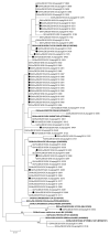
Phylogenetic analysis of norovirus GII.3 genotypes based on Maximum Likelihood estimations of partial ORF2 nucleic acid sequences. Exclusively, bootstrap values (1000 replicates) above 80% are shown. Black circles indicate sequences of nosocomial infections. Labels in bold indicate reference strains, with GenBank accession numbers shown in parenthesis.
Figure 5
Figure 5
Pairwise distances within norovirus (a) GII.3 and (b) GII.4 sequences calculated by Jukes Cantor method in MEGA.
Figure 6
Figure 6
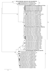
Phylogenetic analysis of norovirus GII.2 polymerase genotypes based on Maximum Likelihood estimations of partial ORF1 nucleic acid sequences. Exclusively, bootstrap values (1000 replicates) above 80% are shown. Black circles indicate sequences of nosocomial infections. Labels in bold indicate reference strains with GenBank accession numbers shown in parenthesis.
Figure 7
Figure 7
Pairwise distances within norovirus (a) GII.2[P2] and (b) GII.2[P16] sequences calculated by Jukes Cantor method in MEGA.

References

    1. Ahmed S.M., Hall A.J., Robinson A.E., Verhoef L., Premkumar P., Parashar U.D., Koopmans M., Lopman B.A. Global Prevalence of Norovirus in Cases of Gastroenteritis: A Systematic Review and Meta-Analysis. Lancet Infect. Dis. 2014;14:725–730. doi: 10.1016/S1473-3099(14)70767-4. - DOI - PMC - PubMed
    1. Patel M.M., Widdowson M.-A., Glass R.I., Akazawa K., Vinjé J., Parashar U.D. Systematic Literature Review of Role of Noroviruses in Sporadic Gastroenteritis. Emerg. Infect. Dis. 2008;14:1224–1231. doi: 10.3201/eid1408.071114. - DOI - PMC - PubMed
    1. Koo H.L., Neill F.H., Estes M.K., Munoz F.M., Cameron A., DuPont H.L., Atmar R.L. Noroviruses: The Most Common Pediatric Viral Enteric Pathogen at a Large University Hospital After Introduction of Rotavirus Vaccination. J. Pediatr. Infect. Dis. Soc. 2013;2:57–60. doi: 10.1093/jpids/pis070. - DOI - PMC - PubMed
    1. Payne D.C., Vinjé J., Szilagyi P.G., Edwards K.M., Staat M.A., Weinberg G.A., Hall C.B., Chappell J., Bernstein D.I., Curns A.T., et al. Norovirus and Medically Attended Gastroenteritis in U.S. Children. N. Engl. J. Med. 2013;368:1121–1130. doi: 10.1056/NEJMsa1206589. - DOI - PMC - PubMed
    1. Hemming M., Räsänen S., Huhti L., Paloniemi M., Salminen M., Vesikari T. Major Reduction of Rotavirus, but Not Norovirus, Gastroenteritis in Children Seen in Hospital after the Introduction of RotaTeq Vaccine into the National Immunization Programme in Finland. Eur. J. Pediatr. 2013;172:739–746. doi: 10.1007/s00431-013-1945-3. - DOI - PMC - PubMed

Publication types

MeSH terms