. 2021 Sep 29;13(10):1960.
doi: 10.3390/v13101960.

Alveolar-like Macrophages Attenuate Respiratory Syncytial Virus Infection

Affiliations

Alveolar-like Macrophages Attenuate Respiratory Syncytial Virus Infection

Bárbara N Porto et al. Viruses. .

Abstract

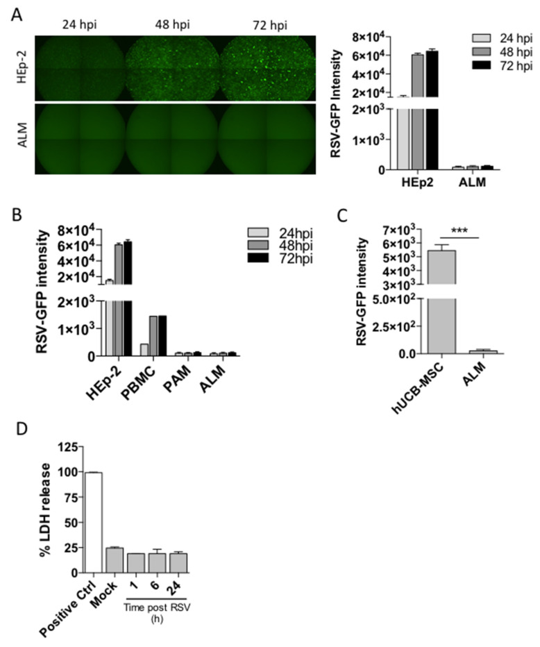
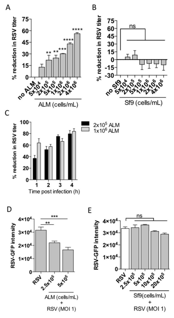
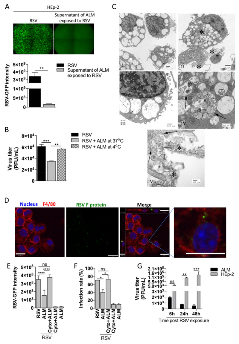
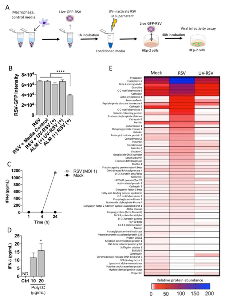
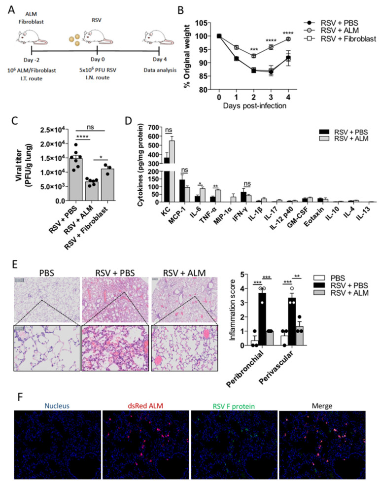
Respiratory Syncytial Virus (RSV) is the leading cause of acute lower respiratory infections in young children and infection has been linked to the development of persistent lung disease in the form of wheezing and asthma. Despite substantial research efforts, there are no RSV vaccines currently available and an effective monoclonal antibody targeting the RSV fusion protein (palivizumab) is of limited general use given the associated expense. Therefore, the development of novel approaches to prevent RSV infection is highly desirable to improve pediatric health globally. We have developed a method to generate alveolar-like macrophages (ALMs) from pluripotent stem cells. These ALMs have shown potential to promote airway innate immunity and tissue repair and so we hypothesized that ALMs could be used as a strategy to prevent RSV infection. Here, we demonstrate that ALMs are not productively infected by RSV and prevent the infection of epithelial cells. Prevention of epithelial infection was mediated by two different mechanisms: phagocytosis of RSV particles and release of an antiviral soluble factor different from type I interferon. Furthermore, intratracheal administration of ALMs protected mice from subsequent virus-induced weight loss and decreased lung viral titres and inflammation, indicating that ALMs can impair the pathogenesis of RSV infection. Our results support a prophylactic role for ALMs in the setting of RSV infection and warrant further studies on stem cell-derived ALMs as a novel cell-based therapy for pulmonary viral infections.

Keywords: Respiratory Syncytial Virus; alveolar macrophages; respiratory infection; stem cells.


Conflict of interest statement

The authors declare no conflict of interest. The funders had no role in the design of the study; in the collection, analyses, or interpretation of data; in the writing of the manuscript, or in the decision to publish the results.

Figures

Figure 1
Figure 1
ALMs are not productively infected by RSV and are resistant to RSV-induced cell death. ALMs (2 × 10⁵/well) or HEp-2 cells (6 × 10³/well) were exposed to RSV-GFP at an MOI of 1. (A) GFP fluorescence was visualized by fluorescence microscopy (left panel) and quantified (right panel) at 24 h intervals for 72 h. (B) HEp-2 cells (6 × 10³/well), human peripheral blood mononuclear cells (2 × 10⁵/well), primary alveolar macrophages (2 × 10⁵/well) and ALMs (2 × 10⁵/well) were exposed to RSV-GFP and productive infection was quantified by fluorescence at 24 h intervals for 72 h. (C) Umbilical cord mesenchymal stromal cells (2 × 10⁵/well) or ALMs (2 × 10⁵/well) were exposed to RSV-GFP for 72 h and productive infection was quantified by fluorescence. (D) ALMs (3 × 10⁵/well) were exposed to RSV (MOI 1) for 1, 6, or 24 h. Afterwards, cell death was assessed by LDH release in ALMs supernatants. Data are representative of 2 independent experiments performed in triplicates and represent mean ±SEM. Data were analyzed with unpaired Student’s t-test. *** p = 0.0002.
Figure 2
Figure 2
ALMs reduce RSV infectivity. Increasing numbers of (A) ALMs or (B) Sf9 cells were incubated with RSV (MOI 1) for 4 h at 37 °C under 5% CO2. Afterwards, the viral titre in the ALMs supernatants was assessed by plaque assay and the percent viral reduction in comparison to controls (no ALM) was calculated. (C) ALMs at the indicated numbers were incubated with RSV for 1–4 h and the reduction in viral infectivity was measured over time. (D) HEp-2 cells (6 × 10³/well) were seeded in black, flat, clear-bottom 96-well plates and allowed to attach overnight at 37 °C. The next day, ALMs (2.5 × 10⁵–5 × 10⁵/mL) and RSV (MOI 1) were added to the cultures for 48 h. GFP fluorescence was quantified as a readout of RSV replication. (E) HEp-2 cells (6 × 10³/well) were seeded in black 96-well plates overnight at 37 °C. The next day, Sf9 cells (2.5 × 10⁵–20 × 105/mL) and RSV (MOI 1) were added to the cultures for 48 h. Afterwards, GFP fluorescence was quantified. Data are representative of 2 independent experiments performed in triplicates and represent mean ±SEM. Data were analyzed with one-way ANOVA with Tukey’s post hoc test. ** p < 0.01; *** p < 0.001; **** p < 0.0001. ns = not significant.
Figure 3
Figure 3
ALMs phagocytose RSV. (A) ALMs (1 × 10⁶/mL) were incubated with RSV-GFP (MOI 1) for 1 h at 37 °C with 5% CO2. Afterwards, the supernatants were collected and used to infect HEp-2 cells for 48 h. As a control, HEp-2 cells were directly infected with RSV-GFP (MOI 1) for 48 h. GFP fluorescence (top) was used to visualize RSV replication and GFP intensity was quantified (bottom) as a readout of RSV replication in HEp-2 cells. (B) ALMs (1 × 10⁶/mL) were incubated with RSV-GFP (MOI 1) for 1 h at either 37 °C or 4 °C. Afterwards, the supernatants were collected and used to infect HEp-2 cells for 48 h. After this period, RSV titre was measured in HEp-2 cells by plaque assay. (C) Electron micrographs of ALMs exposed to active RSV: (i) Unexposed macrophages. Note the limited phagocytosed material. Scale bar = 0.5 µm. (ii) RSV-exposed macrophages. Asterisks indicate accumulations of degraded RSV in both phagosomes and in close proximity to the plasma membrane. Scale bar = 2 µm. (iii). Unexposed macrophage. Note that the phagocytic vacuoles contain limited material when compared to the infected cells. Scale bar = 0.5 µm. (iv) RSV-exposed macrophage. Phagosomes (arrows) contain cellular and viral debris. Scale bar = 0.5 µm. (v) RSV-exposed macrophage. Ingestion through phagocytosis. Phagosomal membranes are seen ingesting degraded viral material (arrows). Degraded virus is seen in the extracellular space in close proximity to the cell surface. Scale bar = 0.1 µm. (D) ALMs (1 × 10⁶/mL) were exposed to RSV (MOI 1) for 4 h at 37 °C on coverslips. Afterwards, cells were fixed with 4% PFA and stained with Hoechst 33342 (1:4000), anti-F4/80 (1:100) and anti-RSV F protein (1:250) antibodies. Overlay of the fluorescence images are shown in the penultimate panel. The last panel shows the merged image magnified 4 times. Images are representative of 2 independent experiments. Images were taken with a Leica DMi8 confocal microscope. Scale bars = 15 μm. (E,F) ALMs (1 × 10⁶/mL) were pretreated with the actin polymerization inhibitor Cytochalasin D (Cyto–10 μM) for 1 h at 37 °C. Afterwards, ALMs were incubated with RSV-GFP (MOI 1) for 1 h. Then, ALMs were pelleted and the supernatants were added to HEp-2 cells for 48 h. After this period, (E) GFP intensity was quantified and (F) the infection rate was measured in HEp-2 cells. (G) ALMs and HEp-2 cells were exposed to RSV-GFP (MOI 1) for 6, 24 or 48 h. Afterwards, cells were harvested and lysed. The intracellular content was subjected to plaque assay to measure viral titre. Data are representative of 2 independent experiments performed in triplicates and represent mean ±SEM. Data were analyzed with unpaired Student’s t-test (A), one-way ANOVA with Tukey’s post hoc test (B,E,F) or 2-way ANOVA (G). * p < 0.05; ** p < 0.01; *** p < 0.001; **** p < 0.0001. ns = not significant.
Figure 4
Figure 4
ALMs confer antiviral effects through a secreted factor. (A) Technical schematic indicating the work flow to measure secreted factor in ALMs supernatants. (B) ALMs (1 × 10⁶/mL) were exposed to RSV or UV-RSV at an MOI of 1 for 1 h. Afterwards, the supernatants were collected and UV-irradiated to neutralize residual active RSV. This was used as a conditioned media, which was then added onto previously seeded HEp-2 cells with competent RSV-GFP (MOI 1) and RSV replication was measured by GFP fluorescence. Control indicating HEp-2 cells with EMEM, media control indicates UV-irradiated EMEM, UV-RSV (+) indicates media containing UV-inactivated RSV, ALM (+) UV-RSV (+) indicates conditioned media from ALMs exposed to UV inactivated RSV, ALM (+) RSV (+) indicated conditioned media from ALMs exposed to RSV. (C,D) (C) ALMs (1 × 10⁶/mL) were stimulated with RSV (MOI 1) or mock-stimulated for 1, 4 or 24 h at 37 °C under 5% CO2. (D) ALMs were stimulated with TLR3 ligand Poly I:C (10 and 20 μg/mL) for 24 h at 37 °C under 5% CO2. Afterwards, supernatants were collected and IFN-β concentrations were determined using ELISA. (E) ALMs (1 × 10⁶/mL) were exposed to mock, RSV or UV-RSV at an MOI of 1 for 4 h. Afterwards, the supernatants were collected, concentrated and prepared for proteomic analysis as described in the Section 2. Data are representative of 2 independent experiments performed in triplicates and represent mean ±SEM. Data were analyzed with one-way ANOVA with Tukey’s post hoc test (B,D). * p < 0.05; **** p < 0.0001.
Figure 5
Figure 5
ALMs protect mice from RSV infection. (A) On day -2, BALB/c mice received an intratracheal injection with 1 × 10⁶ ALMs or 1 × 10⁶ fibroblasts as an inert cellular control. On day 0, mice were intranasally infected with RSV (5 × 10⁶ PFU). Analysis were performed on day 4 post-infection. (B) Percent of weight loss post-infection relative to initial body weight (day 0) (n = 8). (C) RSV viral titres obtained from lung homogenates measured by plaque assay (PFU/g of lung tissue) (n = 7 for RSV and RSV + ALM groups and n = 3 for RSV + fibroblast group). (D) Cytokines and chemokines were measured in the lung BAL of mice (n = 3) by multiplex Luminex® technology and expressed as pg/mg protein. (E) Representative hematoxylin and eosin (H&E)-stained lung tissue images with their respective peribronchial and perivascular inflammation scores for each experimental group. (F) Immunofluorescence of lung tissue samples obtained from animals infected with RSV and treated with ALMs stained to identify dsRed-expressing ALMs and GFP- RSV. Data are representative of 2 independent experiments and are expressed as mean ±SEM. Data were analyzed with two-way ANOVA with Tukey’s multiple comparisons test (B,C,E) or with unpaired Student’s t-test (D). * p < 0.05; ** p < 0.01; *** p < 0.001;**** p < 0.0001. ns = not significant.

References

    1. Meissner H.C. Viral Bronchiolitis in Children. N. Engl. J. Med. 2016;374:62–72. doi: 10.1056/NEJMra1413456. - DOI - PubMed
    1. Nair H., Nokes D.J., Gessner B.D., Dherani M., Madhi S.A., Singleton R.J., O’Brien K., Roca A., Wright P.F., Bruce N., et al. Global burden of acute lower respiratory infections due to respiratory syncytial virus in young children: A systematic review and meta-analysis. Lancet. 2010;375:1545–1555. doi: 10.1016/S0140-6736(10)60206-1. - DOI - PMC - PubMed
    1. Mohapatra S.S., Boyapalle S. Epidemiologic, Experimental, and Clinical Links between Respiratory Syncytial Virus Infection and Asthma. Clin. Microbiol. Rev. 2008;21:495–504. doi: 10.1128/CMR.00054-07. - DOI - PMC - PubMed
    1. Blanken M.O., Rovers M., Molenaar J.M., Winkler-Seinstra P.L., Meijer A., Kimpen J.L., Bont L. Respiratory Syncytial Virus and Recurrent Wheeze in Healthy Preterm Infants. N. Engl. J. Med. 2013;368:1791–1799. doi: 10.1056/NEJMoa1211917. - DOI - PubMed
    1. Hussell T., Bell T.J. Alveolar macrophages: Plasticity in a tissue-specific context. Nat. Rev. Immunol. 2014;14:81–93. doi: 10.1038/nri3600. - DOI - PubMed

Publication types

MeSH terms

Substances

Grants and funding