Review
. 2021 Sep 30;13(10):1973.
doi: 10.3390/v13101973.

Cell Fusion and Syncytium Formation in Betaherpesvirus Infection

Affiliations
Review

Cell Fusion and Syncytium Formation in Betaherpesvirus Infection

Jiajia Tang et al. Viruses. .

Abstract

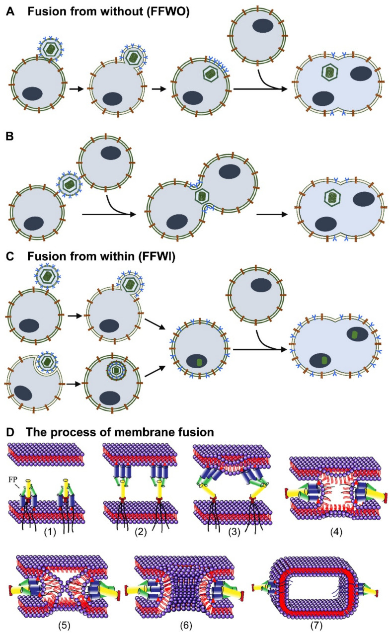
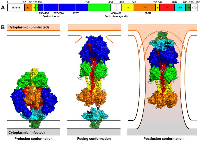
Cell-cell fusion is a fundamental and complex process that occurs during reproduction, organ and tissue growth, cancer metastasis, immune response, and infection. All enveloped viruses express one or more proteins that drive the fusion of the viral envelope with cellular membranes. The same proteins can mediate the fusion of the plasma membranes of adjacent cells, leading to the formation of multinucleated syncytia. While cell-cell fusion triggered by alpha- and gammaherpesviruses is well-studied, much less is known about the fusogenic potential of betaherpesviruses such as human cytomegalovirus (HCMV) and human herpesviruses 6 and 7 (HHV-6 and HHV-7). These are slow-growing viruses that are highly prevalent in the human population and associated with several diseases, particularly in individuals with an immature or impaired immune system such as fetuses and transplant recipients. While HHV-6 and HHV-7 are strictly lymphotropic, HCMV infects a very broad range of cell types including epithelial, endothelial, mesenchymal, and myeloid cells. Syncytia have been observed occasionally for all three betaherpesviruses, both during in vitro and in vivo infection. Since cell-cell fusion may allow efficient spread to neighboring cells without exposure to neutralizing antibodies and other host immune factors, viral-induced syncytia may be important for viral dissemination, long-term persistence, and pathogenicity. In this review, we provide an overview of the viral and cellular factors and mechanisms identified so far in the process of cell-cell fusion induced by betaherpesviruses and discuss the possible consequences for cellular dysfunction and pathogenesis.

Keywords: Herpesviridae; cell–cell fusion; cytomegalovirus; envelope glycoproteins; glycoprotein B; glycoprotein H; glycoprotein L; herpesvirus; polykaryocyte; syncytium formation.


Conflict of interest statement

The authors declare no conflict of interest. The funders had no role in the design of the study; in the collection, analyses, or interpretation of data; in the writing of the manuscript; or in the decision to publish the results.

Figures

Figure 1
Figure 1
Simplified view of the different types of virus-induced membrane fusion. (A) Fusion from without (FFWO): The virus envelope carries glycoproteins, mediating the fusion of the viral envelope with the plasma membrane. The viral envelope is retained on the surface of the infected cell. Envelope glycoproteins interact with receptors on neighboring cells and mediate cell–cell fusion. (B) FFWO can also occur when a viral particle fuses simultaneously with two cells. (C) Fusion from within (FFWI): The virus enters the cell through fusion with the plasma membrane or through endocytosis. Viral gene expression leads to the synthesis of envelope glycoproteins that may be transported to the cellular surface. Viral glycoproteins interact with receptors on adjacent cells and induce cell–cell fusion. (D) Schematic of the membrane fusion process. (1) Activation of the fusion machinery and exposure of specific fusion peptides (FP). (2) Insertion of the FP into the adjacent membrane. (3) Refolding of the FP and induction of membrane deformation. (4) Formation of a transient hemifusion diaphragm. (5) Opening of a fusion pore that completes merging of both membranes. (6) Expansion of the fusion pore. (7) Situation after membrane fusion (postfusion).
Figure 2
Figure 2
Linear representation and structural conformations of HCMV gB. (A) Distribution of HCMV gB structural domains within the gB primary domain. Amino acid indices at the domain boundaries are indicated above the sequence representation. Fusion loops and furin cleavage site are indicated as violet lines. Two known residues that effect HCMV gB fusion activity are marked as grey lines. (B) gB protein structures indicating conformations during the stages of cell–cell fusion. The prefusion conformation (PDB 7KDP) is compressed on the infected cell membrane. During cell–cell fusion, gB extends to bind to an additional uninfected cell membrane. The fusing conformation (PDB 5CXF) is modeled with the MPR-TM domain (PDB 7KDP) modeled by protein alignment to the conserved IV domain. In the postfusion conformation, gB (PDB 5CFX) is modeled with the MPR-TM domain (PDB 7KDP) as shown in [92]. 2021. The surface representation has been generated using PyMOL and the structural domains are mapped and color-coded as described [90,92]: N-terminal signaling sequence (N-term) and cytoplasmic domain (CTD) in white; domain I = blue, II = green, III = yellow, IV = orange, V = red; membrane proximal region (MPR) in cyan, and transmembrane region (TM) in dark green.

Similar articles

Cited by

References

    1. Hernandez J.M., Podbilewicz B. The hallmarks of cell-cell fusion. Development. 2017;144:4481–4495. doi: 10.1242/dev.155523. - DOI - PubMed
    1. Brukman N.G., Uygur B., Podbilewicz B., Chernomordik L.V. How cells fuse. J. Cell Biol. 2019;218:1436–1451. doi: 10.1083/jcb.201901017. - DOI - PMC - PubMed
    1. Lu X., Kang Y. Cell fusion as a hidden force in tumor progression. Cancer Res. 2009;69:8536–8539. doi: 10.1158/0008-5472.CAN-09-2159. - DOI - PMC - PubMed
    1. Platt J.L., Cascalho M. Cell Fusion in Malignancy: A Cause or Consequence? A Provocateur or Cure? Cells. 2019;8:587. doi: 10.3390/cells8060587. - DOI - PMC - PubMed
    1. Ku J.W.K., Chen Y., Lim B.J.W., Gasser S., Crasta K.C., Gan Y.H. Bacterial-induced cell fusion is a danger signal triggering cGAS-STING pathway via micronuclei formation. Proc. Natl. Acad. Sci. USA. 2020;117:15923–15934. doi: 10.1073/pnas.2006908117. - DOI - PMC - PubMed

Publication types

MeSH terms

LinkOut - more resources