. 2021 Oct 8;13(10):2021.
doi: 10.3390/v13102021.

Spike Proteins of SARS-CoV-2 Induce Pathological Changes in Molecular Delivery and Metabolic Function in the Brain Endothelial Cells

Affiliations

Spike Proteins of SARS-CoV-2 Induce Pathological Changes in Molecular Delivery and Metabolic Function in the Brain Endothelial Cells

Eun Seon Kim et al. Viruses. .

Abstract

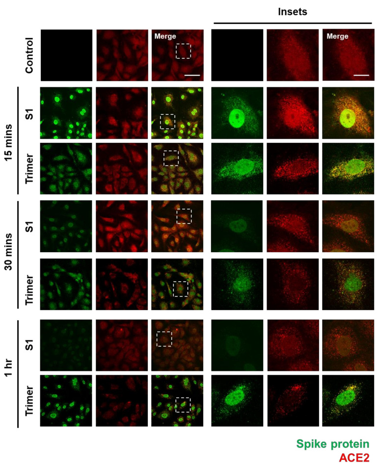
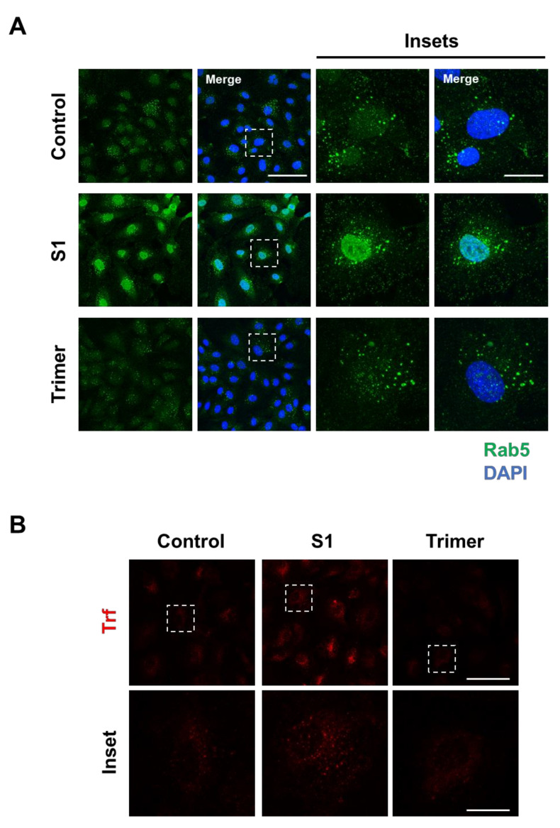
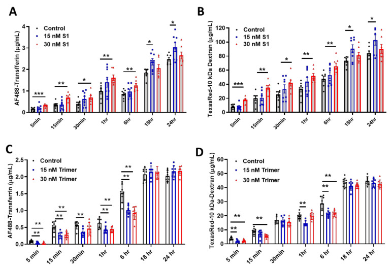
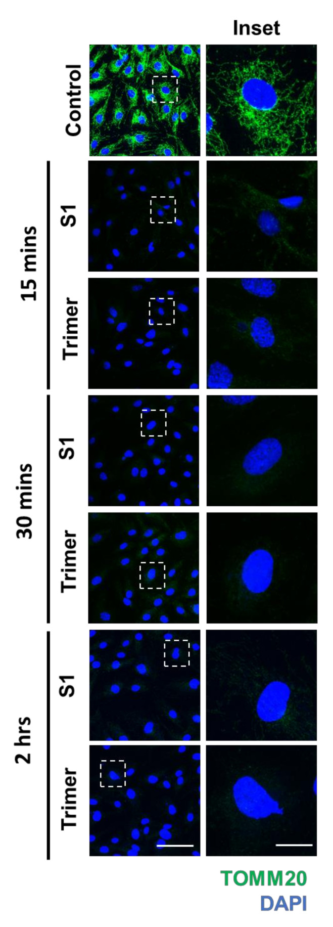
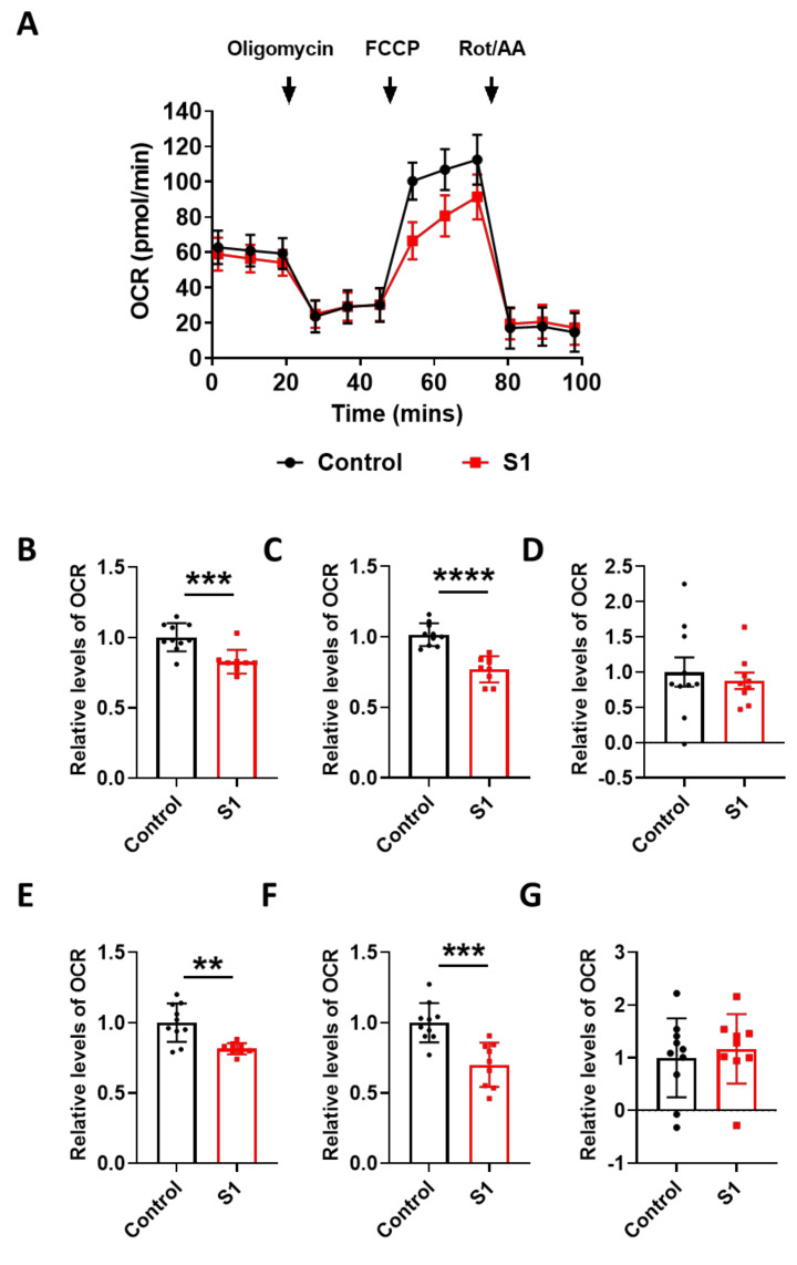
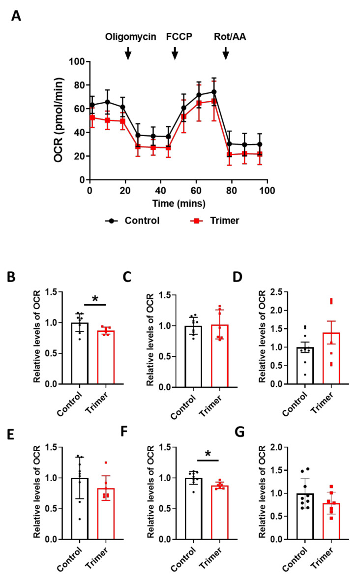
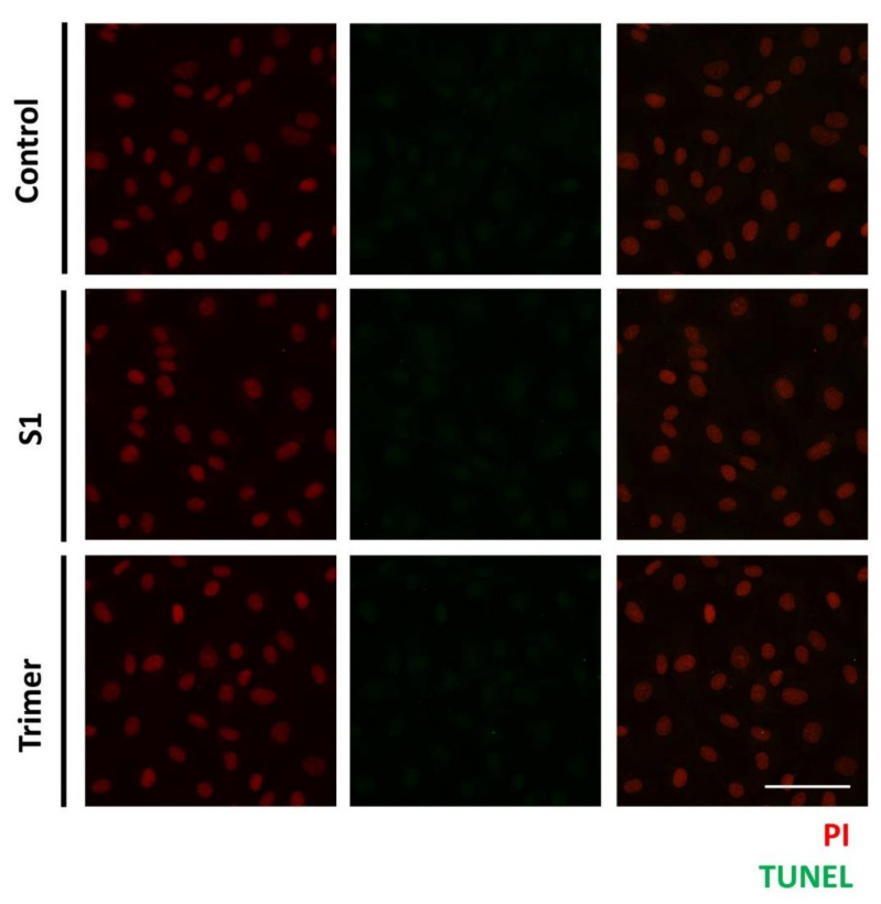
Severe acute respiratory syndrome coronavirus 2 (SARS-CoV-2), which causes the coronavirus disease (COVID-19), is currently infecting millions of people worldwide and is causing drastic changes in people's lives. Recent studies have shown that neurological symptoms are a major issue for people infected with SARS-CoV-2. However, the mechanism through which the pathological effects emerge is still unclear. Brain endothelial cells (ECs), one of the components of the blood-brain barrier, are a major hurdle for the entry of pathogenic or infectious agents into the brain. They strongly express angiotensin converting enzyme 2 (ACE2) for its normal physiological function, which is also well-known to be an opportunistic receptor for SARS-CoV-2 spike protein, facilitating their entry into host cells. First, we identified rapid internalization of the receptor-binding domain (RBD) S1 domain (S1) and active trimer (Trimer) of SARS-CoV-2 spike protein through ACE2 in brain ECs. Moreover, internalized S1 increased Rab5, an early endosomal marker while Trimer decreased Rab5 in the brain ECs. Similarly, the permeability of transferrin and dextran was increased in S1 treatment but decreased in Trimer, respectively. Furthermore, S1 and Trimer both induced mitochondrial damage including functional deficits in mitochondrial respiration. Overall, this study shows that SARS-CoV-2 itself has toxic effects on the brain ECs including defective molecular delivery and metabolic function, suggesting a potential pathological mechanism to induce neurological signs in the brain.

Keywords: BBB; COVID-19; SARS-CoV-2; endothelial cells; metabolism.


Conflict of interest statement

The authors declare no conflict of interest.

Figures

Figure 1
Figure 1
The spike proteins are internalized into the human brain ECs through ACE2. An amount of 15 nM of receptor binding domain (RBD) of spike protein (S1) and active trimer (Trimer) were treated on the primary human brain ECs for different time points up to 1 h, respectively. Cells were stained with spike protein (green) and ACE2 (red) antibody. Insets indicate magnified images of dashed lines. Scale bar, 75 μm (left panel); 20 μm (right panel).
Figure 2
Figure 2
Endosomal trafficking is altered by the treatment of the spike proteins in the human brain ECs. (A) Human primary brain ECs were treated with 15 nM of S1 domain of spike protein (S1) or active trimer (Trimer) for 2 h, respectively. Cells were stained with Rab 5 (green). DAPI (blue) indicates nucleus. Insets indicate magnified images of squares of dashed lines. Scale bars 75 μm (left panel); 20 μm (right panel). (B) 10 μg/mL of human transferrin conjugated with Texasred (Trf, red) and 15 nM of S1 or Trimer were treated in the human primary brain ECs for 2 h, respectively. Scale bars, 75 μm (upper panel); 20 μm (bottom panel). Insets indicate magnified images of squares of dashed lines.
Figure 3
Figure 3
The permeability of molecular delivery is impaired by the treatment of the spike proteins in the human brain ECs. (A,B) Permeability tests were performed with the treatment with 15 nM (blue) or 30 nM (red) of S1 domain of spike protein (S1) each along with of human AF488-Transferrin (10 μg/mL) (A) and TexasRed-10 kDa-Dextran (250 μg/mL) (B) at different time points on the upper chamber. Data were quantified by unpaired student t-tests and means with SD (N ≥ 7). (C,D) 15 nM (blue) or 30 nM (red) of active trimer domain of spike protein (Trimer) was treated each along with AF488-Transferrin (10 μg/mL) (C) and TexasRed-10 kDa-Dextran (250 μg/mL) (D). Data were quantified by unpaired student t-tests, and error bars show means with SD (N ≥ 7). * p < 0.05; ** p < 0.01; *** p < 0.001.
Figure 4
Figure 4
The spike proteins induce a decrease in the mitochondrial protein in the brain ECs. Primary human brain ECs were treated with 15 nM of S1 domain of spike protein (S1) and active trimer (Trimer) for 15, 30 min, and 2 h, respectively. Both are stained with antibody against translocase of the outer mitochondrial membrane complex subunit (TOMM) 20, a marker for the mitochondria (green). DAPI (blue) indicates nucleus. Insets indicate magnified images of squares of dashed lines. Scale bar, 75 μm (left panel); 20 μm (right panel).
Figure 5
Figure 5
S1 domain spike protein reduces overall mitochondrial respiration of the human brain ECs. (A) Mito stress test was performed with the treatment of S1 (15 nM), followed by injection: 2.5 μM oligomycin; 2 μM FCCP; 0.5 μM Rotenone/Antimycin. (BG) Parameters are calculated by Wave software program (Agilent) and normalized by nontreated control indicating basal respiration (B), maximal respiration (C), proton leak (D), ATP production (E), spare respiratory capacity (F), and non-mitochondrial respiration (G). Data were quantified by unpaired student t-tests, and error bars show means with SD (N ≥ 9). ** p < 0.01; *** p < 0.001, **** p < 0.0001.
Figure 6
Figure 6
Active trimer spike protein alters basal respiration and ATP production of mitochondria in the human brain ECs. (A) Mito stress test was performed with the treatment of active trimer (Trimer) (15 nM), followed by injection: 2.5 μM oligomycin; 2 μM FCCP; 0.5 μM Rotenone/Antimycin. (BG) Parameters are calculated by Wave software program (Agilent) and normalized by non-treated control indicating basal respiration (B), maximal respiration (C), spare respiratory capacity (D), proton leak (E), ATP production (F), and non-mitochondrial respiration (G). Data were quantified by unpaired student t-tests, and error bars show means with SD (N ≥ 7). * p < 0.01.
Figure 7
Figure 7
The spike proteins do not induce apoptosis in the human brain ECs. Human brain ECs were treated with 15 nM of S1 and active trimer for 24 h, respectively. DNA fragmentation, as the indicator for apoptotic changes, was assessed with Terminal deoxynucleotidyl transferase dUTP nick end labeling (TUNEL) assay. DNA fragmentation was labeled with AF488 conjugated anti-BrdU antibody (green), which was detected neither control nor the spike proteins (S1 and Trimer) treatment groups. Nucleus was counterstained with propidium iodide (PI, red). Scale bar, 75 μm.

References

    1. Xiong J., Lipsitz O., Nasri F., Lui L.M.W., Gill H., Phan L., Chen-Li D., Iacobucci M., Ho R., Majeed A., et al. Impact of COVID-19 pandemic on mental health in the general population: A systematic review. J. Affect. Disord. 2020;277:55–64. doi: 10.1016/j.jad.2020.08.001. - DOI - PMC - PubMed
    1. WHO Coronavirus Disease (COVID-19) Dashboard: World Health Organization. 2021. [(accessed on 5 October 2021)]. Available online: https://covid19.who.int/
    1. Mo P., Xing Y., Xiao Y., Deng L., Zhao Q., Wang H., Xiong Y., Cheng Z., Gao S., Liang K., et al. Clinical characteristics of refractory COVID-19 pneumonia in Wuhan, China. Clin. Infect. Dis. 2020:ciaa270. doi: 10.1093/cid/ciaa270. - DOI - PMC - PubMed
    1. Wang W., Tang J., Wei F. Updated understanding of the outbreak of 2019 novel coronavirus (2019-nCoV) in Wuhan, China. J. Med. Virol. 2020;92:441–447. doi: 10.1002/jmv.25689. - DOI - PMC - PubMed
    1. Shereen M.A., Khan S., Kazmi A., Bashir N., Siddique R. COVID-19 infection: Origin, transmission, and characteristics of human coronaviruses. J. Adv. Res. 2020;24:91–98. doi: 10.1016/j.jare.2020.03.005. - DOI - PMC - PubMed

Publication types

Substances