. 2021 Oct 11;13(10):2047.
doi: 10.3390/v13102047.

In Silico Structure-Based Design of Antiviral Peptides Targeting the Severe Fever with Thrombocytopenia Syndrome Virus Glycoprotein Gn

Affiliations

In Silico Structure-Based Design of Antiviral Peptides Targeting the Severe Fever with Thrombocytopenia Syndrome Virus Glycoprotein Gn

Shuo-Feng Yuan et al. Viruses. .

Abstract

Severe fever with thrombocytopenia syndrome virus (SFTSV) is an emerging tick-borne bunyavirus in Asia that causes severe disease. Despite its clinical importance, treatment options for SFTSV infection remains limited. The SFTSV glycoprotein Gn plays a major role in mediating virus entry into host cells and is therefore a potential antiviral target. In this study, we employed an in silico structure-based strategy to design novel cyclic antiviral peptides that target the SFTSV glycoprotein Gn. Among the cyclic peptides, HKU-P1 potently neutralizes the SFTSV virion. Combinatorial treatment with HKU-P1 and the broad-spectrum viral RNA-dependent RNA polymerase inhibitor favipiravir exhibited synergistic antiviral effects in vitro. The in silico peptide design platform in this study may facilitate the generation of novel antiviral peptides for other emerging viruses.

Keywords: SFSTV; antiviral; bunyavirales; peptide; tick; treatment.


Conflict of interest statement

J.F-W.C. has received travel grants from Pfizer Corporation Hong Kong and Astellas Pharma Hong Kong Corporation Limited and was an invited speaker for Gilead Sciences Hong Kong Limited and Luminex Corporation. The other authors declared no conflict of interests. The funding sources had no role in study design, data collection, analysis or interpretation, or writing of the report. The corresponding authors had full access to all the data in the study and had final responsibility for the decision to submit it for publication.

Figures
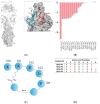
Figure 1
Figure 1
Antibody-based cyclic peptide design strategy. (A) Structural representation of MAb 4-5 (cartoon) and SFTSV Gn (surface) complex (PDB code: 5Y11). Interface residues of MAb 4-5 within 4.0 Å of SFTSV Gn in protein–protein interaction are represented by sticks. The pentapeptide selected for extension and cyclization are highlighted in red. (B) Residue-wise energy breakdown of the SFTSV Gn-MAb binding interface. Attractive and repulsive energies are shown in red and blue, respectively. (C) Diagram of the pentapeptide extension and cyclization. (D) The amino acid sequences of the four newly designed cyclic lariat peptides. The isopeptide bond-forming residues are highlighted in red.
Figure 2
Figure 2
Evaluation of the cyclic peptides’ anti-SFTSV activity and HKU-P1′s binding competition with the biotinylated cyclic peptide. (A) Anti-SFTSV activity of cyclic peptides HKU-P1 to HKU-P4. Out of the four peptides, HKU-P1, HKU-P2, and HKU-P4 displayed anti-SFTSV effects. (B) Sodium dodecyl sulfate-polyacrylamide gel electrophoresis showing the purity of His-tagged SFTSV Gn (lane 2). Lane 1 represents protein molecular weight marker. (C) Detection of the biotin-HKU-P1 binding to SFTSV Gn-coated immunoplate. (D) HKU-P1 inhibited the binding of biotin-HKU-P1 to the SFTSV Gn protein. ** p < 0.01 when compared the HKU-P1 and negative control peptide at each concentration by Student’s T-test.
Figure 3
Figure 3
The anti-SFTSV activity and mode of action of cyclic peptide HKU-P1. (A) Evaluation of the in vitro anti-SFTSV activity and cell toxicity of HKU-P1. The SFTSV intracellular viral RNA load at 1 day post infection (MOI = 0.01) in Huh-7 cells with HKU-P1 was quantified by RT-qPCR. Cell viability was determined at the same condition using CellTiter-Glo assay. SFTSV viral RNA load was normalized as % of the mock-treated control group. HKU-P1 displayed dose-dependent reduction of the SFTSV viral RNA load with >80% (p = 0.010) viral RNA load reduction being achieved at 1000 µg/mL of HKU-P1. The half-maximal inhibitory concentration (IC50) was calculated using non-linear regression analysis in GraphPad Prism. (B) Immunofluorescence staining of SFTSV-NP in virus-infected Huh-7 cells treated with HKU-P1. (C) Time-of-drug-addition assay was performed to determine the step in the SFTSV replication cycle that is interrupted by HKU-P1. HKU-P1 was either pre-incubated with Huh-7 cells at 1h pre-infection (Pre), co-incubated with SFTSV during virus entry (Neut), or added at 1 h post-infection (Post). The viral RNA load was normalized as % of mock-treated control group. Significant viral load reduction was observed in the neutralization group. This was compatible with the postulated mechanism of the cyclic peptides as they were designed as SFTSV-Gn binders. ** denotes p < 0.01 (compared to the mock-treated control group with Student’s t-test). Data are presented as mean values ± standard deviations (n = 3). All experiments were performed in triplicate and repeated twice for confirmation.
Figure 4
Figure 4
Predicted binding mode of HKU-P1 to SFTSV Gn. (A) Conformational landscape analysis of HKU-P1 bound to SFTSV Gn, showing computed binding energy against RMSD to native pentapeptide. Each point represents a conformational sampling attempt succeeded in cyclization. Colors indicate the number of intramolecular hydrogen bonds. (B) Intramolecular hydrogen bonding in top conformation of HKU-P1; hydrogen bonds were indicated with yellow dashed lines. (C) Overlay of top conformation and native pentapeptide. The RMSD value of aligned part was labelled. (D) Binding mode of HKU-P1 top conformation to the SFTSV Gn surface. HKU-P1 top conformation and SFTSV Gn were represented by sticks and surfaces, respectively. Hydrogen bonding residues of SFTSV Gn were shown in green sticks and labelled, and hydrogen bonds were indicated with yellow dashed lines.
Figure 4
Figure 4
Predicted binding mode of HKU-P1 to SFTSV Gn. (A) Conformational landscape analysis of HKU-P1 bound to SFTSV Gn, showing computed binding energy against RMSD to native pentapeptide. Each point represents a conformational sampling attempt succeeded in cyclization. Colors indicate the number of intramolecular hydrogen bonds. (B) Intramolecular hydrogen bonding in top conformation of HKU-P1; hydrogen bonds were indicated with yellow dashed lines. (C) Overlay of top conformation and native pentapeptide. The RMSD value of aligned part was labelled. (D) Binding mode of HKU-P1 top conformation to the SFTSV Gn surface. HKU-P1 top conformation and SFTSV Gn were represented by sticks and surfaces, respectively. Hydrogen bonding residues of SFTSV Gn were shown in green sticks and labelled, and hydrogen bonds were indicated with yellow dashed lines.

References

    1. Maes P., Alkhovsky S.V., Bào Y., Beer M., Birkhead M., Briese T., Buchmeier M.J., Calisher C.H., Charrel R.N., Choi I.R., et al. Taxonomy of the family Arenaviridae and the order Bunyavirales: Update. Arch. Virol. 2018;163:2295–2310. doi: 10.1007/s00705-018-3843-5. - DOI - PubMed
    1. Li J., Li S., Yang L., Cao P., Lu J. Severe fever with thrombocytopenia syndrome virus: A highly lethal bunyavirus. Crit. Rev. Microbiol. 2020;47:112–125. doi: 10.1080/1040841X.2020.1847037. - DOI - PubMed
    1. Leventhal S., Wilson D., Feldmann H., Hawman D. A Look into Bunyavirales Genomes: Functions of Non-Structural (NS) Proteins. Viruses. 2021;13:314. doi: 10.3390/v13020314. - DOI - PMC - PubMed
    1. Liu S., Chai C., Wang C., Amer S., Lv H., He H., Sun J., Lin J. Systematic review of severe fever with thrombocyto-penia syndrome:virology, epidemiology, and clinical characteristics. Rev. Med. Virol. 2014;24:90–102. doi: 10.1002/rmv.1776. - DOI - PMC - PubMed
    1. Hofmann H., Li X.X., Zhang X.A., Liu W., Kuhl A., Kaup F., Soldan S.S., Gonzalez-Scarano F., Weber F., He Y.X., et al. Severe Fever with Thrombocytopenia Virus Glycoproteins Are Targeted by Neutralizing Antibodies and Can Use DC-SIGN as a Receptor for pH-Dependent Entry into Human and Animal Cell Lines. J. Virol. 2013;87:4384–4394. doi: 10.1128/JVI.02628-12. - DOI - PMC - PubMed

Publication types

MeSH terms

LinkOut - more resources