Review
. 2021 Oct 14;13(10):2066.
doi: 10.3390/v13102066.

Glycan Recognition in Human Norovirus Infections

Affiliations
Review

Glycan Recognition in Human Norovirus Infections

Victoria R Tenge et al. Viruses. .

Abstract

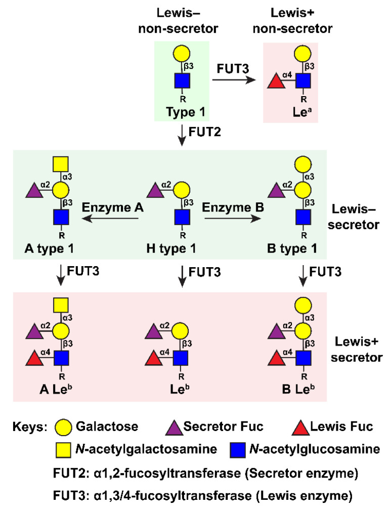
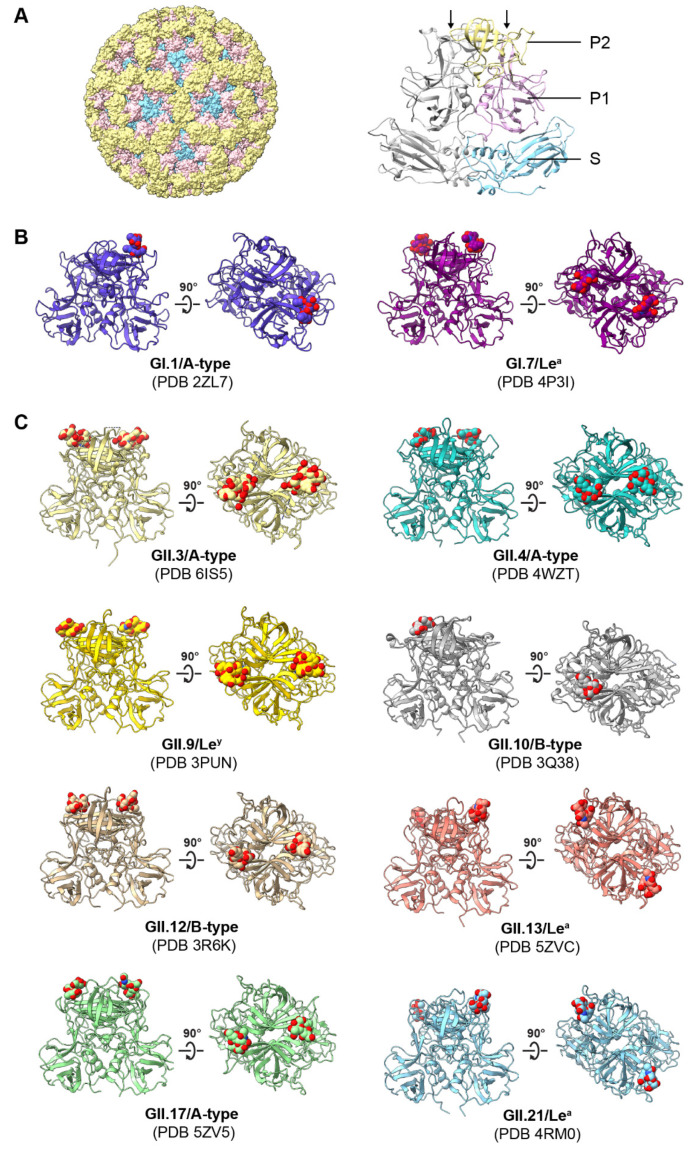
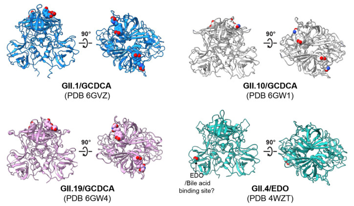
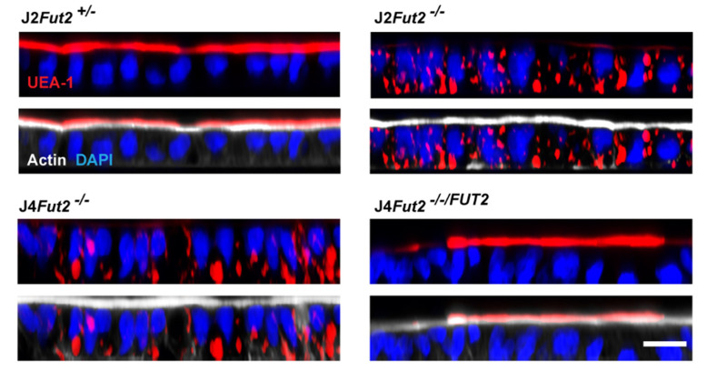
Recognition of cell-surface glycans is an important step in the attachment of several viruses to susceptible host cells. The molecular basis of glycan interactions and their functional consequences are well studied for human norovirus (HuNoV), an important gastrointestinal pathogen. Histo-blood group antigens (HBGAs), a family of fucosylated carbohydrate structures that are present on the cell surface, are utilized by HuNoVs to initially bind to cells. In this review, we describe the discovery of HBGAs as genetic susceptibility factors for HuNoV infection and review biochemical and structural studies investigating HuNoV binding to different HBGA glycans. Recently, human intestinal enteroids (HIEs) were developed as a laboratory cultivation system for HuNoV. We review how the use of this novel culture system has confirmed that fucosylated HBGAs are necessary and sufficient for infection by several HuNoV strains, describe mechanisms of antibody-mediated neutralization of infection that involve blocking of HuNoV binding to HBGAs, and discuss the potential for using the HIE model to answer unresolved questions on viral interactions with HBGAs and other glycans.

Keywords: glycoconjugates; histo-blood group antigens; host–virus interactions; human intestinal enteroids/organoids; human noroviruses; structure.


Conflict of interest statement

M.K.E. is named as an inventor on patents related to cloning of the Norwalk virus genome and HuNoV cultivation, and is a consultant to and received research funding from Takeda Vaccines, Inc. R.L.A. is named as an inventor on patents related to HuNoV cultivation and has received research funding from Takeda Vaccines, Inc. The funders had no role in the design of the study; in the collection, analyses, or interpretation of data; in the writing of the manuscript; or in the decision to publish the results.

Figures

Figure 1
Figure 1
HBGA-chain modification pathways on the type-1 structure. Adapted from Hu et al., 2018 under the terms of the Creative Commons Attribution 4.0 International license (https://creativecommons.org/licenses/by/4.0/) [23]. Accessed on 25 August 2021.
Figure 2
Figure 2
HBGAs bind to HuNoV P domain. (A) Crystal structure of the Norwalk virus-like particle (left) comprised of 90 VP1 dimers (right) (PDB ID: 1IHM). The S domain, P1, and P2 subdomains are colored in blue, pink, and yellow, respectively. The second chain of the VP1 dimer is shown in gray. Arrows point to the P-loops in P2. (B,C) HBGA binding sites located on P domain dimers of GI and GII HuNoVs. The P domains (ribbon representation) and HBGAs (spheres) are shown as side views of the P domain dimers or rotated 90 degrees to show a top view. HBGA carbon coloration matches the ribbon diagram and heteroatoms are colored by element: oxygen, red; nitrogen, blue.
Figure 3
Figure 3
Bile-acid binding sites located on P domain dimers of GII HuNoVs. The P domains and bile acid are shown in ribbon representation and as spheres, respectively. The binding site of bile acid GCDCA on P domain dimers was reported by Kilic et al. [85]. An ethylene glycol (EDO, sphere representation) molecule binds to a GII.4 P domain at the bile acid binding site proposed by Creutznacher et al. [84]. GCDCA/EDO carbon coloration matches the ribbon diagram; heteroatoms are colored by element: oxygen, red; nitrogen, blue.
Figure 4
Figure 4
FUT2 is necessary for α1, 2-fucose expression on the apical surface. Previously unpublished images. HBGA expression was analyzed by UEA-1 lectin (red) in HIE lines as described in [48]. In all image panels, the nuclei are marked with DAPI (blue). In the bottom panels, the brush border is indicated by actin expression using phalloidin (white) for each line.
Figure 5
Figure 5
Jejunal HIE line J2 expressed α2,6-sialic acid glycans. Previously unpublished images. Monolayers of J2 (left), Chinese hamster ovary cells (CHO; middle), and Caco-2 cells (right) were fixed, permeabilized, and stained with Cy3 labeled Sambucus nigra lectin (orange), following protocols previously used for UEA-1 staining [48]. Caco-2 cells, that express α2,6-sialic acids, and CHO cells, that do not express α2,6-sialic acids, were included as positive and negative controls, respectively.

Similar articles

Cited by

References

    1. Paulson J.C., Colley K.J. Glycosyltransferases: Structure, localization, and control of cell type-specific glycosylation. J. Biol. Chem. 1989;264:17615–17618. doi: 10.1016/S0021-9258(19)84610-0. - DOI - PubMed
    1. Gagneux P., Aebi M., Varki A. Evolution of Glycan Diversity. In: Varki A., Cummings R.D., Esko J.D., Stanley P., Hart G.W., Aebi M., Darvill A.G., Kinoshita T., Packer N.H., Prestegard J.H., et al., editors. Essentials of Glycobiology. Cold Spring Harbor Laboratory Press; New York, NY, USA: 2015–2017. pp. 253–264. [Internet] - DOI
    1. Rini J.M., Esko J.D. Glycosyltransferases and Glycan-Processing Enzymes. In: Varki A., Cummings R.D., Esko J.D., Stanley P., Hart G.W., Aebi M., Darvill A.G., Kinoshita T., Packer N.H., Prestegard J.H., et al., editors. Essentials of Glycobiology. Cold Spring Harbor Laboratory Press; New York, NY, USA: 2015–2017. pp. 65–75. [Internet] - DOI - PubMed
    1. Ruvoen-Clouet N., Magalhaes A., Marcos-Silva L., Breiman A., Figueiredo C., David L., Le Pendu J. Increase in Genogroup II.4 Norovirus Host Spectrum by CagA-Positive Helicobacter pylori Infection. J. Infect. Dis. 2014;210:183–191. doi: 10.1093/infdis/jiu054. - DOI - PubMed
    1. Qu D., Wang G., Yu L., Tian F., Chen W., Zhai Q. The effects of diet and gut microbiota on the regulation of intestinal mucin glycosylation. Carbohydr. Polym. 2021;258:117651. doi: 10.1016/j.carbpol.2021.117651. - DOI - PubMed

Publication types

MeSH terms

LinkOut - more resources