Case Reports
. 2022 Jun;24(2):166-171.
doi: 10.7461/jcen.2021.E2021.08.004. Epub 2021 Oct 26.

Transvenous coil embolization of hypoglossal canal dural arteriovenous fistula using detachable coils: A case report

Affiliations
Case Reports

Transvenous coil embolization of hypoglossal canal dural arteriovenous fistula using detachable coils: A case report

Su Min Kye et al. J Cerebrovasc Endovasc Neurosurg. 2022 Jun.

Abstract

The hypoglossal canal (HC) is an unusual location of the posterior fossa dural arteriovenous fistula (AVF), which usually occurs in the transverse or sigmoid sinus. Herein, we report a case of HC dural AVF successfully treated with transvenous coil embolization using detachable coils in a 68-year-old woman who presented with headache and left pulsatile tinnitus for 2 months. Brain magnetic resonance imaging (MRI) and cerebral angiography revealed left HC dural AVF. The pulsatile bruit disappeared immediately after the procedure. Follow-up MRI showed complete disappearance of the fistula. Precise localization of the fistula through careful consideration of the anatomy and transvenous coil embolization using a detachable coil can facilitate the treatment for HC dural AVF.

Keywords: Coil Embolization; Dural Arteriovenous Fistula; Hypoglossal canal.


Figures

Fig. 1.
Fig. 1.
Magnetic resonance angiography shows abnormal signal hyperintensity around the left hypoglossal canal (the “magic wand appearance”) (arrow) (A, B). A bone imaging computed tomography shows bone erosion and expansion of left hypoglossal canal (arrow) (C).
Fig. 2.
Fig. 2.
Cerebral angiography reveals a dural arteriovenous fistula in the anterior condylar vein within the left hypoglossal canal (HC) fed by neuromeningeal branches of the bilatreral ascending pharyngeal artery (APA), occipital artery (A, B, C) and vertebral artery (D), which drained into internal jugular vein (IJV), cavernous sinus (CS) through inferior petrosal sinus (IPS), and vertebral venous plexus (VVP). Schematic illustration shows detailed anatomy of dural arteriovenous fistula (E).
Fig. 3.
Fig. 3.
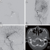
Microcatheter angiography demonstrated dilated venous pouch connected with vertebral venous plexus (arrow) and inferior petrosal sinus (arrowhead) (A). Final angiography showed complete occlusion of dural arteriovenous fistula and contrast stagnation around the hypoglossal canal (arrow) (B, C). CT shows coil mass in the hypoglossal canal (D). CT, computed tomography.
Fig. 4.
Fig. 4.
Follow-up magnetic resonance image (A), and magnetic resonance angiography (B) showed complete disappearance of dural arteriovenous fisula.

Similar articles

Cited by

References

    1. Barnwell SL, Halbach VV, Dowd CF, Higashida RT, Hieshima GB. Dural arteriovenous fistulas involving the inferior petrosal sinus: angiographic findings in six patients. AJNR Am J Neuroradiol. 1990 May;11(3):511–6. - PMC - PubMed
    1. Blomquist MH, Barr JD, Hurst RW. Isolated unilateral hypoglossal neuropathy caused by dural arteriovenous fistula. AJNR Am J Neuroradiol. 1998 May;19(5):951–3. - PMC - PubMed
    1. Choi JW, Kim BM, Kim DJ, Kim DI, Suh SH, Shin N-Y, et al. Hypoglossal canal dural arteriovenous fistula: incidence and the relationship between symptoms and drainage pattern. J Neurosurg. 2013 Oct;119(4):955–60. - PubMed
    1. Combarros O, Alvarez de Arcaya A, Berciano J. Isolated unilateral hypoglossal nerve palsy: nine cases. J Neurol. 1998 Feb;245(2):98–100. - PubMed
    1. Ernst R, Bulas R, Tomsick T, van Loveren H, Aziz KA. Three cases of dural arteriovenous fistula of the anterior condylar vein within the hypoglossal canal. AJNR Am J Neuroradiol. 1999 Nov-Dec;20(10):2016–20. - PMC - PubMed

Publication types