. 2021 Dec;12(2):9411-9423.
doi: 10.1080/21655979.2021.1988372.

MicroRNA-4423-3p inhibits proliferation of fibroblast-like synoviocytes by targeting matrix metalloproteinase 13 in rheumatoid arthritis

Affiliations

MicroRNA-4423-3p inhibits proliferation of fibroblast-like synoviocytes by targeting matrix metalloproteinase 13 in rheumatoid arthritis

Weihong Xu et al. Bioengineered. 2021 Dec.

Abstract

Rheumatoid arthritis (RA) is a chronic inflammatory autoimmune disease that is increasing in incidence worldwide. RA is regulated by a variety of microRNAs (miRNAs/miR). Moreover, analysis of public data has revealed that miR-4423-3p is significantly downregulated in peripheral blood mononuclear cells of RA patients. This study investigated the role of miR-4423-3p in RA. The levels of miR-4423-3p and matrix metalloproteinase 13 (MMP13) in RA patients and the regulatory relationship between miR-4423-3p and MMP13 were analyzed using public data. A dual-luciferase reporter assay was performed to verify that miR-4423-3p targets MMP13 in human fibroblast-like synoviocyte (HFLS) RA cells (HFLS-RA). Following the overexpression of miR-4423-3p, miR-4423-3p inhibitor, and MMP13 in HFLS-RA, viability, proliferation, cell cycle, apoptosis, and invasion/migration assays were used to detect the effects of miR-4423-3p targeting MMP13 on cell biological processes. The results revealed that miR-4423-3p was downregulated in peripheral blood mononuclear cells of RA patients and MMP13 was upregulated in synovial tissue of RA patients. miR-4423-3p targets the 3' untranslated region of MMP13 and downregulates MMP13 expression. After overexpression of miR-4423-3p, cell proliferation, migration, and invasion were inhibited, the cell cycle was prevented and cell apoptosis was promoted. Overexpression of MMP13 promoted cell proliferation, migration, and invasion, while accelerating the cell cycle process and suppressing apoptosis. The findings indicate that in HFLS-RA cells, overexpression of miR-4423-3p inhibited proliferation, migration, and invasion, and promoted apoptosis by negatively regulating MMP13. The overexpression of miR-4423-3p might be a novel strategy for the treatment of RA.

Keywords: Rheumatoid arthritis; fibroblast-like synoviocytes; microRNA; mmp13.


Conflict of interest statement

No potential conflict of interest was reported by the author(s).

Figures

Figure 1.
Figure 1.
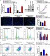
miR-4423-3p inhibits proliferation of HFLS-RA. A: The levels of miR-4423-3p were differentially expressed in healthy people (n = 18) and RA patients (n = 28) in data from the GEO database (GSE124373); ***P < 0.001. B: Intracellular miR-4423-3p expression changes in different groups. C: The MTT assay was used to determine the changes of cell viability in each group. D: Representative images of EdU assay (left) and statistical histograms of EdU+ cells (right). E: Representative images of cell cycle assay (left) and statistical histograms of cell proportions in different phases (right). F: Representative image of apoptosis assay and statistical histogram of the proportion of apoptotic cells in different groups. Mann-Whitney test (a) and student’s t test (b–f) were used for statistical analysis. All experiments were carried out independently at least three times. PI: propidium iodide. **P < 0.01, ***P < 0.001 vs. mimic NC; ns: not significant, #P < 0.05, ##P < 0.01, ###P < 0.001 vs. inhibitor NC
Figure 2.
Figure 2.
miR-4423-3p suppresses migration and invasion of HFLS-RA. A: Representative images of migrated cells (up) and statistical histograms of the number of migrated cells in per field in different groups (bottom). B: Representative images of invasive cells (up) and statistical histograms of the number of invasive cells in per field in different groups (bottom). Student’s t test (a and b) was used for statistical analysis. All experiments were carried out independently at least three times. *P < 0.05, ***P < 0.001 vs. mimic NC; ##P < 0.01 vs. inhibitor NC
Figure 3.
Figure 3.
MMP13 is a direct target of miR-4423-3p. A: Venn diagram of mRNAs targeted by miR-4423-3p. B: Schematic diagram of the 3ʹUTR mutation site of MMP13. C: Dual-luciferase reporter assay verified the targeting relationship between miR-4423-3p and MMP13. Ns: not significant, **P < 0.01 vs. mimic NC. D and E: After overexpression or inhibition of miR-4423-3p in HFLS-RA, mRNA and protein levels of MMP13 were detected by RT-PCR (d) and western blotting (e), respectively. ****P < 0.0001 vs. mimic NC; ###P < 0.001 vs. inhibitor NC. F: Analysis of the difference of MMP13 levels between RA patients (n = 16) and healthy volunteers (n = 7) from GSE77298; ***P < 0.001. Student’s t test (c and d) and Mann-Whitney test (f) were used for statistical analysis. All experiments were carried out independently at least three times
Figure 4.
Figure 4.
Replenishing MMP13 promotes cell proliferation and decreases apoptosis. The miR-4423-3p mimic and MMP13 OE plasmid were co-transfected into HFLS-RA. A: The RNA levels of miR-4423-3p and MMP13 were tested by RT-PCR. B: The protein level of MMP13 was detected by western blotting. C and D: Cell proliferation was analyzed using MTT (c) and EdU (d) assays. E: Cell cycle was detected using PI staining. F: Apoptosis was analyzed using Annexin V-FITC/PI staining. One-way ANOVA (A‒F) was used for statistical analysis. All experiments were carried out independently at least three times. PI: propidium iodide; mimic NC + Vector: mimic NC and pCDH-EF1α-Flag-T2A-puro vector were co-transfected into HFLS-RA; miR-4423-3p mimic + Vector: miR-4423-3p mimic and pCDH-EF1α-Flag-T2A-puro vector co-transfected into HFLS-RA; miR-4423-3p mimic + MMP13 OE: miR-4423-3p mimic and MMP13 OE plasmid co-transfected into HFLS-RA. **P < 0.01, ***P < 0.001, ****P < 0.0001 vs. mimic NC + Vector; ns: not significant, ##P < 0.01, ###P < 0.001, ####P < 0.0001 vs. miR-4423-3p mimic + Vector
Figure 5.
Figure 5.
miR-4423-3p suppresses migration and invasion of HFLS-RA by targeting MMP13. A: Representative images of migrated cells (left) and statistical histograms of the number of migrated cells in per field in different groups (right). B: Representative images of invasive cells (left) and statistical histograms of the number of invasive cells in per field in different groups (right). One-way ANOVA (a and b) was used for statistical analysis. All experiments were carried out independently at least three times. mimic NC + Vector: mimic NC and pCDH-EF1α-Flag-T2A-puro vector co-transfected into HFLS-RA; miR-4423-3p mimic + Vector: miR-4423-3p mimic and pCDH-EF1α-Flag-T2A-puro vector co-transfected into HFLS-RA; miR-4423-3p mimic + MMP13 OE: miR-4423-3p mimic and MMP13 OE plasmid co-transfected into HFLS-RA. ***P < 0.001 vs. mimic NC + Vector; ####P < 0.0001 vs. miR-4423-3p mimic + Vector
Figure 6.
Figure 6.
Detection of apoptosis, cell cycle and EMT-related genes. A: The mRNA levels of Bax, Bcl-2, CDK2, Cyclin D1, E-cadherin, N-cadherin, and Vimentin were tested by RT-PCR. B: The protein levels of Bax, Bcl-2, CDK2, Cyclin D1, E-cadherin, N-cadherin, and vimentin were tested by western blotting. One-way ANOVA (a and b) was used for statistical analysis. All experiments were carried out independently at least three times. mimic NC + Vector: mimic NC and pCDH-EF1α-Flag-T2A-puro vector co-transfected into HFLS-RA; miR-4423-3p mimic + Vector: miR-4423-3p mimic and pCDH-EF1α-Flag-T2A-puro vector co-transfected into HFLS-RA; miR-4423-3p mimic + MMP13 OE: miR-4423-3p mimic and MMP13 OE plasmid co-transfected into HFLS-RA. ns: not significant, **P < 0.01, ***P < 0.001, ***P < 0.001 vs. mimic NC + Vector; ##P < 0.01, ###P < 0.001, ####P < 0.0001 vs. miR-4423-3p mimic + Vector

References

    1. Imas JJ, Ruiz Zamarreño C, Zubiate P, et al.Optical Biosensors for the Detection of Rheumatoid Arthritis (RA) Biomarkers: a Comprehensive Review.Basel, Switzerland Sensors.2020.20(21):6289. - PMC - PubMed
    1. Wysocki T, Olesińska M, Paradowska-Gorycka A.. Current Understanding of an Emerging Role of HLA-DRB1 Gene in Rheumatoid Arthritis-From Research to Clinical Practice. Cells. 2020;9(5):1127. - PMC - PubMed
    1. Littlejohn EA, Monrad SU.. Early Diagnosis and Treatment of Rheumatoid Arthritis. Prim Care. 2018;45(2):237–255. - PubMed
    1. Behl T, Kaur I, Sehgal A, et al. The Lipid Paradox as a Metabolic Checkpoint and Its Therapeutic Significance in Ameliorating the Associated Cardiovascular Risks in Rheumatoid Arthritis Patients. Int J Mol Sci. 2020;21(24):9505. - PMC - PubMed
    1. Donlin LT, Jayatilleke A, Giannopoulou EG, et al. Modulation of TNF-induced macrophage polarization by synovial fibroblasts. J Immunol. 2014;193(5):2373–2383. - PMC - PubMed

Publication types