. 2021 Oct 25;40(1):337.
doi: 10.1186/s13046-021-02129-9.

Super-enhancer-associated TMEM44-AS1 aggravated glioma progression by forming a positive feedback loop with Myc

Affiliations

Super-enhancer-associated TMEM44-AS1 aggravated glioma progression by forming a positive feedback loop with Myc

Erbao Bian et al. J Exp Clin Cancer Res. .

Abstract

Background: Long non-coding RNAs (lncRNAs) have been considered as one type of gene expression regulator for cancer development, but it is not clear how these are regulated. This study aimed to identify a specific lncRNA that promotes glioma progression.

Methods: RNA sequencing (RNA-seq) and quantitative real-time PCR were performed to screen differentially expressed genes. CCK-8, transwell migration, invasion assays, and a mouse xenograft model were performed to determine the functions of TMEM44-AS1. Co-IP, ChIP, Dual-luciferase reporter assays, RNA pulldown, and RNA immunoprecipitation assays were performed to study the molecular mechanism of TMEM44-AS1 and the downstream target.

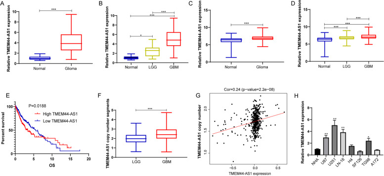
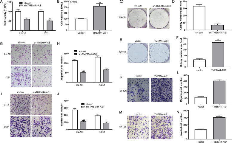
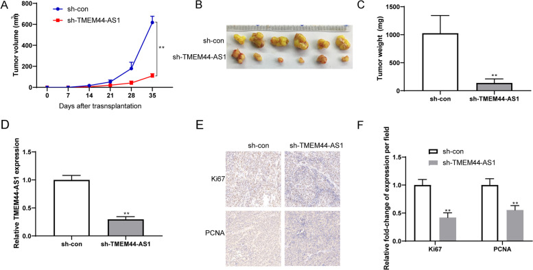
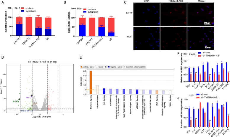
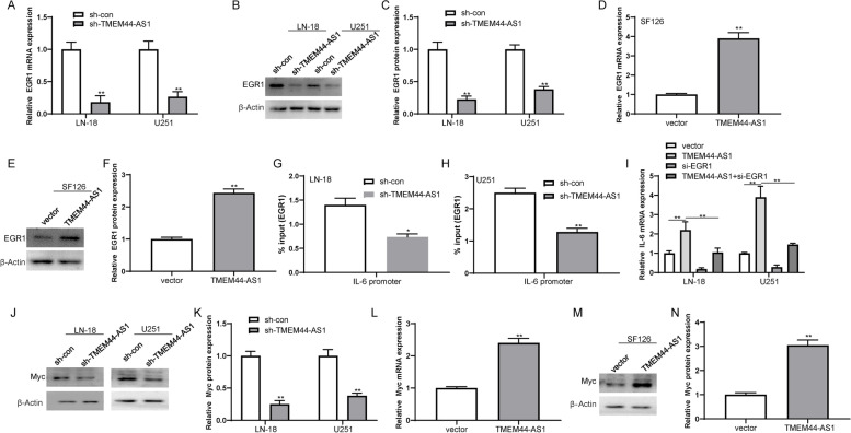
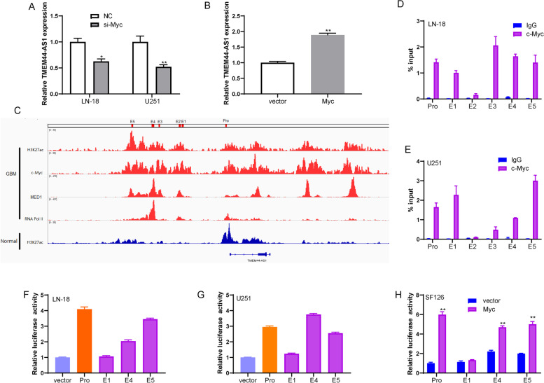
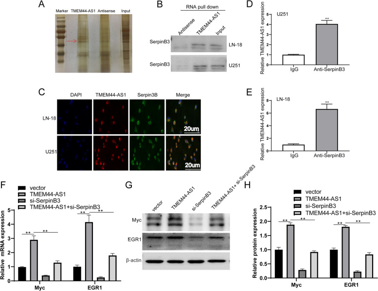
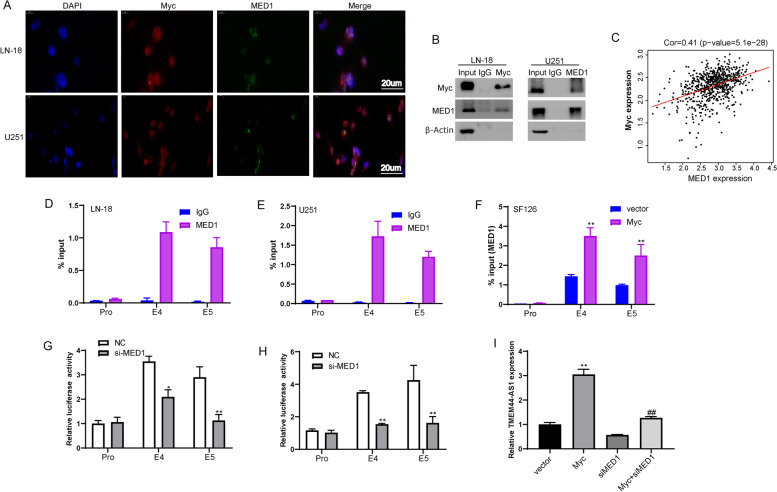
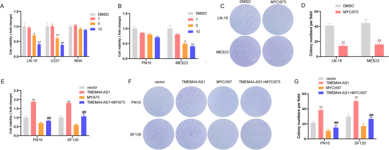
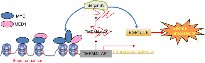
Results: We identified a novel lncRNA TMEM44-AS1, which was aberrantly expressed in glioma tissues, and that increased TMEM44-AS1 expression was correlated with malignant progression and poor survival for patients with glioma. Expression of TMEM44-AS1 increased the proliferation, colony formation, migration, and invasion of glioma cells. Knockdown of TMEM44-AS1 in glioma cells reduced cell proliferation, colony formation, migration and invasion, and tumor growth in a nude mouse xenograft model. Mechanistically, TMEM44-AS1 is directly bound to the SerpinB3, and sequentially activated Myc and EGR1/IL-6 signaling; Myc transcriptionally induced TMEM44-AS1 and directly bound to the promoter and super-enhancer of TMEM44-AS1, thus forming a positive feedback loop with TMEM44-AS. Further studies demonstrated that Myc interacts with MED1 regulates the super-enhancer of TMEM44-AS1. More importantly, a novel small-molecule Myc inhibitor, Myci975, alleviated TMEM44-AS1-promoted the growth of glioma cells.

Conclusions: Our study implicates a crucial role of the TMEM44-AS1-Myc axis in glioma progression and provides a possible anti-glioma therapeutic agent.

Keywords: Glioma; Myc; Super-enhancer; TMEM44-AS1; lncRNA.


Conflict of interest statement

The authors declare that they have no competing interests.

Figures

Fig. 1
Fig. 1
Expression of TMEM44-AS1 in various tissues and cells. (A) Relative TMEM44-AS1 expression in glioma and normal brain tissues from our cohort. (***P < 0.001). (B) The TMEM44-AS1 expression is shown according to the histopathologic grades. (*P < 0.05, ***P < 0.001). (C) Relative TMEM44-AS1 expression in glioma and normal brain tissues from GTEX(n = 1136) and the TCGA database(n = 689). (***P < 0.001). (D) The TMEM44-AS1 expression is shown according to the histopathologic grades of TCGA gliomas. LGG (n = 523); GBM (n = 166). (***P < 0.001). (E) Kaplan-Meier curves of overall survival time of patients based on TMEM44-AS1 expression in glioma samples obtained from the TCGA database. (F) TMEM44-AS1 copy number segments in LGG and GBM were tested using the TCGA database. (***P < 0.001). (G) The correlation between TMEM44-AS1 copy number and TMEM44-AS1 expression in glioma from the TCGA database (n = 528). (H) Relative TMEM44-AS1 in NHA, U87, U251, LN-18, H4, SF126, T98G, A172 cell lines (*P < 0.05, **P < 0.01)
Fig. 2
Fig. 2
TMEM44-AS1 promotes glioma cell proliferation, colony formation, migration and invasion. (A) Cell viability of LN-18 and U251 cells transfected with sh-TMEM44-AS1 or sh-con was evaluated with CCK8 assay, respectively. *P < 0.05, **P < 0.01. (B) CCK-8 assay of SF126 cells transfected with TMEM44-AS1 plasmid or vector. **P < 0.01. (C-D) Colony formation assay of LN-18 cells transfected with sh-TMEM44-AS1 or sh-con. **P < 0.01. (E-F) Colony formation assay of SF126 cells transfected with TMEM44-AS1 or empty vector. **P < 0.01. (G-J) Representative images of transwell migration and invasion assays performed in LN-18 and U251 transfected sh-TMEM44-AS1 or sh-con cells. **P < 0.01. (K-N) Representative images of transwell migration and invasion assays in SF126 cells after transfection of sh-TMEM44-AS1 or sh-con. **P < 0.01. Data are presented as mean ± SD. The independent biological experiments were repeated at least three times
Fig. 3
Fig. 3
TMEM44-AS1 promotes glioma cell growth in vivo. (A-C) Glioma cells stably transfected with either sh-TMEM44-AS1 or sh-con were inoculated into the flanks of nude mice to construct a transplanted tumor model. Representative images of tumor volume growth curve and tumor weight were shown. (D) RT-qPCR was conducted to detect the expression levels of TMEM44-AS1 in tumor samples. **P < 0.01. (E) Ki67 and PCNA immunohistochemical (IHC) staining of tumor tissues of mice injected with sh-con or sh-TMEM44-AS1 cells. (F) The Ki67 and PCNA positive cells were qualified. **p < 0.01 vs. sh-con
Fig. 4
Fig. 4
TMEM44-AS1 represses Myc translocation. (A-B) Subcellular fractionation analysis was performed in LN-18 and U251 cells. Cytoplasmic fraction (blue), and nuclear fraction (red) and probed for TMEM44-AS1. GAPDH was used as a cytoplasmic marker, U6 as a nuclear marker, and MALAT1 as the positive control. (C) RNA FISH assays for TMEM44-AS1. Nuclei were stained with DAPI. Scale bar = 20 μm. (D) Volcan representation of genes differentially expressed in LN-18 knockdown TMEM44-AS1 vs. LN-18 sh-con cells (green, downregulation; red, upregulation). (E) Ingenuity pathway analysis revealed that genes differentially expressed upon TMEM44-AS1 knockdown are markedly related to several cancer-associated signaling pathways. (F-G) Expression of p38MAPK and IL-6 signaling pathway associated with genes in LN-18 and U251 cells transfected with sh-TMEM44-AS1 or sh-con. *P < 0.05, **P < 0.01
Fig. 5
Fig. 5
TMEM44-AS1 regulated EGR1/IL-6 and Myc signaling in glioma cells. (A-C) The expression of EGR1 upon knockdown of TMEM44-AS1 in LN-18 and U251 cells. **P < 0.01. (D-F) Expression of EGR1 in SF126 cells was examined transfected with vector or TMEM44-AS1 plasmid. **P < 0.01.(G-H) ChIP analyses of TMEM44-AS1 knockdown LN-18 and U251 cells were conducted on indicated IL-6 primer using the EGR1 antibodies. Enrichment is determined relative to input controls. *P < 0.05, **P < 0.01 vs. sh-con. (I) IL-6 expression in SF126 cells co-transfected with TMEM44-AS1 plasmid and si-EGR1. **p < 0.01 vs. vector; **P < 0.01 vs. TMEM44-AS1. (J-K) Expression of Myc protein in LN-18 and U251 cells transfected with sh-TMEM44-AS1 or sh-con. **P < 0.01. (L-N) Expression of Myc in SF126 cells transfected with vector or TMEM44-AS1 expression plasmid. **P < 0.01. Data are from three biological replicates represented as mean ± SD
Fig. 6
Fig. 6
Myc bind to TMEM44-AS1 super-enhancer and promoter region, and promoted its transcription. (A) RT-qPCR assays measuring the expression of TMEM44-AS1 upon knockdown of Myc in LN-18 and U251 cells. *P < 0.05, **P < 0.01. (B) Expression of TMEM44-AS1 in SF126 cells was examined transfected with vector or Myc plasmid. **P < 0.01. (C) ChIP-seq profiles of H3K27ac, Myc, MED1, and RNA pol II in GBM cells and H3K27ac in normal brain tissues. (D-E) Myc ChIP followed by RT-qPCR analyzed the occupation of Myc on the super-enhancer and promoter of TMEM44-AS1. The co-immunoprecipitated DNA was amplified by PCR using indicated primer. (F-G) Enhancer and promoter activity measured by luciferase reporter assays in LN-18 and U251 cells. Three constituent enhancers (E1, E4 and E5) within the SE were separately cloned into luciferase reporter vector pGL3-enhancer and the promoter was cloned into luciferase reporter vector pGL3-promoter. (H) Luciferase assay analysis of SF126 cells transfected with TMEM44-AS1 promoter, or constituent enhancers (E1, E4 and E5) constructs together with Myc or vector
Fig. 7
Fig. 7
The interaction of TMEM44-AS1 and SerpinB3 activates Myc. (A) TMEM44-AS1 pulldown assay was performed and the associated proteins were separated with SDS-PAGE and silver staining. (B) SerpinB3 in TMEM44-AS1 RNA pulldown were analyzed by western blot. (C) Co-localization of TMEM44-AS1 and SerpinB3 in glioma cells. TMEM44-AS1 probes (red) and SerpinB3 antibody (green) were used for staining. Scale bars, 20 μm. (D-E) RNA immunoprecipitation (RIP) using the SerpinB3 antibody followed by RT-qPCR for TMEM44-AS1. (F-H) Expression of Myc and EGR1 in LN-18 and U251 cells co-transfected with TMEM44-AS1 plasmid and si-SerpinB3. **p < 0.01 vs. vector; **P < 0.01 vs. TMEM44-AS1
Fig. 8
Fig. 8
Myc affects MED1 occupancy at the TMEM44-AS1 super-enhancer in glioma cells. (A) Immunofluorescence staining to detect the co-localization Myc (red), and MED1 (green), and DAPI (blue) in LN-18 and U251 cells. (B) Co-IP assays were performed using LN-18 and U251 cells lysate with Myc or MED1 antibodies and detected by western blot with indicated antibodies. (C) Correlation between Myc and MED1 expression in the TCGA glioma dataset. (D-E) MED1 ChIP followed by qPCR analyzed the occupation of MED1 on the super-enhancer and promoter of TMEM44-AS1. The co-immunoprecipitated DNA was amplified by PCR using indicated primer. (F) The enrichment of MED1 on the enhancers of TMEM44-AS1 was analyzed with ChIP assays using the MED1 antibody after SF126 cells were transfected with vector or Myc plasmid. **P < 0.01 vs. vector. The independent biological experiments were repeated at least three times. (G-H) The luciferase reporter assays were used to detect the TMEM44-AS1 promoter, or constituent enhancers (E4 and E5) in LN-18 and U251 cells transfected with si-MED1 or NC. *P < 0.05, **P < 0.01 vs. NC. The independent biological experiments were repeated at least three times. (I) TMEM44-AS1 expression in LN-18 and U251 cells co-transfected with Myc plasmid and si-MED1. **p < 0.01 vs. vector; ##P < 0.01 vs. Myc
Fig. 9
Fig. 9
Myci975 reversed TMEM44-AS1-promoted the growth of glioma cells .(A) CCK-8 assay of LN-18, U251, and NHA cells treated with DMSO or Myci975 at an indicated concentration. *P < 0.05, **P < 0.01 vs. DMSO. (B) CCK-8 assay of PN16 and MES23 treated with DMSO or Myci975 at an indicated concentration. *P < 0.05, **P < 0.01 vs. DMSO. (C-D) Colony formation assay of LN-18 and MES23 cells treated with DMSO or Myci975 at an indicated concentration. **P < 0.01 vs. DMSO. (E) CCK-8 assay of PN16 and SF126 cells transfected with TMEM44-AS1 plasmid following Myci975 treatment. **P < 0.01 vs. vector, ##P < 0.01 vs. TMEM44-AS1. (F-G) Colony formation assay of PN16 and SF126 cells transfected with TMEM44-AS1 plasmid following Myci975 treatment. **P < 0.01 vs. vector, ##P < 0.01 vs. TMEM44-AS1
Fig. 10
Fig. 10
Depiction of the proposed signaling network involved in the regulation of super-enhancer associated-TMEM44-AS1 in glioma progression

References

    1. Wen PY, Kesari S. Malignant gliomas in adults. N Engl J Med. 2008;359(5):492–507. doi: 10.1056/NEJMra0708126. - DOI - PubMed
    1. Louis DN, Perry A, Reifenberger G, von Deimling A, Figarella-Branger D, Cavenee WK, Ohgaki H, Wiestler OD, Kleihues P, Ellison DW. The 2016 World Health Organization classification of tumors of the central nervous system: a summary. Acta Neuropathol. 2016;131(6):803–820. doi: 10.1007/s00401-016-1545-1. - DOI - PubMed
    1. F. Graus, Bruna J., Pardo J., Escudero D., Vilas D., Barcelo I., Brell M., Pascual C., Crespo J. A., Erro E., Garcia-Romero J. C., Estela J., Martino J., Garcia-Castano A., Mata E., Lema M., Gelabert M., Fuentes R., Perez P., Manzano A., Aguas J., Belenguer A., Simon A., Henriquez I., Murcia M., Vivanco R., Rojas-Marcos I., Munoz-Carmona D., Navas I., de Andres P., Mas G., Gil M., Verger E. Patterns of care and outcome for patients with glioblastoma diagnosed during 2008-2010 in Spain. Neuro Oncol.2013; 15(6):797-805. - PMC - PubMed
    1. Mikheeva SA, Mikheev AM, Petit A, Beyer R, Oxford RG, Khorasani L, Maxwell JP, Glackin CA, Wakimoto H, Gonzalez-Herrero I, Sanchez-Garcia I, Silber JR, Horner PJ, Rostomily RC. TWIST1 promotes invasion through mesenchymal change in human glioblastoma. Mol Cancer. 2010;9:194. doi: 10.1186/1476-4598-9-194. - DOI - PMC - PubMed
    1. Huang Z, Zhou JK, Peng Y, He W, Huang C. The role of long noncoding RNAs in hepatocellular carcinoma. Mol Cancer. 2020;19(1):77. doi: 10.1186/s12943-020-01188-4. - DOI - PMC - PubMed

MeSH terms