. 2021 Oct 25;20(1):138.
doi: 10.1186/s12943-021-01444-1.

CircCD44 plays oncogenic roles in triple-negative breast cancer by modulating the miR-502-5p/KRAS and IGF2BP2/Myc axes

Affiliations

CircCD44 plays oncogenic roles in triple-negative breast cancer by modulating the miR-502-5p/KRAS and IGF2BP2/Myc axes

Jie Li et al. Mol Cancer. .

Erratum in

Abstract

Background: Emerging studies have revealed the potent functions of circRNAs in breast cancer tumorigenesis. However, the biogenesis, biofunction and mechanism of circRNAs in triple-negative breast cancer (TNBC) are largely unknown.

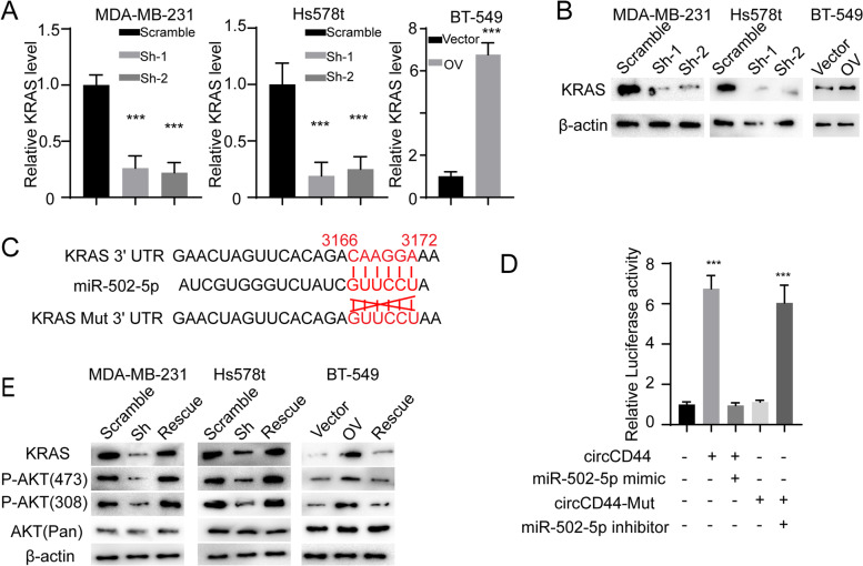
Methods: High-throughput RNA sequencing was applied to identify dysregulated circRNAs in TNBCs and paired normal tissues. RNA pulldown and luciferase assays were performed to investigate the interaction between circular CD44 (circCD44, also annotated as hsa_circ_0021735) and miR-502-5p. RNA pulldown and RIP assays were used to investigate the interaction between circCD44 and IGF2BP2. Cell viability, colony formation, migration/invasion assays and in vivo tumorigenesis were used to investigate circCD44 biological functions.

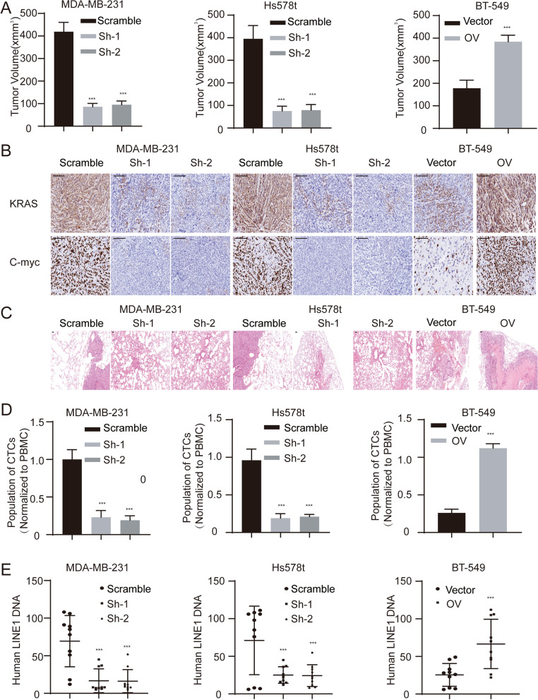
Results: CircCD44 is an uncharacterized circRNA, which is highly expressed in TNBC, and its expression is negatively correlated with the prognosis of TNBC patients. CircCD44 promotes TNBC proliferation, migration, invasion and tumorigenesis at least partially by sponging miR-502-5p and interacting with IGF2BP2.

Conclusion: Our data suggested that overexpressed circCD44 promotes TNBC progression. CircCD44 is potentially a novel diagnostic and therapeutic marker for TNBC patients.

Keywords: IGF2BP2; KRAS; MYC; TNBC; circCD44; circRNAs.


Conflict of interest statement

There are no potential conflicts of interest to disclose.

Figures

Fig. 1
Fig. 1
CircCD44 was upregulated in triple-negative breast cancers (TNBCs). A Schematic graph of the scanning strategy. Five paired normal tissues and TNBCs were included and subjected to circRNA sequencing to identify the dysregulated circRNAs, as we previously reported [13]. B Heatmap of candidate RNA sequences in TNBCs and paired normal tissues. Blue, downregulated circRNAs; red, upregulated circRNAs. C Schematic diagram of circCD44 formation from CircBase; validation strategy for circCD44 and Sanger sequencing of the junction of circCD44. D Left: FISH detection of circCD44 using a junction-specific probe in the indicated shRNA-transfected MDA-MB-231 cells; scale bar: 20 μm. Right: qRT–PCR analysis of circCD44 in different cell fractions. GAPDH and Malat1 were used as positive controls for the cytoplasm and nuclei. E qRT–PCR analysis of CD44 and circCD44 using random primers or oligo dT primers. F Relative RNA level of circCD44 in 36 TNBCs and 27 non-TNBCs and paired normal tissues. All RNA levels were normalized to the paired normal tissue of TNBC; ***, P < 0.001, NS, non-significant. G Relative circCD44 level in breast cancer cell lines and normal breast epithelial cells; ***, P < 0.001. H Thirty-six TNBC patients were divided into two groups with a cutoff of the mean level of circCD44 detected by qRT–PCR in the whole cohort. Kaplan–Meier survival analysis was then applied. Data are representative of at least 2–3 experiments with similar results
Fig. 2
Fig. 2
CircCD44 promotes the proliferation and chemoresistance of TNBCs. A Establishment of circCD44-stable knockdown and overexpression cell lines using two specific shRNAs or circCD44 plasmid. The relative RNA level was detected in each cell line; ***, P < 0.001. B CCK-8 assay of different cell lines with the indicated modifications; ***, P < 0.001. C Representative images of colony formation assays in different cell lines with the indicated modifications. Statistical analysis is shown in (E); ***, P < 0.001. D Representative image of the EdU assay in different cell lines with the indicated modifications. Statistical analysis is shown in (F). Scale bar: 50 μm; ***, P < 0.001. G The indicated cells were treated with Cyc, and the rate of apoptosis was detected; ***, P < 0.001. H. Immunoblot of the caspase cascade in different cell lines with the indicated modifications. C-C3, cleaved Caspase 3; C-C9, cleaved Caspase 9; C3, Caspase 3; C9, Caspase 9. Data are representative of at least 2–3 experiments with similar results
Fig. 3
Fig. 3
CircCD44 promotes the migration and invasion of TNBCs. A Representative image of the wound healing assay (upper) and relative wound closure (lower) in TNBCs with the indicated modifications, scale bar: 100 μm; ***, p < 0.001. B Representative image of the migration assay and invasion chamber assay in TNBCs with the indicated modifications; scale bar: 50 μm. C Statistical analysis of migration assay and invasion chamber assay in TNBCs with the indicated modifications; ***, P < 0.001. D Immunoblot of EMT markers in indicated cells. Data are representative of at least 2–3 experiments with similar results
Fig. 4
Fig. 4
CircCD44 acts as an endogenous competing RNA by sponging miRNA miR-502–5p. A Schematic diagram of circCD44, miR-502–5p and the mutant allele. We identified miR-502–5p as a potential target miRNA by searching CircNET, RNAhybrid and miRNAda tools. The interaction site between circCD44 and miR-502–5p was obtained with an online tool (https://circinteractome.nia.nih.gov/). The circCD44 mut allele was established by replacing A with U, U with A, C with G and G with C. B Relative RNA level of miR-502–5p in TNBC cell lines with the indicated modifications using miRNA detection primers; ***, P < 0.001. C FISH of circCD44 and miR-502–5p in the MDA-MB-231 cell line, scale bar: 20 μm. D BT-549 cells were transfected with circCD44 and the mutant allele. RNA pulldown was applied using a junction-specific probe, the complex was subjected to qRT–PCR assay, and then miR-502–5p was detected and normalized; ***, P < 0.001. E The relative RNA level of miR-502–5p was determined in the whole cohort of our in-house TNBC database; ***, P < 0.001. F The correlation between miR-502–5p and circCD44 was evaluated, and regression analysis was applied (r = 0.72, P < 0.001). Data are representative of at least 2–3 experiments with similar results
Fig. 5
Fig. 5
CircCD44 inhibits miR-502–5p-mediated KRAS degradation. A The relative KRAS RNA level was detected in different cells with the indicated modifications; ***, P < 0.001. B Immunoblot of KRAS in cells with the indicated modifications. C Schematic image of the binding site between miR-502–5p and circCD44 and the mutant allele. The binding site was obtained from the online tool, TargetScan, and the mutant allele was established as illustrated. D HEK293T cells were transfected with different plasmids or microRNAs as indicated. Relative luciferase activity was detected and normalized to the control; ***, p < 0.001. E miR-502–5p inhibitor was transfected into circCD44 knockdown cells, and miR-502–5p mimic was transfected into circCD44-overexpressing cells (these two cell lines were named “Rescue”). Immunoblot for KRAS and downstream AKT signaling components in TNBCs with the indicated modifications was detected. Data are representative of at least 2–3 experiments with similar results
Fig. 6
Fig. 6
CircCD44 directly binds to IGF2BP2. A RNA pulldown and LC–MS assay. An RNA pulldown assay was applied in the MDA-MB-231 cell line using junction-specific probes, and the samples were subjected to LC–MS. IGF2BP2 was identified and indicated. B The indicated cells were subjected to an RNA pulldown assay, and IGF2BP2 was detected. C An RIP assay was applied using IGF2BP2 antibody in TNBCs, and the complex was subjected to qRT-PCT assay. The relative RNA level of circCD44 was detected, ***, p < 0.001. D Molecular docking predicting the potential binding motif of circCD44 and IGF2BP2; gray, circCD44; blue, IGF2BP2. E Detailed amino acid interacting residues of IGF2BP2 and circCD44 predicted in (D). F Schematic diagram of WT and the mutant allele. circCD44 Mut was generated by changing interacting residues U to A, A to U, C to G and G to C. An IGF2BP2 mutant was established by replacing the interacting residue with IMKDNKHSDCDRQDT. G MDA-MB-231 cells were transfected with WT/Mut circCD44 and subjected to an RNA pulldown assay. IGF2BP2 was detected with immunoblotting. H Cells transfected with WT/Mut IGF2BP2 were subjected to an RIP assay, and the relative RNA level of circCD44 was measured; ***, p < 0.001. Data are representative of at least 2–3 experiments with similar results
Fig. 7
Fig. 7
CircCD44 prolonged C-Myc mRNA half-life in an IGF2BP2-dependent manner. A Relative C-Myc RNA level in cells with the indicated modification; ***, p < 0.001. B Immunoblot for C-Myc in cells with the indicated modifications. C mRNA half-life of C-Myc in TNBCs with different modifications; RNA was collected at different times and subjected to qRT–PCR assay, and the relative C-myc RNA was detected and normalized, ***, P < 0.001. D Left: BT549 cells were transfected with IGF2BP2-specific shRNAs, and IGF2BP2 WT/Mut was then transfected to re-express different IGF2BP2 alleles. Right: BT549 cells were transfected with circCD44 WT/Mut. The relative C-Myc RNA level was detected; ***, p < 0.001. E Immunoblot for IGF2BP2 and C-Myc in BT549 cells with the indicated modifications. F Half-life of C-Myc mRNA in BT549 cells with the indicated modifications; ***, P < 0.001. G Immunoblot for IGF2BP2 in cells with circCD44 knockdown or overexpression. H RIP-IB assay. m6A antibody was used for RIP assay, and m6A, C-Myc and IGF2BP2 were measured in cells with the indicated modifications. C-Myc was transfected into all cells to avoid endogenous expression variation. I The RIP assay in (H) was applied, and the complex was subjected to qRT–PCR assay. The relative RNA level of C-Myc was detected, ***, p < 0.001. Data are representative of at least 2–3 experiments with similar results
Fig. 8
Fig. 8
CircCD44 promotes tumorigenesis and progression of TNBCs in vivo. A Tumor volume of subcutaneous xenografts with the indicated modifications; ***, P < 0.001. B Subcutaneous xenograft tumors were collected, and KRAS and C-Myc were measured using IHC; scale bar: 200 μm. C Pulmonary tissue was harvested and subjected to HE staining, and a representative image of pulmonary metastatic tumors is shown; scale bar: 200 μm. D Blood was collected from mice, and the cycling tumor cells were identified using flow cytometry. The level of CTCs was normalized to that of peripheral blood monocytes; ***, P < 0.001. E Blood was collected, and human LINE1 DNA was detected and normalized to mouse MEFs; ***, P < 0.001. Data are representative of at least 2–3 experiments with similar results

References

    1. Bray F, et al. Global cancer statistics 2018: GLOBOCAN estimates of incidence and mortality worldwide for 36 cancers in 185 countries. CA Cancer J Clin. 2018;68(6):394–424. - PubMed
    1. Masuda N, et al. Adjuvant Capecitabine for breast Cancer after preoperative chemotherapy. N Engl J Med. 2017;376(22):2147–59. - PubMed
    1. Mougalian SS, et al. Use of neoadjuvant chemotherapy for patients with stage I to III breast cancer in the United States. Cancer. 2015;121(15):2544–52. - PubMed
    1. Pascual J, Turner NC. Targeting the PI3-kinase pathway in triple-negative breast cancer. Ann Oncol. 2019;30(7):1051–60. - PubMed
    1. Bao X, et al. Integrated analysis of single-cell RNA-seq and bulk RNA-seq unravels tumour heterogeneity plus M2-like tumour-associated macrophage infiltration and aggressiveness in TNBC. Cancer Immunol Immunother. 2021;70(1):189–202. - PMC - PubMed

Publication types

MeSH terms

LinkOut - more resources