

Endothelial mechanotransduction in cardiovascular development and regeneration: emerging approaches and animal models
- PMID: 34696883
- PMCID: PMC9113082
- DOI: 10.1016/bs.ctm.2021.07.002
Endothelial mechanotransduction in cardiovascular development and regeneration: emerging approaches and animal models
Abstract
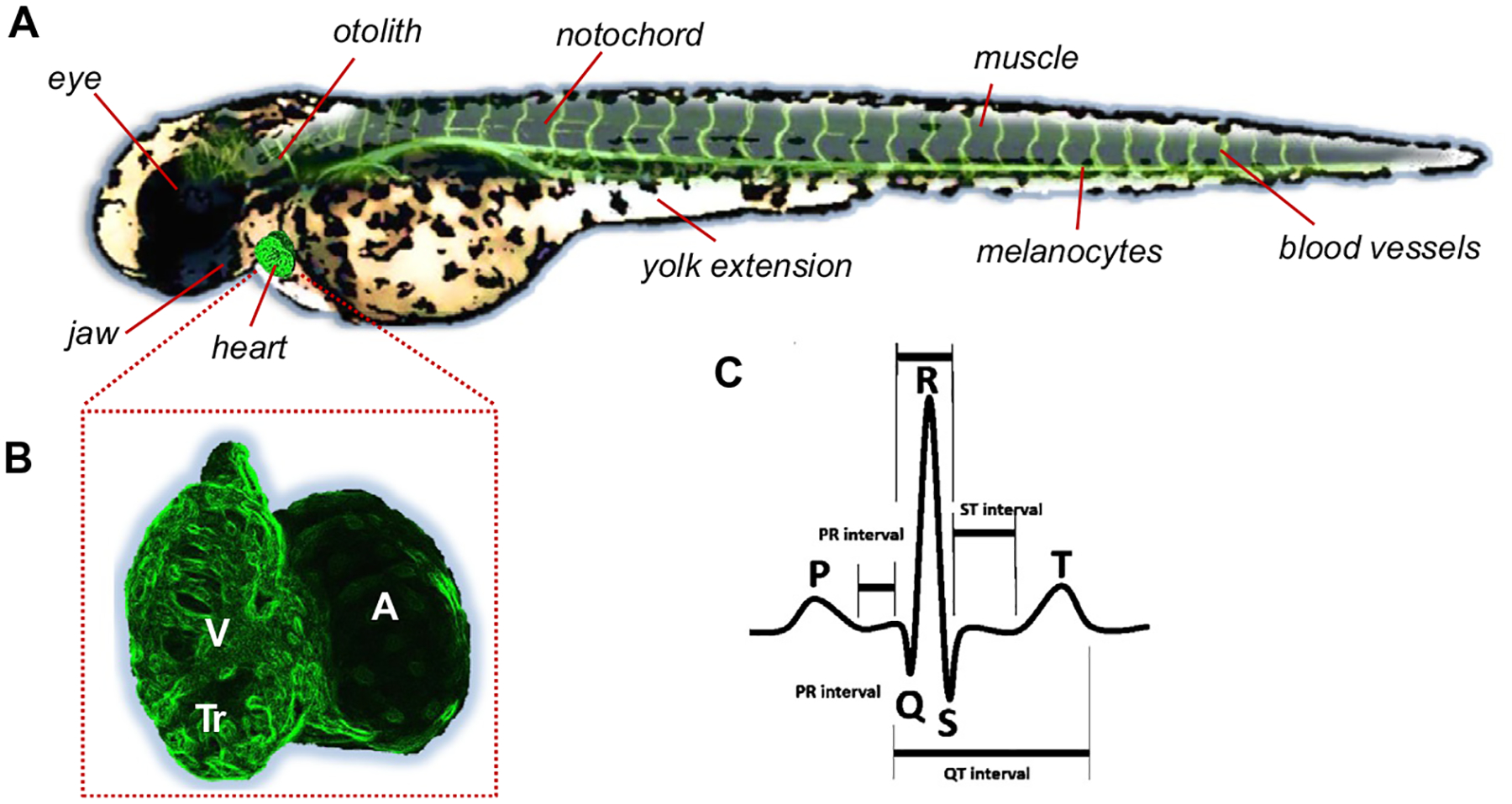
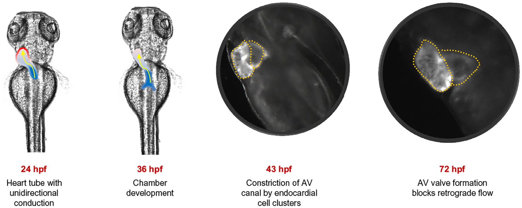
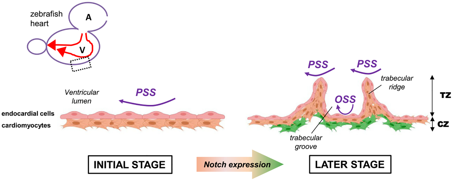
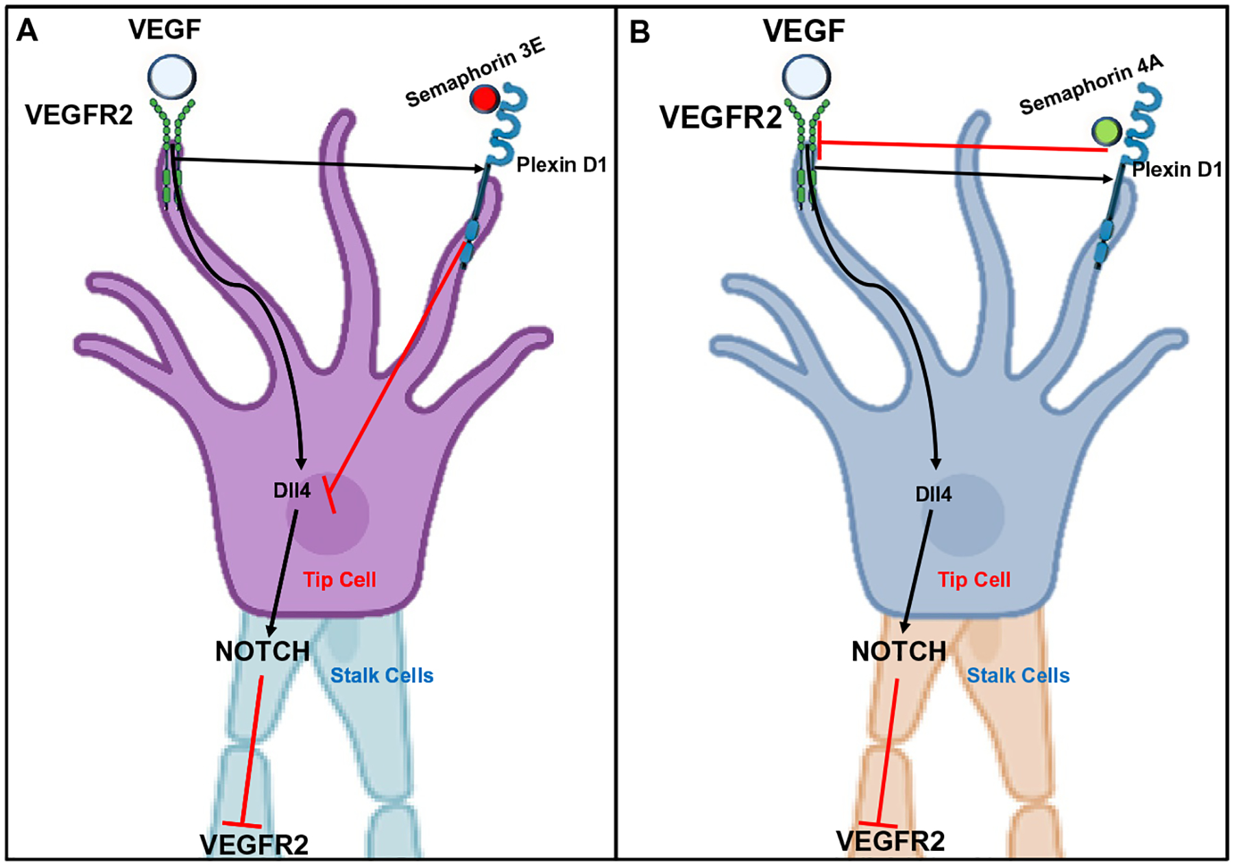
Living cells are exposed to multiple mechanical stimuli from the extracellular matrix or from surrounding cells. Mechanoreceptors are molecules that display status changes in response to mechanical stimulation, transforming physical cues into biological responses to help the cells adapt to dynamic changes of the microenvironment. Mechanical stimuli are responsible for shaping the tridimensional development and patterning of the organs in early embryonic stages. The development of the heart is one of the first morphogenetic events that occur in embryos. As the circulation is established, the vascular system is exposed to constant shear stress, which is the force created by the movement of blood. Both spatial and temporal variations in shear stress differentially modulate critical steps in heart development, such as trabeculation and compaction of the ventricular wall and the formation of the heart valves. Zebrafish embryos are small, transparent, have a short developmental period and allow for real-time visualization of a variety of fluorescently labeled proteins to recapitulate developmental dynamics. In this review, we will highlight the application of zebrafish models as a genetically tractable model for investigating cardiovascular development and regeneration. We will introduce our approaches to manipulate mechanical forces during critical stages of zebrafish heart development and in a model of vascular regeneration, as well as advances in imaging technologies to capture these processes at high resolution. Finally, we will discuss the role of molecules of the Plexin family and Piezo cation channels as major mechanosensors recently implicated in cardiac morphogenesis.
Keywords: Light sheet microscopy; Mechanotransduction; Notch; Shear stress; Trabeculation; Zebrafish.
Copyright © 2021 Elsevier Inc. All rights reserved.
Figures
Similar articles
-
A method to quantify mechanobiologic forces during zebrafish cardiac development using 4-D light sheet imaging and computational modeling.PLoS Comput Biol. 2017 Oct 30;13(10):e1005828. doi: 10.1371/journal.pcbi.1005828. eCollection 2017 Oct. PLoS Comput Biol. 2017. PMID: 29084212 Free PMC article.
-
Light-sheet Fluorescence Microscopy to Capture 4-Dimensional Images of the Effects of Modulating Shear Stress on the Developing Zebrafish Heart.J Vis Exp. 2018 Aug 10;(138):57763. doi: 10.3791/57763. J Vis Exp. 2018. PMID: 30148501 Free PMC article.
-
Extracellular mechanical forces drive endocardial cell volume decrease during zebrafish cardiac valve morphogenesis.Dev Cell. 2022 Mar 14;57(5):598-609.e5. doi: 10.1016/j.devcel.2022.02.011. Epub 2022 Mar 3. Dev Cell. 2022. PMID: 35245444
-
Hemodynamics driven cardiac valve morphogenesis.Biochim Biophys Acta. 2016 Jul;1863(7 Pt B):1760-6. doi: 10.1016/j.bbamcr.2015.11.014. Epub 2015 Nov 30. Biochim Biophys Acta. 2016. PMID: 26608609 Review.
-
The molecular mechanism of mechanotransduction in vascular homeostasis and disease.Clin Sci (Lond). 2020 Sep 18;134(17):2399-2418. doi: 10.1042/CS20190488. Clin Sci (Lond). 2020. PMID: 32936305 Review.
Cited by
-
Force-sensing protein expression in response to cardiovascular mechanotransduction.EBioMedicine. 2024 Dec;110:105412. doi: 10.1016/j.ebiom.2024.105412. Epub 2024 Oct 30. EBioMedicine. 2024. PMID: 39481337 Free PMC article. Review.
-
Vascular mechanotransduction.Physiol Rev. 2023 Apr 1;103(2):1247-1421. doi: 10.1152/physrev.00053.2021. Epub 2023 Jan 5. Physiol Rev. 2023. PMID: 36603156 Free PMC article. Review.
-
Spatiotemporal modulation of nitric oxide and Notch signaling by hemodynamic-responsive Trpv4 is essential for ventricle regeneration.Cell Mol Life Sci. 2024 Jan 27;81(1):60. doi: 10.1007/s00018-023-05092-0. Cell Mol Life Sci. 2024. PMID: 38279064 Free PMC article.
References
Publication types
MeSH terms
Grants and funding
LinkOut - more resources
Full Text Sources
Research Materials
