. 2021 Dec 1;41(48):9919-9931.
doi: 10.1523/JNEUROSCI.1329-21.2021. Epub 2021 Oct 25.

Dynamic Rhythmogenic Network States Drive Differential Opioid Responses in the In Vitro Respiratory Network

Affiliations

Dynamic Rhythmogenic Network States Drive Differential Opioid Responses in the In Vitro Respiratory Network

Nicholas J Burgraff et al. J Neurosci. .

Abstract

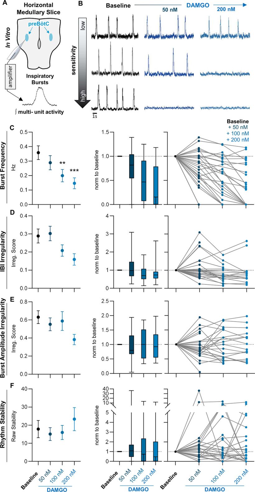
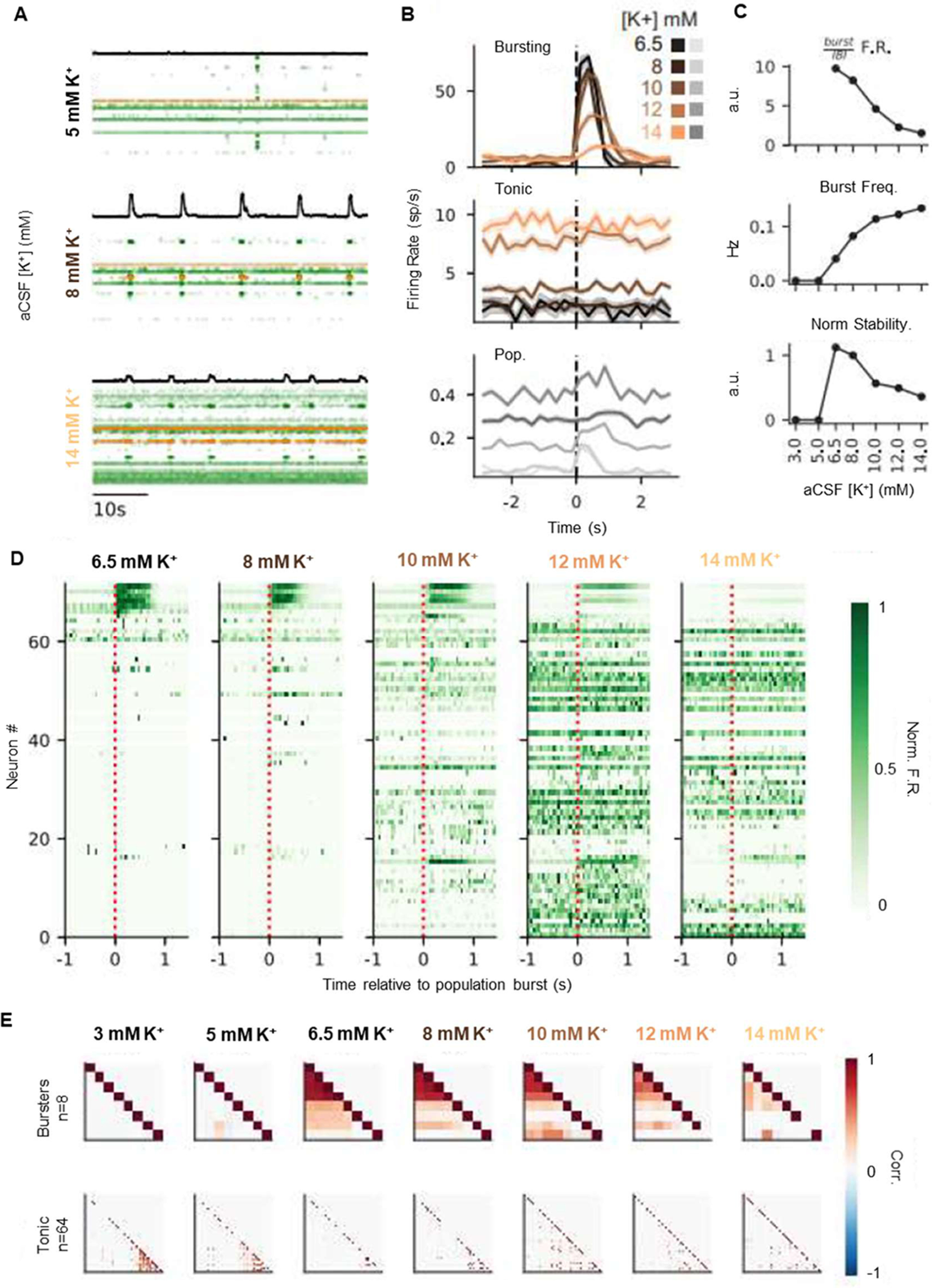
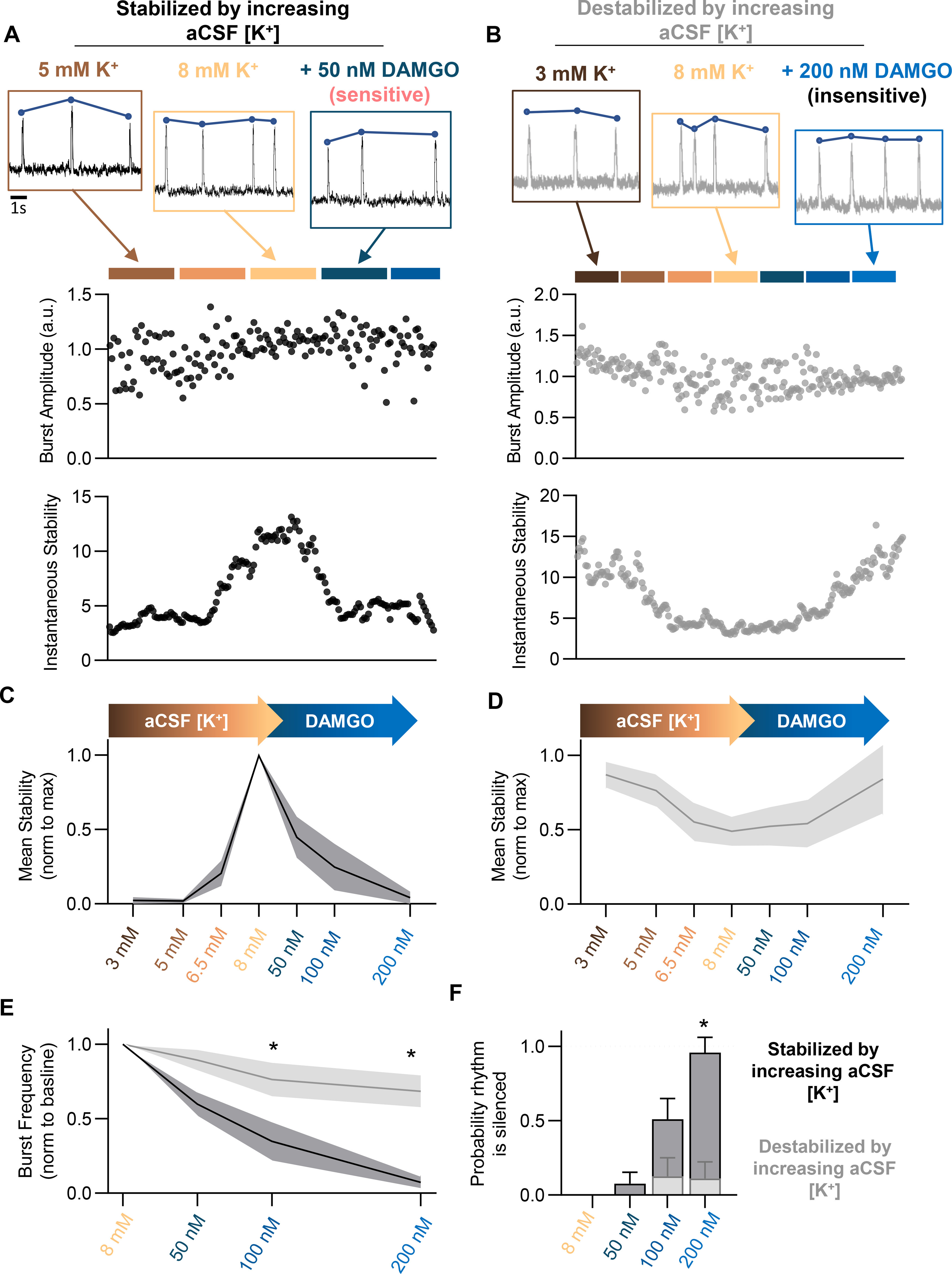
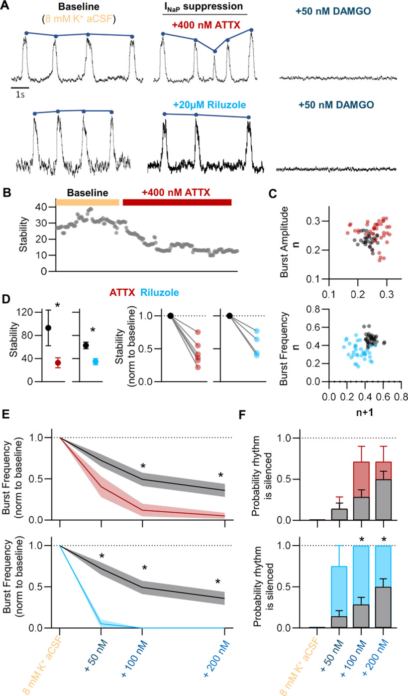
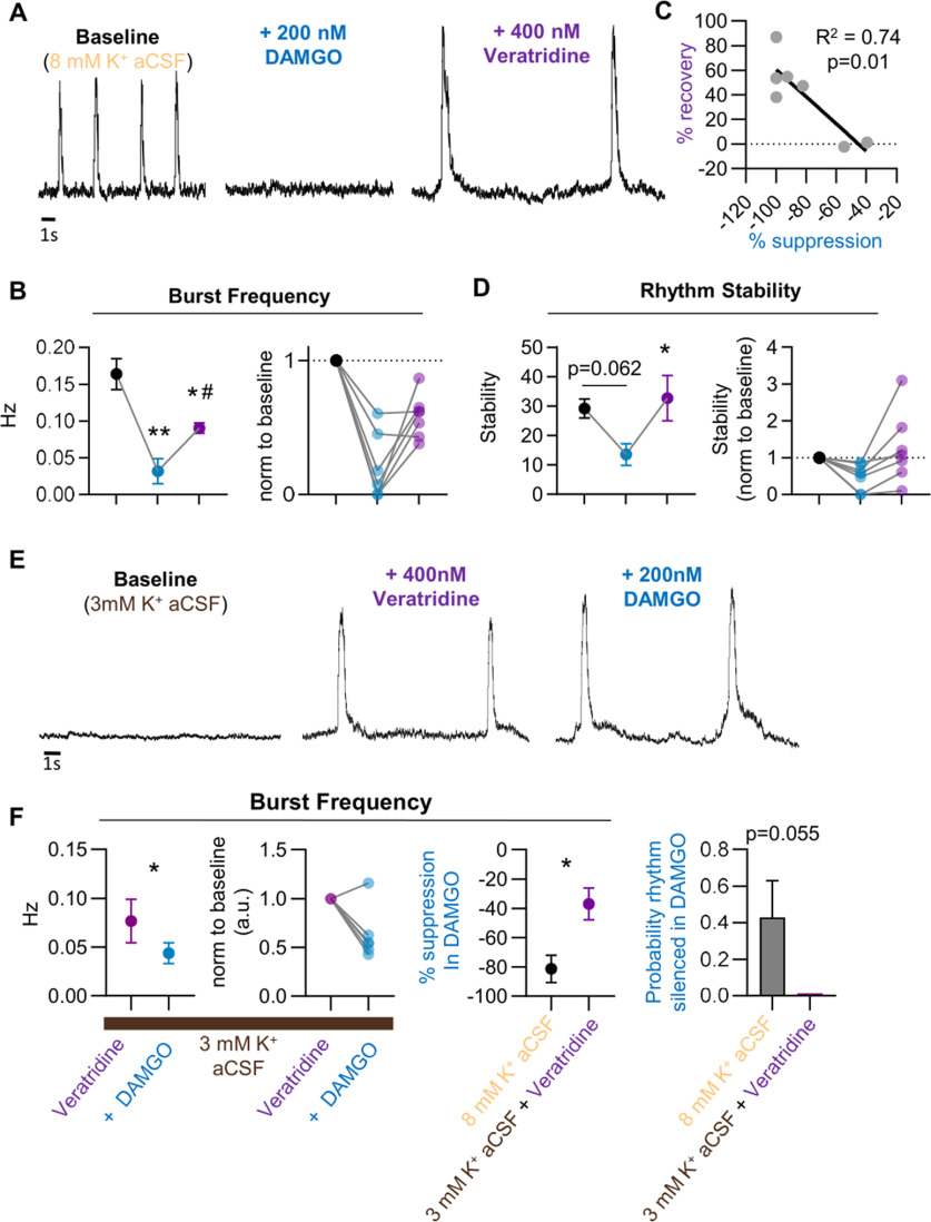
Death from opioid overdose is typically caused by opioid-induced respiratory depression (OIRD). A particularly dangerous characteristic of OIRD is its apparent unpredictability. The respiratory consequences of opioids can be surprisingly inconsistent, even within the same individual. Despite significant clinical implications, most studies have focused on average dose-r esponses rather than individual variation, and there remains little insight into the etiology of this apparent unpredictability. The preBötzinger complex (preBötC) in the ventral medulla is an important site for generating the respiratory rhythm and OIRD. Here, using male and female C57-Bl6 mice in vitro, we demonstrate that the preBötC can assume different network states depending on the excitability of the preBötC and the intrinsic membrane properties of preBötC neurons. These network states predict the functional consequences of opioids in the preBötC, and depending on network state, respiratory rhythmogenesis can be either stabilized or suppressed by opioids. We hypothesize that the dynamic nature of preBötC rhythmogenic properties, required to endow breathing with remarkable flexibility, also plays a key role in the dangerous unpredictability of OIRD.SIGNIFICANCE STATEMENT Opioids can cause unpredictable, life-threatening suppression of breathing. This apparent unpredictability makes clinical management of opioids difficult while also making it challenging to define the underlying mechanisms of OIRD. Here, we find in brainstem slices that the preBötC, an opioid-sensitive subregion of the brainstem, has an optimal configuration of cellular and network properties that results in a maximally stable breathing rhythm. These properties are dynamic, and the state of each individual preBötC network relative to the optimal configuration of the network predicts how vulnerable rhythmogenesis is to the effects of opioids. These insights establish a framework for understanding how endogenous and exogenous modulation of the rhythmogenic state of the preBötC can increase or decrease the risk of OIRD.

Keywords: network; opioid; preBötzinger; respiratory; rhythm; stability.


Figures

Figure 1.
Figure 1.
The functional consequences of opioids in the preBötC are variable in vitro. A, Horizontal brainstem slice preparations were used to compare the effects of opioids on the inspiratory rhythm across different preBötC networks. Integrated population bursts represent fictive inspiratory activity. B, C, Horizontal slices showed a wide range of responses to bath application of 50–200 nm DAMGO with some slices silenced by 50 nm DAMGO (bottom), whereas others showed little to no response up to 200 nm (top). C, Despite the variation in individual responses, the mean effect was a dose-dependent decrease in burst frequency with increasing doses of DAMGO. D, E, The IRS of the interburst interval (D) and peak amplitude (E) across successive population bursts increased in some slices and decreased in others following bath application of DAMGO from 50 to 200 nm DAMGO. F, Changes in rhythm stability following DAMGO application similarly showed variation across slices with some slices increasing stability, and others decreasing stability with subsequent DAMGO application. Left, Graphs show means ± SE. Middle, Graphs show median, interquartile rage, and minimum/maximum. Right, Graphs show individual replicates. **,***p < 0.05, significantly different from control value. Repeated-measures mixed-model analysis with Sidak's multiple comparisons was used to determine significant differences across doses.
Figure 2.
Figure 2.
Peak stability occurs at variable levels of network excitability. A, The level of aCSF [K+] needed to elicit bursting within the preBötC is variable among slices. Forty-eight percent of slices required 8 mm [K+] to elicit network-wide bursting, whereas 17% required 6.5 mm, 10% required 5 mm, and 24% required 3 mm. B, Representative tracings from preBötC population recordings following stepwise increases in aCSF [K+] from 3 to 8 mm. Top, Tracing shows a rhythm that increases stability as aCSF [K+] is increased. Middle, Tracing shows a rhythm that increases and then decreasing stability in response to increased aCSF [K+]. Bottom, Tracing shows a rhythm that is destabilized by increasing aCSF [K+]. The point of maximum stability (stabilitymax) is noted for each example. C, Following alignment of stabilitymax for each rhythm, levels of aCSF [K+] before reaching peak stability show lower rhythm stability, whereas increases in aCSF [K+] beyond stabilitymax result in decreasing rhythm stability. Averaging across slices results in a stability curve, where increasing aCSF [K+] initially increases rhythm stability followed by a decrease in stability as aCSF [K+] is further increased.
Figure 3.
Figure 3.
Changes in rhythm stability are associated with a trade-off between bursting and tonic spiking activity. A, Rasters of 72 simultaneously recorded single units at 5, 8, and 14 mm [K+]. Black trace is integrated contralateral population recording. Each row is a single neuron, and each dot is a recorded spike. Rows are ordered by recording depth. Cells highlighted in orange represent the tonic and bursting cell examples in B. B, Burst aligned spiking at each K+ concentration for a bursting (top) and tonic (middle) neuron (highlighted in A). Traces are average spikes/s over 10 min recordings for each K+ level, binned at 250 ms. Pooled spiking activity across all neurons is shown (bottom) in gray, where y-axis is spikes/s/neuron. Shaded regions are ± SE. Burst spiking decreases and tonic spiking increases as K+ increases; nbursts = (9,25,39,45,40) for K+ = (6.5,8,10,12,14) mm. C, Top, Ratio of burst firing rate (500 ms after burst onset) to interburst firing rate (1 s before burst onset) pooled across all neurons, as a function of K+. Middle, Burst frequency increases, and burst stability decreases (bottom) as [K+] increases. D, Burst aligned peristimulus time histograms of spike rate for each recorded neuron (rows), at each K+ concentration. Neurons are ordered by decreasing burst-related firing rate as observed at K+ = 6.5 mm. Spike rates are normalized by the maximal firing rate of each neuron. E, Top, Pairwise correlations between bursting and tonic (bottom) neurons for each K+ level. Neurons are clustered by Ward linkage at 6.5 mm K+ (bursters) or 3 mm (tonic).
Figure 4.
Figure 4.
PreBötC excitability relative to stabilitymax predicts DAMGO sensitivity. Stability of preBötC bursting was assessed while increasing aCSF [K+] from 3 to 8 mm, followed by DAMGO application. AF, Representative recordings and quantified peak amplitude and stability over the course of aCSF [K+] and DAMGO administration. Rhythms that increased stability with increasing aCSF [K+] (A, C; N = 13) exhibited significantly greater susceptibility to OIRD (E, F) compared with rhythms that decreased stability with increasing aCSF [K+] (B, D; N = 9). Rhythms that increased stability with increasing aCSF [K+] became destabilized following DAMGO application (A, C), whereas rhythms decreasing stability with increasing aCSF [K+] became stabilized following DAMGO (B, D); *p < 0.05. Differences in the effect of DAMGO between preBötC networks that increased versus decreased rhythm stability during increasing aCSF [K+] were assessed with a repeated-measures mixed-effect analysis with Sidak's multiple comparisons.
Figure 5.
Figure 5.
INaP attenuation destabilizes the inspiratory rhythm and increases the severity of OIRD. A, Representative population recordings before and after bath application of the NaV1.6 inhibitor, ATTX, or riluzole, followed by subsequent bath application of DAMGO. B, Representative quantified rhythm stability following bath application of ATTX. C, Peak amplitude and frequency Poincaré plots demonstrating increases in amplitude and frequency irregularity following ATTX (400 nm) or riluzole (20 μm) application, respectively. D, Mean (left) and individual (right) changes in rhythm stability following application of ATTX (red) or riluzole (blue). E, Mean burst frequency responses to stepwise increases in [DAMGO] following control conditions (gray) or pretreatment with either 400 nm ATTX (top, red) or 20 μm riluzole (bottom, blue). Brainstem slices pretreated with ATTX or riluzole displayed greater depression in burst frequency following DAMGO application, compared with control values. F, Probability that the inspiratory burst rhythm was silenced following stepwise increases in [DAMGO] during control conditions (gray), or following pretreatment with 400 nm ATTX (top, red) or 20 μm riluzole (bottom, blue); *p < 0.05, significantly different from mean control values. Differences in stability assessed with paired two-tailed t test. Differences in DAMGO sensitivity between control and pretreated conditions assessed as two-way repeated-measures ANOVA with Sidak's multiple comparisons test (condition and [DAMGO] as factors).
Figure 6.
Figure 6.
Slowing Na+ channel inactivation can protect against and partially reverse OIRD. A, Representative preBötC population recording demonstrating cessation of bursting following 200 nm DAMGO application and partial recovery with Veratridine (400 nm). B, Mean burst frequency (left) and individual replicates (right) following bath application of 200 nm DAMGO and subsequent application of 400 nm Veratridine. C, Linear regression analysis shows a correlation between burst frequency recovery with veratridine and the magnitude of frequency suppression following 200 nm DAMGO. D, Following DAMGO administration, suppression of bursting coincided with reduced stability of the rhythm (p = 0.06). Four hundred micromolars Veratridine partially recovered the rhythm and resulted in an increase in rhythm stability (p < 0.05). E, Application of 400 nm Veratridine in 3 mm [K+] aCSF elicits rhythmic bursting from the preBötC that is significantly less sensitive to suppression by 200 nm DAMGO. F, Population burst frequency decreases following 200 nm DAMGO in the presence of 400 nm Veratridine; however, the magnitude of suppression is significantly less compared with frequency suppression following 200 nm DAMGO in control conditions with 8 mm [K+] aCSF; *p < 0.05, significantly different from control; #p < 0.05, significantly different from 200 nm [DAMGO]. Differences in burst frequency and stability (B, D) assessed as one-way repeated-measures ANOVA with Sidak's multiple comparisons test. F, Differences in burst frequency following DAMGO in control and Veratridine pretreated conditions assessed as unpaired two-tailed t test.

Similar articles

Cited by

References

    1. Bachmutsky I, Wei XP, Kish E, Yackle K (2020) Opioids depress breathing through two small brainstem sites. Elife 9:e52694. 10.7554/eLife.52694 - DOI - PMC - PubMed
    1. Baekey DM, Morris KF, Nuding SC, Segers LS, Lindsey BG, Shannon R (2004) Ventrolateral medullary respiratory network participation in the expiration reflex in the cat. J Appl Physiol (1985) 96:2057–2072. 10.1152/japplphysiol.00778.2003 - DOI - PubMed
    1. Baertsch NA, Ramirez JM (2019) Insights into the dynamic control of breathing revealed through cell-type-specific responses to substance P. Elife 8:e51350. 10.7554/eLife.51350 - DOI - PMC - PubMed
    1. Baertsch NA, Severs LJ, Anderson TM, Ramirez JM (2019) A spatially dynamic network underlies the generation of inspiratory behaviors. Proc Natl Acad Sci U S A 116:7493–7502. 10.1073/pnas.1900523116 - DOI - PMC - PubMed
    1. Ballanyi K, Lalley PM, Hoch B, Richter DW (1997) cAMP-dependent reversal of opioid- and prostaglandin-mediated depression of the isolated respiratory network in newborn rats. J Physiol 504:127–134. 10.1111/j.1469-7793.1997.127bf.x - DOI - PMC - PubMed

Publication types

Substances

LinkOut - more resources