

Intraluminal Titanium Alloy Stent to Prevent Tracheal Stenosis in Tracheal Anastomosis
- PMID: 34697148
- PMCID: PMC8627747
- DOI: 10.21873/invivo.12612
Intraluminal Titanium Alloy Stent to Prevent Tracheal Stenosis in Tracheal Anastomosis
Abstract
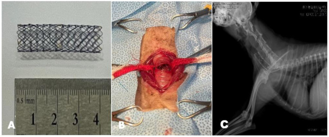
Background/aim: Tracheal stenosis can cause respiratory problems in mature, small-breed dogs. This study aimed to evaluate the placement of an intratracheal titanium alloy stent to prevent tracheal stenosis in canine tracheal anastomosis.
Materials and methods: The self-expandable intratracheal stent was an alloy of nickel and titanium, at the same atomic ratio. Vital signs and respiratory patterns, C-reactive protein, radiography, computed tomography, and endoscopy results after intraluminal stenting were assessed for 3-5 months.
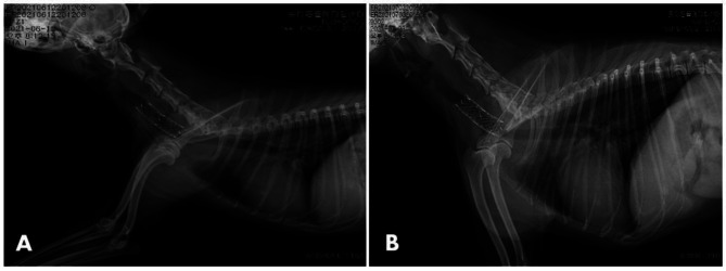
Results: No dogs showed evidence of intraluminal tracheal stenosis or tracheitis in the region of stent insertion on tracheoscopy and computed tomography after tracheal stent placement. After 1-2 weeks of tracheal stent placement, all dogs resolved coughing and dyspnea signs and resumed normal activities.
Conclusion: The intratracheal stent showed no movement or deformation in the trachea, and had flexibility and an appropriate radial force. Therefore, titanium alloy tracheal stents are useful in stenotic operations for tracheal reconstruction.
Keywords: Tracheal stenosis; dog; intratracheal stent; titanium alloy.
Copyright © 2021 International Institute of Anticancer Research (Dr. George J. Delinasios), All rights reserved.
Conflict of interest statement
The Authors declare that they have no competing interests regarding this study.
Figures


References
-
- White R, Williams J. Tracheal collapse in the dog - is there really a role for surgery? A survey of 100 cases. Journal of Small Animal Practice. 2021;35(4):191–196. doi: 10.1111/j.1748-5827.1994.tb01685.x. - DOI
MeSH terms
Substances
LinkOut - more resources
Full Text Sources
Research Materials