. 2021 Nov-Dec;35(6):3537-3545.
doi: 10.21873/invivo.12656.

Hemodynamic Alteration in the Liver in Acute Hepatitis: A Quantitative Evaluation Using Computed Tomographic Perfusion

Affiliations

Hemodynamic Alteration in the Liver in Acute Hepatitis: A Quantitative Evaluation Using Computed Tomographic Perfusion

Akihiro Nishie et al. In Vivo. 2021 Nov-Dec.

Abstract

Background/aim: We aimed to elucidate the hemodynamic alterations in the liver of patients with acute hepatitis (AH) using computed tomography perfusion imaging.

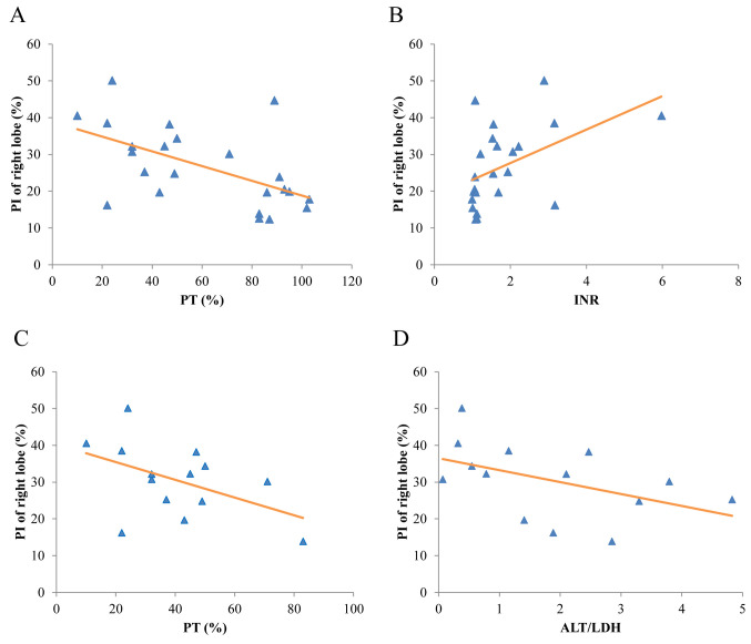
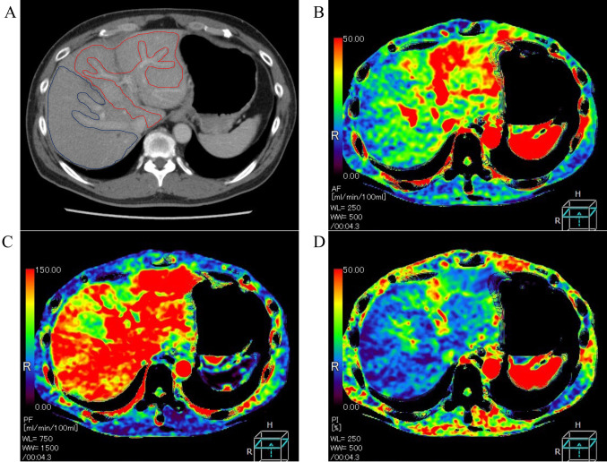
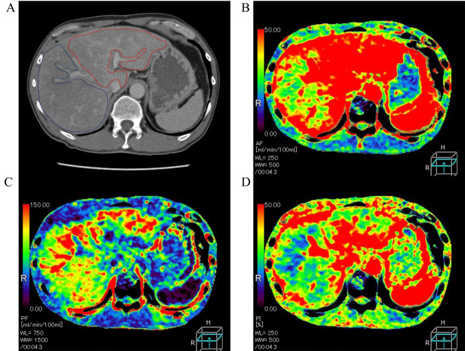
Patients and methods: For 14 patients with AH and nine patients with no disease (ND group), we compared the mean arterial blood flow (AF), portal blood flow (PF) and perfusion index (%) [PI=AF/(AF+PF) ×100] of the right and left liver lobes and investigated their relationship with clinical factors.

Results: The mean PI of the right lobe in the AH group (30.5±10.0%) was significantly higher than that in the ND group (20.8±9.7%) (p=0.031). For all patients of the AH and ND groups, the PI of the right lobe was increased as the prothrombin time decreased (R=-0.56, p=0.006) and as the prothrombin time-international normalized ratio increased (R=0.48, p=0.02).

Conclusion: The PI of the right liver lobe may increase in AH and may be a predictive parameter for the severity of hepatic failure.

Keywords: Acute hepatitis; CT perfusion; hemodynamic alteration.


Conflict of interest statement

Akihiro Nishie was a staff member of joint research departments in Kyushu University with Canon Medical Systems Corporation. He received a research grant from Canon Medical Systems Corporation. The other Authors have no conflicts of interest.

Figures

Figure 1
Figure 1. A: The correlation between the prothrombin time (PT) and the perfusion index of the right liver lobe (PI) for the whole patient cohort [acute hepatitis (AH) and no-disease (ND) groups]. The correlation coefficient was −0.56. PI (%)=−0.20×PT (%)+38.9. B: The correlation between PT-international normalized ratio (INR) and PI for the whole patient cohort. The correlation coefficient was 0.48. PI (%)=4.55 × PT-INR+18.6. C: The correlation between PT and PI of the right liver lobe for patients of the AH group. The correlation coefficient was −0.48. PI (%)=−0.24 × PT (%)+40.3. D: The correlation between alanine aminotransferase/lactate dehydrogenase (ALT/LDH) ratio and the PI patients of the AH group. The correlation coefficient was −0.47. PI (%)=−3.24 × ALT/LDH+36.5.
Figure 2
Figure 2. A 55-year-old man underwent computed tomography perfusion imaging for the evaluation of gastric cancer. He had no underlying liver disease, liver dysfunction, or liver metastasis. A: Portal venous phase image: No abnormality in morphology or attenuation was observed in the liver. Regions of interests placed on the right (blue) and left (red) lobes on each map are shown. B: Map of the arterial blood flow (AF): The AF values of the right and left lobes were 26.4 ml/min/100 ml tissue and 40.4 ml/min/100 ml tissue, respectively. C: Map of the portal blood flow (PF): The PF values of the right and left lobes were 127.4 ml/min/100 ml tissue and 139.8 ml/min/100 ml tissue, respectively. D: Perfusion index (PI) map: The PI values of the right and left lobes were 17.8% and 24.1%, respectively.
Figure 3
Figure 3. A 55-year-old man underwent computed tomography perfusion imaging for the evaluation of acute hepatitis. He had no underling liver disease, and drug induction was suspected as the cause of acute hepatic failure. The prothrombin time (PT) and alanine aminotransferase/lactate dehydrogenase (ALT/LDH) ratio were 10 ms and 0.32, respectively. A: Portal venous phase image: No abnormality in morphology, but slightly heterogeneous attenuation was observed in the liver. Regions of interest placed on the right (blue) and left (red) lobes on each map are indicated. B: Map of the arterial blood flow (AF): The AF values of the right and left lobes were 53 ml/min/100 ml tissue and 84.8 ml/min/100 ml tissue, respectively. C: Map of the portal blood flow (PF): The PF values of the right and left lobes were 79.8 ml/min/100 ml tissue and 56.2 ml/min/100 ml tissue, respectively. D: Perfusion index (PI) map: The PI values of the right and left lobes were 40.6% and 60.6%, respectively. The patient was treated with plasma exchange and arterial steroid injection therapy for the liver. This case had a high PI, a low PT, and a low ALT/LDH.
Figure 4
Figure 4. A 39-year-old man underwent CT perfusion imaging for the evaluation of acute hepatitis. He was a carrier of hepatitis virus B, which was thought to be the cause of his acute hepatic failure. The prothrombin time (PT) and alanine aminotransferase/lactate dehydrogenase (ALT/LDH) ratio were 37 ms and 4.8, respectively. A: Portal venous phase image: No abnormality in morphology was observed in the liver. Regions of interest placed on the right (blue) and left (red) lobes on each map are indicated. B: Map of the arterial blood flow (AF): The AF values of the right and left lobes were 32.6 ml/min/100 ml tissue and 38.2 ml/min/100 ml tissue, respectively. C: Map of the portal blood flow (PF): The PF values of the right and left lobes were 99.5 ml/min/100 ml tissue and 105.5 ml/min/100 ml tissue, respectively. D: Perfusion index (PI) map: The PI values of the right and left lobes were 25.3% and 30.1%, respectively. He was treated with plasma exchange and arterial steroid injection therapy for the liver. This case had a low PI, a relatively low PT, and a high ALT/LDH ratio.

Similar articles

Cited by

  • 'Fulminant hepatic failure' anesthesiologic considerations.
    Vetrugno L, Alessandri F, Toscano A, Voza A, Deana C. Vetrugno L, et al. Curr Opin Anaesthesiol. 2025 Aug 1;38(4):503-512. doi: 10.1097/ACO.0000000000001530. Epub 2025 May 26. Curr Opin Anaesthesiol. 2025. PMID: 40511630 Free PMC article. Review.

References

    1. Takikawa Y, Endo R, Suzuki K, Fujiwara K, Omata M, Fulminant Hepatitis Study Group of Japan Prediction of hepatic encephalopathy development in patients with severe acute hepatitis. Dig Dis Sci. 2006;51(2):359–364. doi: 10.1007/s10620-006-3138-7. - DOI - PubMed
    1. Chun LJ, Tong MJ, Busuttil RW, Hiatt JR. Acetaminophen hepatotoxicity and acute liver failure. J Clin Gastroenterol. 2009;43(4):342–349. doi: 10.1097/MCG.0b013e31818a3854. - DOI - PubMed
    1. Wu Z, Han M, Chen T, Yan W, Ning Q. Acute liver failure: mechanisms of immune-mediated liver injury. Liver Int. 2010;30(6):782–794. doi: 10.1111/j.1478-3231.2010.02262.x. - DOI - PubMed
    1. Hillenbrand P, Parbhoo SP, Jedrychowski A, Sherlock S. Significance of intravascular coagulation and fibrinolysis in acute hepatic failure. Gut. 1974;15(2):83–88. doi: 10.1136/gut.15.2.83. - DOI - PMC - PubMed
    1. Possamai LA, Thursz MR, Wendon JA, Antoniades CG. Modulation of monocyte/macrophage function: a therapeutic strategy in the treatment of acute liver failure. J Hepatol. 2014;61(2):439–445. doi: 10.1016/j.jhep.2014.03.031. - DOI - PubMed