. 2021 Nov 1;25(6):585-592.
doi: 10.4196/kjpp.2021.25.6.585.

Effects of exercise on AKT/PGC1-α/FOXO3a pathway and muscle atrophy in cisplatin-administered rat skeletal muscle

Affiliations

Effects of exercise on AKT/PGC1-α/FOXO3a pathway and muscle atrophy in cisplatin-administered rat skeletal muscle

Jun Hyun Bae et al. Korean J Physiol Pharmacol. .

Abstract

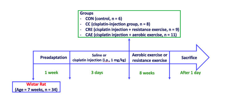
Cisplatin has been reported to cause side effects such as muscle wasting in humans and rodents. The physiological mechanisms involved in preventing muscle wasting, such as the regulation of AKT, PGC1-α, and autophagy-related factor FOXO3a by MuRF 1 and Atrogin-1, remain unclear following different types of exercise and in various skeletal muscle types. Eight-week-old male Wistar rats (n = 34) were assigned to one of four groups: control (CON, n = 6), cisplatin injection (1 mg/kg) without exercise (CC, n = 8), cisplatin (1 mg/kg) + resistance exercise (CRE, n = 9) group, and cisplatin (1 mg/kg) + aerobic exercise (CAE, n = 11). The CRE group performed progressive ladder exercise (starting with 10% of body weight on a 1-m ladder with 2-cm-interval grids, at 85°) for 8 weeks. The CAE group exercised by treadmill running (20 m/min for 60 min daily, 4 times/week) for 8 weeks. Compared with the CC group, the levels of the autophagy-related factors BNIP3, Beclin 1, LC3-II/I ratio, p62, and FOXO3a in the gastrocnemius and soleus muscles were significantly decreased in the CRE and CAE groups. The CRE and CAE groups further showed significantly decreased MuRF 1 and Atrogin-1 levels and increased phosphorylation of AKT, FOXO3a, and PGC1-α. These results suggest that both ladder and aerobic exercise directly affected muscle wasting by modulating the AKT/PGC1-α/FOXO3a signaling pathways regardless of the skeletal muscle type.

Keywords: Autophagy; Cisplatin; Exercise training; Muscle atrophy; Skeletal muscle.


Conflict of interest statement

CONFLICTS OF INTEREST

The authors declare no conflicts of interest.

Figures

Fig. 1
Fig. 1. Experimental procedure of the study.
Fig. 2
Fig. 2. Expression levels of autophagy-related factors in gastrocnemius muscle.
(A, G) Representative bands. (B) Beclin 1/GAPDH, (C) phospho-FOXO3a/total-FOXO3a, (D) p62/GAPDH, (E) BNIP3/GAPDH, (F) LC3-II/I ratio, (H) phospho-mTOR/total-mTOR, (I) phospho-Akt/total-Akt, and (J) PGC1-α/GAPDH. CON, control (n = 6); CC, cisplatin control (n = 8); CRE, resistance exercise with cisplatin treatment (n = 9); CAE, aerobic exercise with cisplatin treatment (n = 11). AKT, protein kinase B; FOXO3a, forkhead box O3; PGC1-α, peroxisome proliferator-activated receptor gamma coactivator 1-alpha; LC3, lipidated microtubule-associated protein 1 light chain 3; p62, sequestosome-1; Beclin 1, autophagy-related 6 homolog; BNIP3, BCL2-interacting protein 3; GAPDH, glyceraldehyde-3-phosphate dehydrogenase. One-way analysis of variance and Tukey’s post-hoc were used for statistical analysis; units are arbitrary. *p < 0.05, **p < 0.01, ***p < 0.001.
Fig. 3
Fig. 3. Expression levels of autophagy-related factors in the soleus muscle.
(A, G) Representative bands. (B) Beclin 1/GAPDH, (C) phospho-FOXO3a/total-FOXO3a, (D) p62/GAPDH, (E) BNIP3/GAPDH, (F) LC3-II/I ratio, (H) phospho-mTOR/total-mTOR, (I) phospho-Akt/total-Akt, and (J) PGC1-α/GAPDH. CON, control (n = 6); CC, cisplatin control (n = 8); CRE, resistance exercise with cisplatin treatment (n = 9); CAE, aerobic exercise with cisplatin treatment (n = 11). AKT, protein kinase B; FOXO3a, forkhead box O3; PGC1-α, peroxisome proliferator-activated receptor gamma coactivator 1-alpha; LC3, lipidated microtubule-associated protein 1 light chain 3; p62, sequestosome-1; Beclin 1, autophagy-related 6 homolog; BNIP3, BCL2-interacting protein 3; GAPDH, glyceraldehyde-3-phosphate dehydrogenase. One-way analysis of variance and Tukey’s post-hoc were used for statistical analysis; units are arbitrary. *p < 0.05, **p < 0.01, ***p < 0.001.
Fig. 4
Fig. 4. Expression levels of muscle atrophy-related proteins including 4EBP1, Atrogin-1, and MuRF 1, in the gastrocnemius and soleus muscles.
(A, E) Representative bands. (B) Phospho-4EBP1/total 4EBP1 in gastrocnemius, (C) Atrogin-1/GAPDH in gastrocnemius, (D) MuRF 1/GAPDH in gastrocnemius, (F) phospho-4EBP1/total 4EBP1 in soleus, (G) Atrogin-1/GAPDH in soleus, and (H) MuRF 1/GAPDH in soleus. CON, control (n = 6); CC, cisplatin control (n = 8); CRE, resistance exercise with cisplatin treatment (n = 9); CAE, aerobic exercise with cisplatin treatment (n = 11). MuRF 1, muscle RING finger protein 1; Atrogin-1, muscle-specific F-box protein; GAPDH, glyceraldehyde-3-phosphate dehydrogenase. One-way analysis of variance and Tukey’s post-hoc were used for statistical analysis; units are arbitrary. *p < 0.05, **p < 0.01, ***p < 0.001.
Fig. 5
Fig. 5. AKT/PGC1-α/FOXO3a signaling pathways affected muscle atrophy and autophagy regulation.
AKT, protein kinase B; FOXO3a, forkhead box O3; PGC1-α, peroxisome proliferator-activated receptor gamma coactivator 1-alpha; MuRF 1, muscle RING finger protein 1; Atrogin-1, muscle-specific F-box protein; LC3, lipidated microtubule-associated protein 1 light chain 3; p62, sequestosome-1; Beclin 1, autophagy-related 6 homolog; BNIP3, BCL2-interacting protein 3.

Similar articles

Cited by

References

    1. Conte E, Bresciani E, Rizzi L, Cappellari O, De Luca A, Torsello A, Liantonio A. Cisplatin-induced skeletal muscle dysfunction: mechanisms and counteracting therapeutic strategies. Int J Mol Sci. 2020;21:1242. doi: 10.3390/ijms21041242. - DOI - PMC - PubMed
    1. Fearon K, Strasser F, Anker SD, Bosaeus I, Bruera E, Fainsinger RL, Jatoi A, Loprinzi C, MacDonald N, Mantovani G, Davis M, Muscaritoli M, Ottery F, Radbruch L, Ravasco P, Walsh D, Wilcock A, Kaasa S, Baracos VE. Definition and classification of cancer cachexia: an international consensus. Lancet Oncol. 2011;12:489–495. doi: 10.1016/S1470-2045(10)70218-7. - DOI - PubMed
    1. Brunet A, Bonni A, Zigmond MJ, Lin MZ, Juo P, Hu LS, Anderson MJ, Arden KC, Blenis J, Greenberg ME. Akt promotes cell survival by phosphorylating and inhibiting a Forkhead transcription factor. Cell. 1999;96:857–868. doi: 10.1016/S0092-8674(00)80595-4. - DOI - PubMed
    1. Mammucari C, Milan G, Romanello V, Masiero E, Rudolf R, Del Piccolo P, Burden SJ, Di Lisi R, Sandri C, Zhao J, Goldberg AL, Schiaffino S, Sandri M. FoxO3 controls autophagy in skeletal muscle in vivo. Cell Metab. 2007;6:458–471. doi: 10.1016/j.cmet.2007.11.001. - DOI - PubMed
    1. Galluzzi L, Baehrecke EH, Ballabio A, Boya P, Bravo-San Pedro JM, Cecconi F, Choi AM, Chu CT, Codogno P, Colombo MI, Cuervo AM, Debnath J, Deretic V, Dikic I, Eskelinen EL, Fimia GM, Fulda S, Gewirtz DA, Green DR, Hansen M, et al. Molecular definitions of autophagy and related processes. EMBO J. 2017;36:1811–1836. doi: 10.15252/embj.201796697. - DOI - PMC - PubMed