. 2021 Oct 25;12(1):6157.
doi: 10.1038/s41467-021-26244-5.

Antibiotic-chemoattractants enhance neutrophil clearance of Staphylococcus aureus

Affiliations

Antibiotic-chemoattractants enhance neutrophil clearance of Staphylococcus aureus

Jennifer A E Payne et al. Nat Commun. .

Abstract

The pathogen Staphylococcus aureus can readily develop antibiotic resistance and evade the human immune system, which is associated with reduced levels of neutrophil recruitment. Here, we present a class of antibacterial peptides with potential to act both as antibiotics and as neutrophil chemoattractants. The compounds, which we term 'antibiotic-chemoattractants', consist of a formylated peptide (known to act as chemoattractant for neutrophil recruitment) that is covalently linked to the antibiotic vancomycin (known to bind to the bacterial cell wall). We use a combination of in vitro assays, cellular assays, infection-on-a-chip and in vivo mouse models to show that the compounds improve the recruitment, engulfment and killing of S. aureus by neutrophils. Furthermore, optimizing the formyl peptide sequence can enhance neutrophil activity through differential activation of formyl peptide receptors. Thus, we propose antibiotic-chemoattractants as an alternate approach for antibiotic development.


Conflict of interest statement

A.Y.P., J.A.E.P., J.T., and M.J.C. have filed for patent protection (PCT/AU2021/050306) of the antibiotic-chemoattractant compounds. The remaining authors declare no competing interests.

Figures

Fig. 1
Fig. 1. Overcoming S. aureus immune evasion with immunotherapeutic antibiotics that bind to bacteria, retains activity against bacteria and can recruit neutrophils.
a Our strategy combines the possibility of direct killing of the bacteria with enhancing the neutrophil response. Vancomycin (yellow) targets and binds directly to S. aureus cell wall, while the fPep helps the immune response (blue) by enhancing the chemotactic gradient, directing immune cells to the bacterial invader and improving phagocytosis. Neutrophil image modified from. b Linkage to vancomycin can be made through three main sites: the vancosamine primary amine (V linked, purple); methylated amine (N-linked, blue); or the carboxyl group (C-linked, green). c Fluorescently labelled BODIPY vancomycin (B2) or the conjugate (fPep = van, B3) imaged by Airyscan super-resolution microscopy localized to the wall of the Gram-positive bacteria B. subtilis, S. aureus-MSSA, and an MRSA clinical isolate. Scale bars are 1 μm; Insert image is nuclear staining by Hoechst 33342, images are representative of 3 biological replicates. The linkage site to vancomycin and the length of the PEG linker was critical to maintain antimicrobial activity against S. aureus as seen in a microbroth dilution assay (d) but had little effect on the chemotaxis of human neutrophils in transwell assay (e). Three different length PEG linkers were trialed: no linker (0), 3 PEG units (3), or 6 PEG units (6) at the three attachment sites on vancomycin, leading to three different C-linked (C13), N-linked (N46), and V-linked (V79) compounds. The growth of a clinical isolate of S. aureus—MRSA was determined relative to the no protein control. Chemotaxis was calculated relative to the no protein control and 100% chemotaxis set as the neutrophil recruitment observed to fMLFG (FP1) at 100 nM for each donor. Error bars are SEM, n = 3 biologically independent experiments.
Fig. 2
Fig. 2. The fPep sequence either free or linked to vancomycin affects neutrophil recruitment.
a Schematic depiction of the process to synthesize and test a library of fPeps centered on the fMLFG sequence. The initial library of 27 fPep were generated by combinatorial solid-phase peptide synthesis on SynPhaseΤΜ lanterns. A transwell assay was used to determine the chemotaxis of human neutrophils to these peptides in a concentration range of 1–1000 nM. The library of fPeps were grouped into 5 profiles (line graphs, B-F, n = 3 biological independent experiments, error bars are SEM) based on the concentration of the fPep that resulted in the greatest recruitment of neutrophils. Peak recruitment was observed at: 100 nM (b), both 10 and 100 nM (c), 10 nM (d), 100 and 1000 nM (e), and 1000 nM (f). The changes in fMLFG sequence are indicated by the altered residue number (1 = fM, 2 = L, 3 = F, 4 = G), with the side chain of the switched amino acid indicated. Representative fPeps from each of these profiles were linked to the C-terminus of vancomycin via click chemistry and retested for the ability to recruit neutrophils using transwell assay (bar graphs, solid bars represent conjugate). Chemotaxis was calculated relative to the no protein control and 100% chemotaxis set as the neutrophil recruitment observed to fMLFG at 100 nM for each donor, n = 3–4 biological independent experiments, error bars are SEM. Dotted line on graphs indicates average recruitment to 0 nM.
Fig. 3
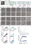
Fig. 3. Linking a fPep to vancomycin enhances the phagocytosis activity of neutrophils.
a The infection-on-a-chip device used to monitor neutrophil migration and phagocytosis over time present in a six-well plate. Within this microfluidic device the egg-like microchambers in the main channel were loaded with S. aureus plus compound, washed, and neutrophils loaded. A chemotactic gradient of compound establishes from the microchamber along a single cell wide connecting channel. Bacteria are labelled with the pH activated dye, pHrodo to allow monitoring of phagocytosis. b Time course of neutrophils migrating into the microchamber containing S. aureus bioparticles labelled with pHrodo, in the presence of free fPep (FP1, or FP11) or conjugated (C1 or C11) at 1000 or 0 nM (control). Images are representative of four donors; scale bars 10 μm. Quantification of neutrophils having migrated into the microchamber (c) along with the area of pHrodo fluorescence (d), or the area of GFP fluorescence as a measure of S. aureus growth (e) determined in the presence of conjugated (triangles, C1 or C11) or free fPep (circles, fMLFG FP1, black; and fMChaFG FP11, teal; at 1000 or 0 nM, control). Data are the average of four donors, with error bars being SEM. f The sequence of the conjugate effects efficiency of neutrophil phagocytosis as determined by the area of pHrodo fluorescence per neutrophil recruited into the microchamber at 2 h. Data from each donor is linked by dotted lines. g fMLFG conjugated to vancomycin (fMLFG = van C1) or free fPep (FP1) were incubated with S. aureus bioparticles and washed before loading into the microchamber. The washed conjugate treated S. aureus recruited similar levels of neutrophils compared to samples with 100 nM fMLFG (open circles). Data are representative of three donors, error bars are SEM.
Fig. 4
Fig. 4. Interaction of fPeps with human FPRs and activation of downstream pathways.
a Competition binding to human neutrophils between the FPR2 antagonist RhB-PB10 and different fPeps. fMLF binds preferentially to FPR1 and was used as a negative control, while fMIVIL binds preferentially to FPR2. The peptides fMLFG and fMChaFG and their corresponding conjugates were examined for their ability to compete with RhB-PB10 binding to neutrophils, n = 5 biologically independent experiments, with error bars of SEM. b CHO cells overexpressing human FPR1 or 2 were assessed for the downstream activation of either ERK1/2 phosphorylation or Ca2+ mobilization ([Ca2+]i)with Emax and pEC50 calculated. Data presented as mean ± SEM and an unpaired t test were used to compare the effects of the fMLFG and fMChaFG in free and conjugated forms. n indicates the number of biological replicates with two-tailed p-values indicated by *p = 0.0002, **p = 0.02, ***p = 0.01 vs. fMLFG, #p = 0.0006, ##p = 0.01, ###p = 0.000048 vs fMChaFG. c Time course of ERK1/2 phosphorylation (n = 4 biological replicates performed in triplicate, error bars are SEM). d Concentration-response curves for pERK1/2 were determined at 5 min, the peak-response time point determine during the time course. The positive controls were the hexapeptide Trp-Lys-Tyr-Met-Val-D-Met (WKYMVm), a specific FPR2 agonist, along with N-formyl-Met-Leu-Phe (fMLF) as a specific FPR1 agonist. Data were normalized to vehicle and the response elicited by 10% FBS, with nonlinear regression curve plotted with error bars of SEM, (n = 3–5 experiments, each performed in duplicate). e Intracellular calcium mobilization was determined in response to fPeps in CHO cells overexpressing either human FPR1 or FPR2. The fluorescence signal was normalized to the cellular response mediated by the positive control, adenosine triphosphate (ATP) at 100 µM, plotted with nonlinear regression curve, with error bars of SEM, (n = 3–7 experiments, each performed in triplicate).
Fig. 5
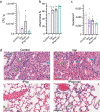
Fig. 5. fPep-vancomycin conjugate C11 reduces bacterial load and inflammation in a mouse pneumonia model.
Eight-week-old female mice were infected by intranasal inhalation of 107 CFU S. aureus to induce pneumonia. One-hour postinfection (hpi) mice were given intranasal therapy at 0.2 mg/mouse equivalent of vancomycin or vehicle control (control). The lungs were collected at 12 hpi, and the bacterial load in the lung tissue of four mice per treatment group was determined (a). The fMChaFG conjugate (C11, fPep = van) resulted in half the bacterial load compared to vancomycin (van) alone (n = 4 mice/group, error bars are SEM). The percentage of alveoli space (b) or the number of nuclei (c) was determined for hematoxylin and eosin-stained histology lung samples (d) from 6 fields of view of the outer lung lobes. Uninfected mouse lung is indicated by dotted line, error bars are SEM. Green arrows indicate neutrophils, blue arrows proteinaceous fluid, and orange arrows indicate cocci bacteria.

Similar articles

Cited by

References

    1. CDC. Antibiotic resistance threats in the United States. (Report, 2013).
    1. O’Neill, J. The Review on antimicrobial resistance. (Report, 2016).
    1. Pfaller, M. A., Carvalhaes, C. G., Smith, C. J., Diekema, D. J. & Castanheira, M. Bacterial and fungal pathogens isolated from patients with bloodstream infection: frequency of occurrence and antimicrobial susceptibility patterns from the SENTRY Antimicrobial Surveillance Program (2012–2017). Diagn. Microbiol. Infect. Dis.97, 115016 (2020). - PubMed
    1. Guerra FE, Borgogna TR, Patel DM, Sward EW, Voyich JM. Epic immune battles of history: neutrophils vs. Staphylococcus aureus. Front. Cell. Infect. Microbiol. 2017;7:286. doi: 10.3389/fcimb.2017.00286. - DOI - PMC - PubMed
    1. Jiang JH, et al. Antibiotic resistance and host immune evasion in Staphylococcus aureus mediated by a metabolic adaptation. Proc. Natl Acad. Sci. USA. 2019;116:3722–3727. doi: 10.1073/pnas.1812066116. - DOI - PMC - PubMed

Publication types

MeSH terms