. 2021 Oct 25;11(1):20974.
doi: 10.1038/s41598-021-00439-8.

Riluzole-induced apoptosis in osteosarcoma is mediated through Yes-associated protein upon phosphorylation by c-Abl Kinase

Affiliations

Riluzole-induced apoptosis in osteosarcoma is mediated through Yes-associated protein upon phosphorylation by c-Abl Kinase

Marian Raghubir et al. Sci Rep. .

Abstract

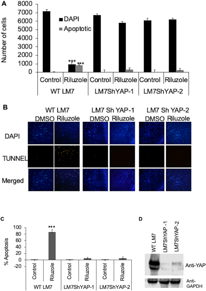
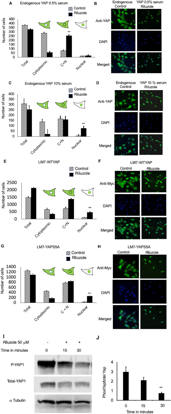
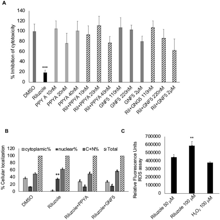
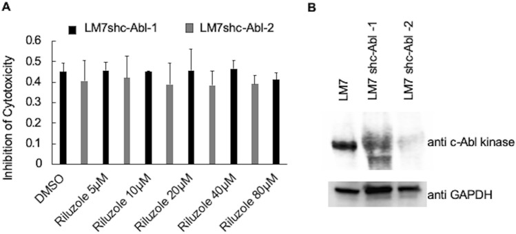
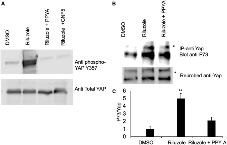
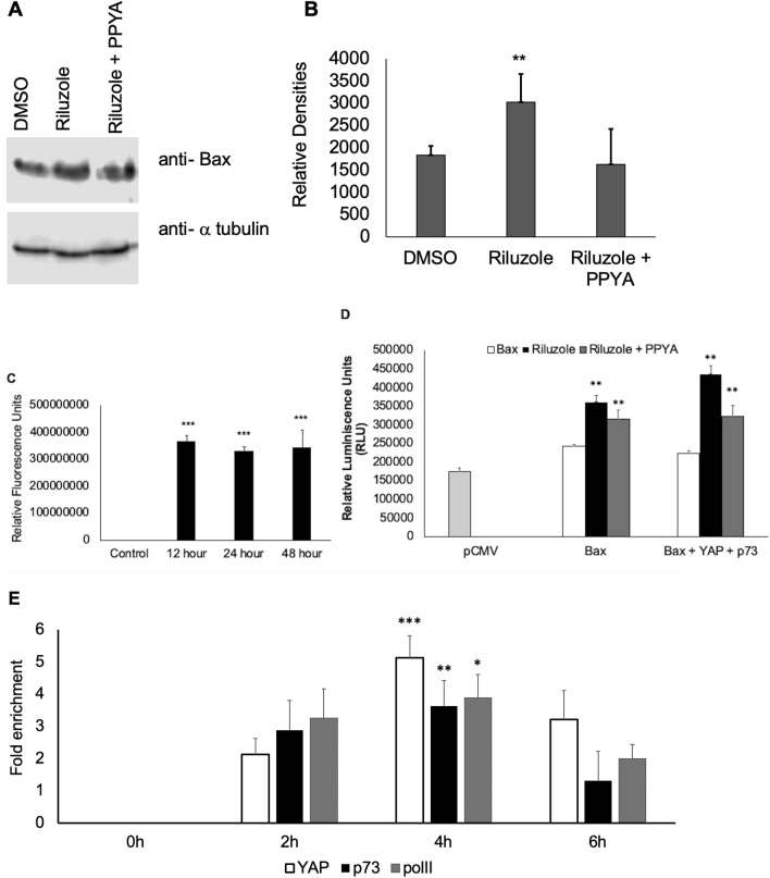
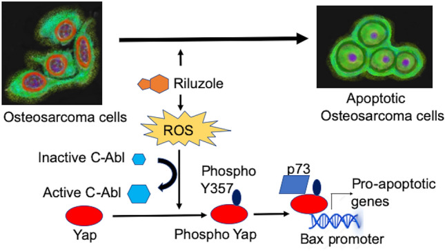
Our lab has previously demonstrated Riluzole to be an effective drug in inhibiting proliferation and inducing apoptosis in both human and mouse osteosarcoma. Yes-associated protein is a transcription co-activator, known to be involved in cell proliferation or apoptosis depending on its protein partner. In the present study we investigated the role of YAP in apoptosis in osteosarcoma, we hypothesized that YAP may be activated by Riluzole to induce apoptosis in osteosarcoma. By knocking down the expression of YAP, we have demonstrated that Riluzole failed to induce apoptosis in YAP deficient osteosarcoma cells. Riluzole caused translocation of YAP from the cytoplasm to the nucleus, indicating YAP's role in apoptosis. Both Riluzole-induced phosphorylation of YAP at tyrosine 357 and Riluzole-induced apoptosis were blocked by inhibitors of c-Abl kinase. In addition, knockdown of c-Abl kinase prevented Riluzole-induced apoptosis in LM7 cells. We further demonstrated that Riluzole promoted interaction between YAP and p73, while c-Abl kinase inhibitors abolished the interaction. Subsequently, we demonstrated that Riluzole enhanced activity of the Bax promoter in a luciferase reporter assay and enhanced YAP/p73 binding on endogenous Bax promoter in a ChIP assay. Our data supports a novel mechanism in which Riluzole activates c-Abl kinase to regulate pro-apoptotic activity of YAP in osteosarcoma.


Conflict of interest statement

The authors declare no competing interests.

Figures

Figure 1
Figure 1
YAP knockdown decreases Riluzole induced apoptosis in LM7 cells. (A) Apoptosis in WT LM7 cells with control shRNA or LM7shYAP-1and LM7shYAP-2 with shYAP were treated with Riluzole (100 μM) or DMSO for control. DAPI positive and TUNEL positive cells were scored by fluorescence microscopy. Triplicate samples were analyzed for each treatment. The experiment was repeated three times, p value ***< 0.001. (B) Representative images of TUNEL assay are shown. (C) Percentage of apoptotic cells was analyzed based on the number of DAPI positive and TUNEL positive cells. (D) Western blot of YAP expression in WT LM7 cells and LM7shYAP cells using mouse anti-YAP antibody and reprobed with rabbit anti-GAPDH as a loading control.
Figure 2
Figure 2
Riluzole alters subcellular localization of YAP. (A). Subcellular localization of endogenous YAP was measured in LM7 cells grown in 0.5% serum, p value < 0.05**. (B) Representative images for LM7 cells grown in 0.5% serum. (C) In 10% serum, p value < 0.05**. (D) Representative images for LM7 cells grown in 10% serum. (E) Subcellular localization of stably expressed Myc-tagged WT-YAP in LM7 cells, p value < 0.05**. (F) Representative images for Myc-tagged WT-YAP in LM7 cells. (G) YAP mutant (YAP S5A) (C + N = cytoplasmic + nuclear), p value < 0.05**. (H). Representative images of Myc-tagged YAP mutant (YAP S5A) LM7 cells. The cells were treated with DMSO or Riluzole (50 μM) for 1 h, fixed and stained with either anti-YAP (AD) or anti-Myc (EH) antibody. The samples were mounted in DAPI containing media. Fluorescence microscopy was performed on blinded samples and YAP localization was analyzed from triplicate samples in each four independent experiments. (I) Whole cell extracts were analyzed by western blot using either anti-phospho 127, anti-YAP or anti tubulin antibodies. (J) Intensity of the bands was measured by densitometry using LiCOR imaging system. The ratio of Phospho YAP to total YAP is represented. The experiment was repeated this experiment three times with triplicate samples in each trial, p value < 0.005**.
Figure 3
Figure 3
(A) Inhibitors of c-Abl Kinase block Riluzole-induced apoptosis. (A) Cytotoxicity was measured using MTT assay in LM7 cells in the presence of 100 μM Riluzole, or PPY A or GNF-5 or Riluzole + PPY A or Riluzole + GNF-5, p value < 0.0001***. (B) Subcellular localization of YAP in the presence of Riluzole (100 μM) or Riluzole + PPY A (20 nM) or Riluzole + GNF-5 (220 nM), p value < 0.05**. (C) Intracellular ROS was measured in LM7 cells treated with Riluzole or H2O2 as a positive control. These experiments were repeated three times with triplicate samples, p value < 0.05**.
Figure 4
Figure 4
c-Abl Kinase is required for Riluzole-induced apoptosis in LM7 Cells. (A) LM7 cells with c-Abl kinase knockdown were tested for Riluzole sensitivity at various doses as indicated in a MTT assay. Data for two independent selected stable cells lines, LM7shc-Abl-1 and LM7shc-Abl-2, is shown. (B) Western blot of c-Abl kinase knockdown and GAPDH is shown as a control.
Figure 5
Figure 5
Riluzole induces phosphorylation of YAP at Y357 to promote binding of YAP with p73. (A) Whole cell extracts of treated LM7 cells with DMSO, Riluzole (50 μM) or Riluzole (50 μM) + PPY A (20 nM) were blotted with rabbit anti-YAP (phosphoY357) antibody and the blot was reprobed with mouse anti-YAP antibody. (B) Co-immunoprecipitation was performed to assess interaction between endogenous YAP and p73 using whole cell extracts. Samples were immunoprecipitated with mouse anti-YAP antibody and the western blot was probed with rabbit anti-p73 antibody, the blot was reprobed with mouse anti-YAP antibody. An asterisk next to the band indicates a nonspecific band. (C) The densities of the immunoprecipitated p73 and YAP were quantified to measure the ratio of p73/YAP. These experiments were repeated three times to confirm the results, p value < 0.05**, IP immunoprecipitation.
Figure 6
Figure 6
Riluzole enhances Bax expression by inducing Bax promoter activity in LM7 cells. (A) Whole cell extracts from LM7 cells were probed with anti-Bax antibody to determine the effect of Riluzole (50 μM) and Riluzole (50 μM) + PPY A (20 nM) on Bax expression, α tubulin is used as a loading control. (B) Bar graph showing densities of bands from 3 independent experiments. (C) Caspase assay represents data from 3 independent experiments, p value = 0.003***. (D) Bax promoter driven luciferase expression was measured in LM7 cells. LM7 cells were treated with DMSO or Riluzole (50 μM) or Riluzole (50 μM) + PPY A (20 nM) for 24 h, p value < 0.05**. (E) Fold enhancement of the Bax promoter activity was measured using Chromatin Immunoprecipitation assay using anti YAP, anti p73 and or Polymerase II antibodies followed by qPCR to detect and quantify fold induction of Bax promoter. Representative data of three independent experiments is shown, p value < 0.5* p value < 0.05**. p value < 0.005***.
Figure 7
Figure 7
Mechanism of action of Riluzole-induced apoptosis in osteosarcoma. A schematic representation of the mechanism of Riluzole action is shown.

References

    1. Srinivas US, Tan BWQ, Vellayappan BA, Jeyasekharan AD. ROS and the DNA damage response in cancer. Redox Biol. 2019;25:101084. doi: 10.1016/j.redox.2018.101084. - DOI - PMC - PubMed
    1. Yang Y, Yang R, Roth M, Piperdi S, Zhang W, Dorfman H, Rao P, Park A, Tripathi S, Freeman C, Zhang Y, Sowers R, Rosenblum J, Geller D, Hoang B, Gill J, Gorlick R. Genetically transforming human osteoblasts to sarcoma: Development of an osteosarcoma model. Genes Cancer. 2017;8(1–2):484–494. doi: 10.18632/genesandcancer.133. - DOI - PMC - PubMed
    1. Kaste SC, Pratt CB, Cain AM, Jones-Wallace DJ, Rao BN. Metastases detected at the time of diagnosis of primary pediatric extremity osteosarcoma at diagnosis: Imaging features. Cancer. 1999;86(8):1602–1608. doi: 10.1002/(SICI)1097-0142(19991015)86:8<1602::AID-CNCR31>3.0.CO;2-R. - DOI - PubMed
    1. Mialou V, Philip T, Kalifa C, Perol D, Gentet JC, Marec-Berard P, Pacquement H, Chastagner P, Defaschelles AS, Hartmann O. Metastatic osteosarcoma at diagnosis: Prognostic factors and long-term outcome—The French pediatric experience. Cancer. 2005;104(5):1100–1109. doi: 10.1002/cncr.21263. - DOI - PubMed
    1. Geller DS, Gorlick R. Osteosarcoma: A review of diagnosis, management, and treatment strategies. Clin. Adv. Hematol. Oncol. 2010;8(10):705–718. - PubMed

Publication types

MeSH terms