

Endonuclease-based genotyping of the RBM as a method to track the emergence or evolution of SARS-CoV-2 variants
- PMID: 34697603
- PMCID: PMC8529542
- DOI: 10.1016/j.isci.2021.103329
Endonuclease-based genotyping of the RBM as a method to track the emergence or evolution of SARS-CoV-2 variants
Abstract
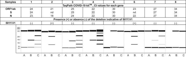
Since the beginning of the COVID-19 pandemics, variants have emerged. Some of them display increased transmissibility and/or resistance to immune response. Most of the mutations involved in the functional adaptation are found in the receptor-binding motif (RBM), close to the interface with the receptor ACE2. We thus developed a fast molecular assay to detect mutations in the RBM coding sequence. After amplification, the amplicon is heat-denatured and hybridized with an amplicon of reference. The presence of a mutation can be detected using a mismatch-specific endonuclease and the cleavage pattern is analyzed by capillary electrophoresis. The method was validated on RNA of severe acute respiratory syndrome coronavirus 2 (SARS-CoV-2) variants produced in vitro before being implemented for clinical samples. The assay showed 97.8% sensitivity and 97.8% specificity. The procedure can be set up for high-throughput identification of the presence of mutations and serve as a first-line screening to select the samples for full genome sequencing.
Keywords: Methodology in biological sciences; Virology.
© 2021 The Authors.
Conflict of interest statement
The authors declare no competing interests
Figures


Similar articles
-
Receptor-Binding-Motif-Targeted Sanger Sequencing: a Quick and Cost-Effective Strategy for Molecular Surveillance of SARS-CoV-2 Variants.Microbiol Spectr. 2022 Jun 29;10(3):e0066522. doi: 10.1128/spectrum.00665-22. Epub 2022 May 31. Microbiol Spectr. 2022. PMID: 35638906 Free PMC article.
-
Emergency SARS-CoV-2 Variants of Concern: Novel Multiplex Real-Time RT-PCR Assay for Rapid Detection and Surveillance.Microbiol Spectr. 2022 Feb 23;10(1):e0251321. doi: 10.1128/spectrum.02513-21. Epub 2022 Feb 23. Microbiol Spectr. 2022. PMID: 35196812 Free PMC article.
-
Developing an Amplification Refractory Mutation System-Quantitative Reverse Transcription-PCR Assay for Rapid and Sensitive Screening of SARS-CoV-2 Variants of Concern.Microbiol Spectr. 2022 Feb 23;10(1):e0143821. doi: 10.1128/spectrum.01438-21. Epub 2022 Jan 5. Microbiol Spectr. 2022. PMID: 34985323 Free PMC article.
-
SARS-CoV-2 Omicron Mutation Is Faster than the Chase: Multiple Mutations on Spike/ACE2 Interaction Residues.Immune Netw. 2021 Dec 23;21(6):e38. doi: 10.4110/in.2021.21.e38. eCollection 2021 Dec. Immune Netw. 2021. PMID: 35036025 Free PMC article. Review.
-
Monoclonal antibodies for COVID-19 therapy and SARS-CoV-2 detection.J Biomed Sci. 2022 Jan 4;29(1):1. doi: 10.1186/s12929-021-00784-w. J Biomed Sci. 2022. PMID: 34983527 Free PMC article. Review.
Cited by
-
Recombinant receptor-binding motif of spike COVID-19 vaccine candidate induces SARS-CoV-2 neutralizing antibody response.Bioimpacts. 2024 Nov 4;15:30520. doi: 10.34172/bi.30520. eCollection 2025. Bioimpacts. 2024. PMID: 40256231 Free PMC article.
-
Discovery of a Potential Allosteric Site in the SARS-CoV-2 Spike Protein and Targeting Allosteric Inhibitor to Stabilize the RBD Down State using a Computational Approach.Curr Comput Aided Drug Des. 2024;20(6):784-797. doi: 10.2174/1573409919666230726142418. Curr Comput Aided Drug Des. 2024. PMID: 37493168
References
-
- Bal A., Destras G., Gaymard A., Stefic K., Marlet J., Eymieux S., Regue H., Semanas Q., d’Aubarede C., Billaud G., et al. Two-step strategy for the identification of SARS-CoV-2 variant of concern 202012/01 and other variants with spike deletion H69–V70, France, August to December 2020. Eurosurveillance. 2021;26:2100008. doi: 10.2807/1560-7917.ES.2021.26.3.2100008. - DOI - PMC - PubMed
-
- Davies H., Dicks E., Stephens P., Cox C., Teague J., Greenman C., Bignell G., O’Meara S., Edkins S., Parker A., et al. High throughput DNA sequence variant detection by conformation sensitive capillary electrophoresis and automated peak comparison. Genomics. 2006;87:427–432. doi: 10.1016/j.ygeno.2005.11.008. - DOI - PubMed
-
- Greaney A.J., Loes A.N., Crawford K.H.D., Starr T.N., Malone K.D., Chu H.Y., Bloom J.D. Comprehensive mapping of mutations in the SARS-CoV-2 receptor-binding domain that affect recognition by polyclonal human plasma antibodies. Cell Host Microbe. 2021;29:463–476.e6. doi: 10.1016/j.chom.2021.02.003. - DOI - PMC - PubMed
LinkOut - more resources
Full Text Sources
Miscellaneous
