. 2021 Oct 12;10(4):39.
doi: 10.3390/antib10040039.

Novel Selection Approaches to Identify Antibodies Targeting Neoepitopes on the C5b6 Intermediate Complex to Inhibit Membrane Attack Complex Formation

Affiliations

Novel Selection Approaches to Identify Antibodies Targeting Neoepitopes on the C5b6 Intermediate Complex to Inhibit Membrane Attack Complex Formation

Lasse Stach et al. Antibodies (Basel). .

Abstract

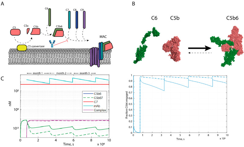
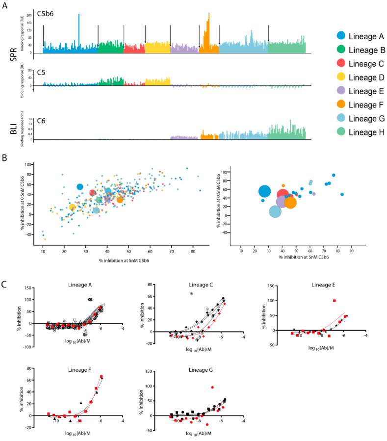
The terminal pathway of complement is implicated in the pathology of multiple diseases and its inhibition is, therefore, an attractive therapeutic proposition. The practicalities of inhibiting this pathway, however, are challenging, as highlighted by the very few molecules in the clinic. The proteins are highly abundant, and assembly is mediated by high-affinity protein-protein interactions. One strategy is to target neoepitopes that are present transiently and only exist on active or intermediate complexes but not on the abundant native proteins. Here, we describe an antibody discovery campaign that generated neoepitope-specific mAbs against the C5b6 complex, a stable intermediate complex in terminal complement complex assembly. We used a highly diverse yeast-based antibody library of fully human IgGs to screen against soluble C5b6 antigen and successfully identified C5b6 neoepitope-specific antibodies. These antibodies were diverse, showed good binding to C5b6, and inhibited membrane attack complex (MAC) formation in a solution-based assay. However, when tested in a more physiologically relevant membrane-based assay these antibodies failed to inhibit MAC formation. Our data highlight the feasibility of identifying neoepitope binding mAbs, but also the technical challenges associated with the identification of functionally relevant, neoepitope-specific inhibitors of the terminal pathway.

Keywords: antibody discovery; complement; neoepitope; terminal pathway; therapeutic antibody.


Conflict of interest statement

E.-M.N., M.F., S.J.K, T.D.B., L.S., E.K.H.D., R.P.B., D.A.G., K.A.W., N.T., A.S., J.L.B. and J.E.C. are employees and shareholders of GSK plc. J.P.H., A.T. and C.L.H. are now employees of Gyroscope Therapeutics, AstraZeneca and Newcastle University (Translational and Clinical Research Institute), respectively.

Figures

Figure 1
Figure 1
Schematics of biology, desired molecule and antibody selection strategy. (A) Schematic representation of membrane attack complex formation, indicating the interaction targeted by the proposed therapeutic. (B) Space filling representations of the atomic models of C6 (PDB: 3T5O), C5 (PDB: 3CU7) and C5b6 (PDB: 4A5W), illustrating the formation of neoepitopes. (C) Dose prediction modelling of an anti-C5b6 antibody with 1 pM affinity and an on-rate of 1 × 10⁷ Ms−1 and onthly dosing of 10 mg/kg. Simulation carried out for both reported C7−C5b6 on-rates; 4 × 10⁵ Ms−1 (dashed line) and 2 × 10⁶ Ms−1 (solid line). Fraction target engagement plotted on the right.
Figure 2
Figure 2
Naïve selections strategy and sorting. (A) Flow diagram of the 6 rounds of selection to isolate anti-C5b6 antibodies from a yeast library. Selection rounds are colour coded as MACS (orange), FACS (blue) and negative selection FACS (grey). NSB refers to a proprietary reagent to select against antibodies displaying nonspecific binding. (B) FACS plots of round 5, where clones were selected that show strong binding to C5b6, but no binding to C7 once bound to C5b6 already. These are coloured in blue. All three plots show the same population, with different parameters plotted on each axis.
Figure 3
Figure 3
Characterisation of naïve selections output (A) Relative binding responses measured by BLI of the antibodies from light chain batch shuffle against three antigens, C5 (red), C6 (amber), C5b6 (green) at 50 nM. Stacked bars with height indicate relative binding responses for each of the 3 analytes. Each bar represents one clone. (B) Level of terminal complement complex production in the presence of low (black) and high (grey) antibody concentrations, visualised in a bar chart. The output was tested in 1% NHS at ~1–2 μM (high), 0.02–0.06 μM, the controls were added as follows: anti-C5 and disabled anti-C5 (0.02/0.01 μM), isotype control at 14/0.44 μM. (C) BLI binding responses from (A) filtered to the 8 clones that were chosen as a hit panel. Side-by-side bars indicating the respective, absolute binding responses. Data in A and C has been normalised for the differences in molecular weight of the analytes. (D) Antibody heavy chain and kappa light chain genes and CDRH3 sequences.
Figure 4
Figure 4
MAC on a chip and a novel SPR assay format. (A) Schematic representation of the SPR assay format. Composite model generated using PDB depositions 5I5K and 4A5W. An anti-C5 tool antibody (blue) is immobilised on a CM5 chip (grey) and C5b6 (red/orange) is captured by the antibody. Analytes can then be tested for binding against C5b6. (B) Reconstitution of the membrane attack complex on an SPR chip surface. Analytes injected as indicated by the legend. Experiment was performed with either captured C5 (inset) or C5b6 (main). (C) Measuring the on−rate of C7 on C5b6. Experimental data in red and fit to a 1:1 binding model in black. Table of kinetic parameters below. (D) Binding of the 8 hit panel clones to anti-C5 captured C5b6. Experimental data coloured as indicated in the legend. Fits to a 1:1 binding model in black. Excluded data shown as dotted lines. Binding kinetics only reported for the true 1:1 Fab-binding.
Figure 5
Figure 5
Development of functional complement assays. (A) Liposome leakage assay development. Fluorescence intensity measured kinetically and C7 injection time point indicated. The assay was run using full MAC (red), a no-C5b6 low control (black), full MAC plus detergent high control (blue) and full MAC plus an anti-MAC tool antibody (green). (B) Liposome assay development using an anti-C7 mAb was used anti-MAC positive control antibody and an isotype control antibody. (C) Dose−dependent effect of the anti−MAC antibody on the kinetics of liposome formation.
Figure 6
Figure 6
Characterisation of the affinity matured output. (A) Relative binding responses of affinity matured clones for the 3 antigens. C5 and C5b6 binding is measured by SPR using the capture method described in Figure 4, binding to C6 is measured by BLI. Clones are coloured by lineage and the parental clone, the first bar for each lineage, is highlighted with an arrow. The C5 and C5b6 binding responses are plotted on the same axis and not normalised for the relative molecular weights of C5 and C5b6. (B) %−inhibition in the C9-oligomerisation assay with 0.5 nM C5b6 and 2.5 nM C7 (y−axis) and 5 nM C5b6 and 10 nM C7 (x−axis). Each circle represents one clone, coloured by lineage. Parental clones are shown as larger circles. Right: A selection of 24 promising clones, from 5 lineages, to be further analysed in a dose−response HTRF assay. (C) Dose response curves of the 24 clones tested in the HTRF assay using 5 nM C5b6 and 10 nM C7, grouped by lineage, with parental clones highlighted in red.

Similar articles

Cited by

References

    1. Dunkelberger J.R., Song W.C. Complement and its role in innate and adaptive immune responses. Cell Res. 2010;20:34–50. doi: 10.1038/cr.2009.139. - DOI - PubMed
    1. Ricklin D., Hajishengallis G., Yang K., Lambris J.D. Complement: A key system for immune surveillance and homeostasis. Nat. Immunol. 2010;11:785–797. doi: 10.1038/ni.1923. - DOI - PMC - PubMed
    1. Taylor P.R., Carugati A., Fadok V.A., Cook H.T., Andrews M., Carroll M.C., Savill J.S., Henson P.M., Botto M., Walport M.J. A hierarchical role for classical pathway complement proteins in the clearance of apoptotic cells in vivo. J. Exp. Med. 2000;192:359–366. doi: 10.1084/jem.192.3.359. - DOI - PMC - PubMed
    1. Zipfel P.F., Skerka C. Complement regulators and inhibitory proteins. Nat. Rev. Immunol. 2009;9:729–740. doi: 10.1038/nri2620. - DOI - PubMed
    1. Wong E.K.S., Kavanagh D. Diseases of complement dysregulation-an overview. Semin. Immunopathol. 2018;40:49–64. doi: 10.1007/s00281-017-0663-8. - DOI - PMC - PubMed

LinkOut - more resources