

Cytoskeletal Actin Structure in Osteosarcoma Cells Determines Metastatic Phenotype via Regulating Cell Stiffness, Migration, and Transmigration
- PMID: 34698103
- PMCID: PMC8928956
- DOI: 10.3390/cimb43030089
Cytoskeletal Actin Structure in Osteosarcoma Cells Determines Metastatic Phenotype via Regulating Cell Stiffness, Migration, and Transmigration
Abstract
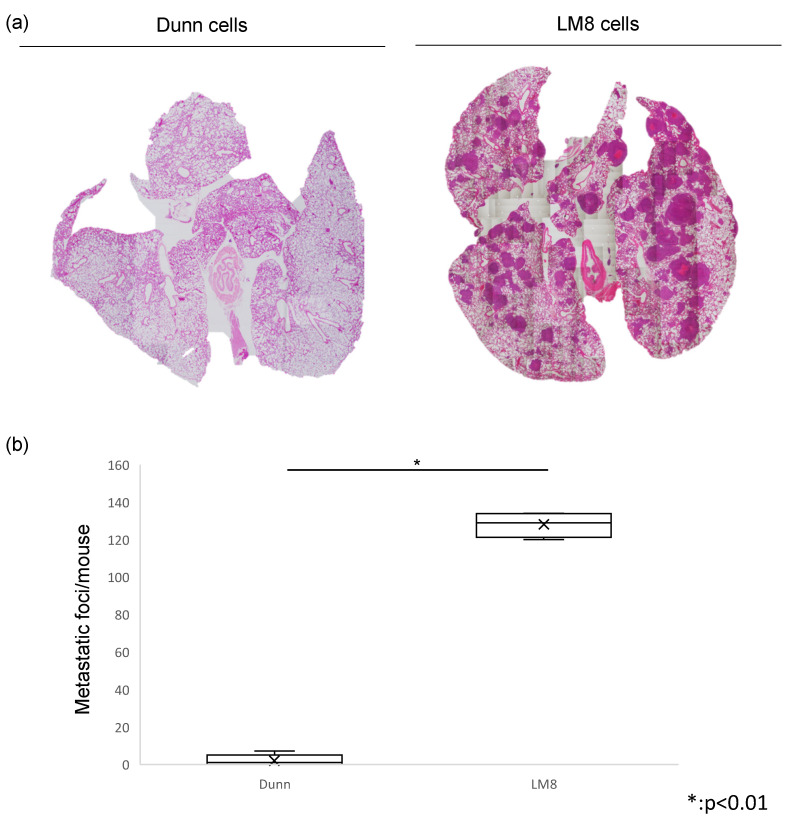
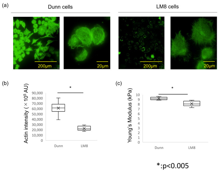
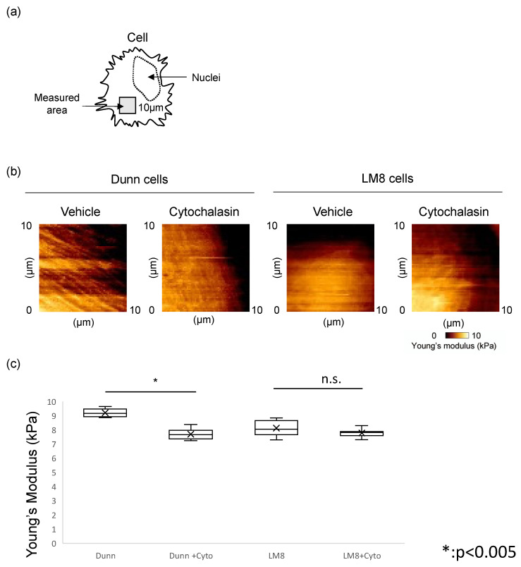
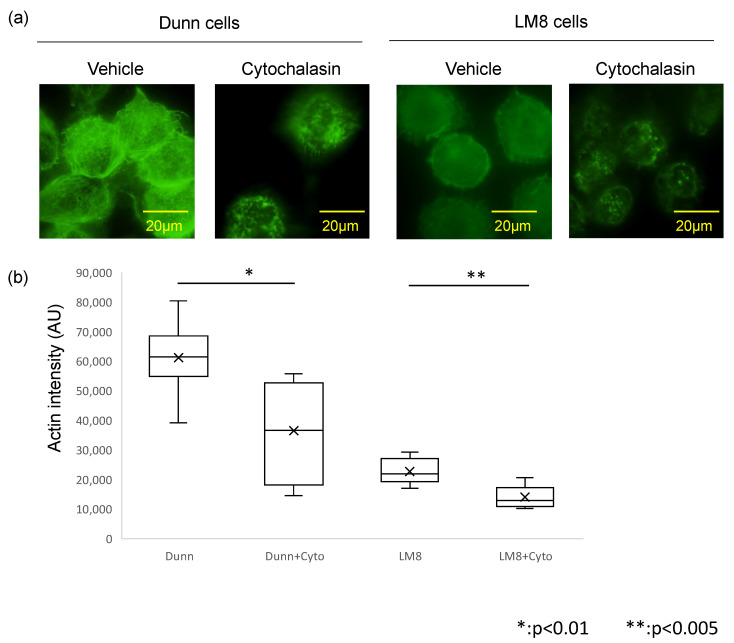
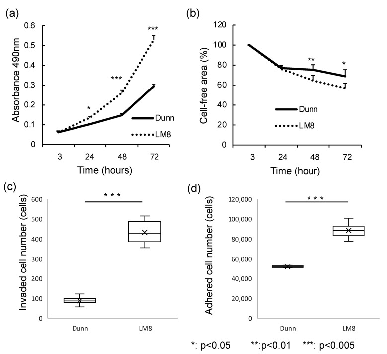
Osteosarcoma is the most common primary malignant bone tumor. The cause of death due to osteosarcoma is typically a consequence of metastasis to the lung. Controlling metastasis leads to improved prognosis for osteosarcoma patients. The cell stiffness of several tumor types is involved in metastatic potential; however, it is unclear whether the metastatic potential of osteosarcoma depends on cell stiffness. In this study, we analyzed the cell stiffness of the low metastatic Dunn cell line and its highly metastatic LM8 subline, and compared actin organization, cell proliferation, and metastasis. Actin cytoskeleton, polymerization, stiffness, and other cellular properties were analyzed. The organization of the actin cytoskeleton was evaluated by staining F-actin with Alexa Fluor 488 phalloidin. Cell stiffness was measured using Atomic Force Microscopy (AFM). Cell proliferation, migration, invasion, and adhesion were also evaluated. All experiments were performed using mouse osteosarcoma cell lines cultured in the absence and presence of cytochalasin. In LM8 cells, actin polymerization was strongly suppressed and actin levels were significantly lower than in Dunn cells. Stiffness evaluation revealed that LM8 cells were significantly softer than Dunn. Young's modulus images showed more rigid fibrillar structures were present in Dunn cells than in LM8 cells. LM8 cells also exhibited a significantly higher proliferation. The migration and invasion potential were also higher in LM8 cells, whereas the adhesion potential was higher in Dunn cells. The administration of cytochalasin resulted in actin filament fragmentation and decreased actin staining intensity and cell stiffness in both LM8 and Dunn cells. Cells with high metastatic potential exhibited lower actin levels and cell stiffness than cells with low metastatic potential. The metastatic phenotype is highly correlated to actin status and cell stiffness in osteosarcoma cells. These results suggest that evaluation of actin dynamics and cell stiffness is an important quantitative diagnostic parameter for predicting metastatic potential. We believe that these parameters represent new reliable quantitative indicators that can facilitate the development of new drugs against metastasis.
Keywords: actin cytoskeleton; atomic force microscopy; cell stiffness; metastasis; osteosarcoma cell line.
Conflict of interest statement
The authors declare that they have no conflict of interest.
Figures
Similar articles
-
A Novel Approach to Reducing Lung Metastasis in Osteosarcoma: Increasing Cell Stiffness with Carbenoxolone.Curr Issues Mol Biol. 2023 May 17;45(5):4375-4388. doi: 10.3390/cimb45050278. Curr Issues Mol Biol. 2023. PMID: 37232747 Free PMC article.
-
Dynamic analysis of lung metastasis by mouse osteosarcoma LM8: VEGF is a candidate for anti-metastasis therapy.Clin Exp Metastasis. 2013 Apr;30(4):369-79. doi: 10.1007/s10585-012-9543-8. Epub 2012 Oct 18. Clin Exp Metastasis. 2013. PMID: 23076771 Free PMC article.
-
Mesenchymal mode of migration participates in pulmonary metastasis of mouse osteosarcoma LM8.Clin Exp Metastasis. 2010 Dec;27(8):619-30. doi: 10.1007/s10585-010-9352-x. Epub 2010 Sep 26. Clin Exp Metastasis. 2010. PMID: 20872237
-
Actin dynamics during tumor cell dissemination.Int Rev Cell Mol Biol. 2021;360:65-98. doi: 10.1016/bs.ircmb.2020.09.004. Epub 2020 Nov 24. Int Rev Cell Mol Biol. 2021. PMID: 33962751 Free PMC article. Review.
-
Characterization of LIMA1 and its emerging roles and potential therapeutic prospects in cancers.Front Oncol. 2023 May 19;13:1115943. doi: 10.3389/fonc.2023.1115943. eCollection 2023. Front Oncol. 2023. PMID: 37274282 Free PMC article. Review.
Cited by
-
The CBP/β-Catenin Antagonist, ICG-001, Inhibits Tumor Metastasis via Blocking of the miR-134/ITGB1 Axis-Mediated Cell Adhesion in Nasopharyngeal Carcinoma.Cancers (Basel). 2022 Jun 25;14(13):3125. doi: 10.3390/cancers14133125. Cancers (Basel). 2022. PMID: 35804897 Free PMC article.
-
Microenvironment matters: insights from the FOSTER consortium on microenvironment-driven approaches to osteosarcoma therapy.Cancer Metastasis Rev. 2025 Apr 10;44(2):44. doi: 10.1007/s10555-025-10257-3. Cancer Metastasis Rev. 2025. PMID: 40210800 Free PMC article. Review.
-
RARRES2 is involved in the "lock-and-key" interactions between osteosarcoma stem cells and tumor-associated macrophages.Sci Rep. 2024 Jan 27;14(1):2267. doi: 10.1038/s41598-024-52738-5. Sci Rep. 2024. PMID: 38280909 Free PMC article.
-
3D-printed β-TCP scaffold as a bone-mimicking environment for an engineered model of osteosarcoma: In vitro properties and transcriptomic insights.Mater Today Bio. 2025 Apr 12;32:101766. doi: 10.1016/j.mtbio.2025.101766. eCollection 2025 Jun. Mater Today Bio. 2025. PMID: 40290888 Free PMC article.
-
Targeting metastasis in paediatric bone sarcomas.Mol Cancer. 2025 May 29;24(1):153. doi: 10.1186/s12943-025-02365-z. Mol Cancer. 2025. PMID: 40442778 Free PMC article. Review.
References
-
- Kaya M., Wada T., Kawaguchi S., Nagoya S., Yamashita T., Abe Y., Hiraga H., Isu K., Shindoh M., Higashino F., et al. Increased pre-therapeutic serum vascular endothelial growth factor in patients with early clinical relapse of osteosarcoma. Br. J. Cancer. 2002;86:864–869. doi: 10.1038/sj.bjc.6600201. - DOI - PMC - PubMed
MeSH terms
Substances
LinkOut - more resources
Full Text Sources
Miscellaneous
