

Oligomerization-driven MLKL ubiquitylation antagonizes necroptosis
- PMID: 34698396
- PMCID: PMC8634140
- DOI: 10.15252/embj.2019103718
Oligomerization-driven MLKL ubiquitylation antagonizes necroptosis
Abstract
Mixed lineage kinase domain-like (MLKL) is the executioner in the caspase-independent form of programmed cell death called necroptosis. Receptor-interacting serine/threonine protein kinase 3 (RIPK3) phosphorylates MLKL, triggering MLKL oligomerization, membrane translocation and membrane disruption. MLKL also undergoes ubiquitylation during necroptosis, yet neither the mechanism nor the significance of this event has been demonstrated. Here, we show that necroptosis-specific multi-mono-ubiquitylation of MLKL occurs following its activation and oligomerization. Ubiquitylated MLKL accumulates in a digitonin-insoluble cell fraction comprising organellar and plasma membranes and protein aggregates. Appearance of this ubiquitylated MLKL form can be reduced by expression of a plasma membrane-located deubiquitylating enzyme. Oligomerization-induced MLKL ubiquitylation occurs on at least four separate lysine residues and correlates with its proteasome- and lysosome-dependent turnover. Using a MLKL-DUB fusion strategy, we show that constitutive removal of ubiquitin from MLKL licences MLKL auto-activation independent of necroptosis signalling in mouse and human cells. Therefore, in addition to the role of ubiquitylation in the kinetic regulation of MLKL-induced death following an exogenous necroptotic stimulus, it also contributes to restraining basal levels of activated MLKL to avoid unwanted cell death.
Keywords: DUB-fusion; MLKL; membranes; necroptosis; ubiquitylation.
© 2021 The Authors.
Conflict of interest statement
SNY, AB, UN, CF, SEG, JMM, JMH and JS contribute to, or have contributed to, a project with Anaxis Pty Ltd to develop necroptosis inhibitors.
Figures
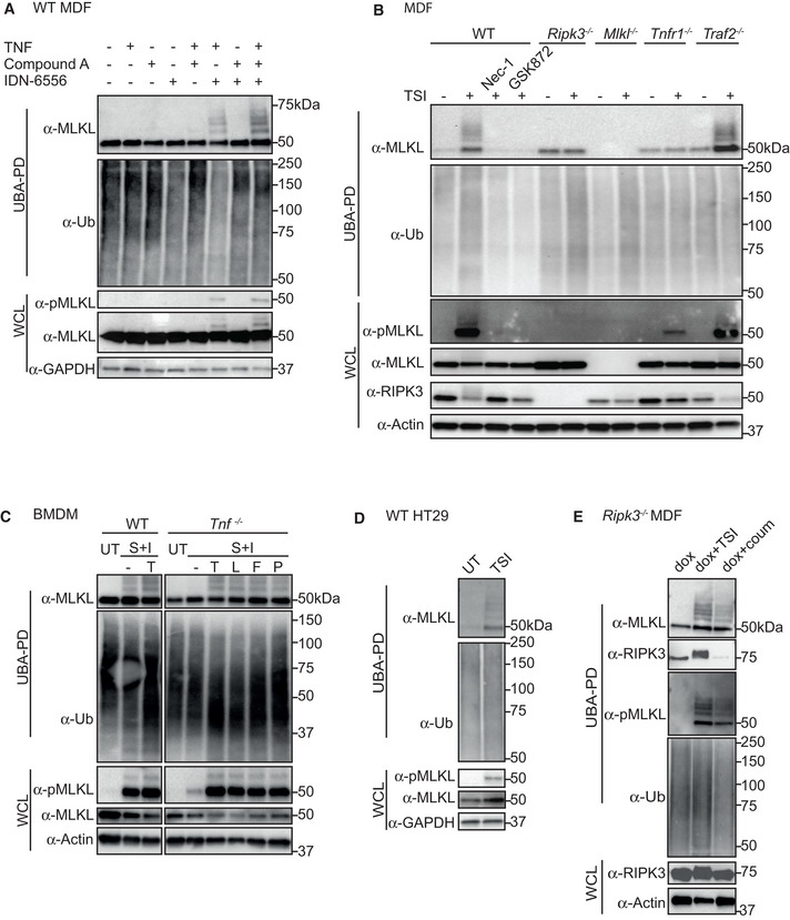
WT MDFs were treated ± TSI individually or in combination for 3 h. Whole cell lysate (WCL) and UBA pulldown (UBA PD) fractions were analysed by Western blot and probed with antibodies as indicated. Representative of three independent experiments. Samples of UBA pulldown in following figures were analysed in the same way unless otherwise indicated.
WT, Ripk3 −/−, Mlkl −/−, Tnfr1 −/− and Traf2 −/− MDFs were untreated (UT) or treated with TSI for 3 h. Nec‐1 and GSK872 were added to inhibit RIPK1 and RIPK3 kinase activities, respectively.
WT and Tnf −/− BMDMs were treated ± death ligands including TNF (T), LPS (L), Fas ligand (F) and Poly I:C (P) in addition to S and I for 3 h.
WT HT29 cells were untreated (UT) or treated with TSI for 16 h.
RIPK3‐gyrase was inducibly expressed in Ripk3 −/− MDFs by doxycycline (dox) for 5 h, and cells were then treated ± combination of TSI, or coumermycin (coum) for 3 h.
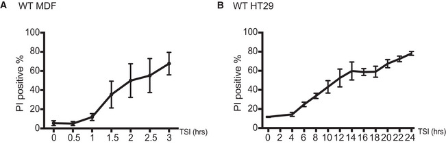
- A, B
MDFs (A) and HT29 (B) cells were treated with TSI. Cell death was measured by PI staining and flow cytometry. Data are plotted as mean ± SEM of at least three independent experiments.
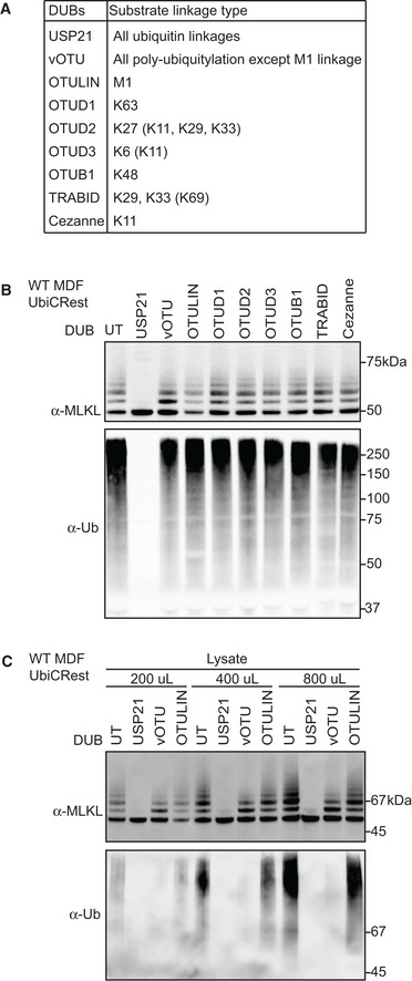
Deubiquitylating enzymes (DUBs) and their ubiquitin substrates. Less efficiently cleaved substrates are indicated in brackets.
UBA pulldown from WT MDFs treated with TSI for 3 h were subjected to the DUBs shown in (A). Beads eluates were analysed by Western blot and probed with antibodies as indicated. Representative of three independent experiments.
1.4‐ml cleared cell lysate from 4 × 10⁶ TSI‐treated WT MDFs was split into three parts of the indicated volume, followed by UBA pulldown and DUB incubation. Bead eluates were analysed by Western blot and probed with the indicated antibodies. Representative of three independent experiments.
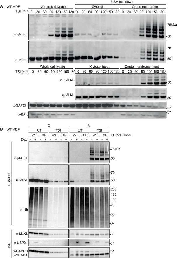
WT MDFs were treated with TSI for indicated time. Cells were fractionated into cytosol (C) and crude membrane (M), followed by UBA pulldown. All fractions were analysed by Western blot and probed with antibodies as indicated. Representative of three independent experiments.
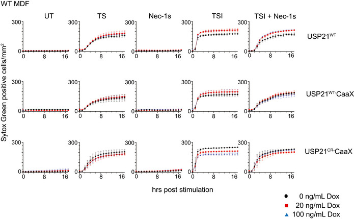
WT USP21‐CaaX and USP21C221R‐CaaX (CR) were inducibly expressed in WT MDFs by doxycycline for 5 h. Ubiquitylated proteins were enriched followed by TSI stimulation and cellular fractionation. All fractions were analysed by Western blot and probed with antibodies as indicated. Representative of three independent experiments.
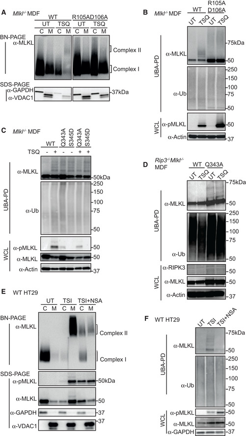
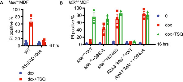
WT and R105AD106A mutant MLKL were inducibly expressed in Mlkl −/− MDFs by doxycycline, at the same time cells were untreated (UT) or treated with TSQ for 6 h. Cells were fractionated into cytosol (C) and crude membrane (M). Fractions were analysed by BN‐ or SDS–PAGE, Western blot and probed with the indicated antibodies. Representative of three independent experiments.
Cell lysates from (A) were subjected to UBA pulldown and analysed as described above.
WT, Q343A and S345D mutant MLKL were inducibly expressed in Mlkl −/− MDFs by doxycycline; at the same time, cells were treated ± TSQ for 16 h, followed by UBA pulldown. Representative of three independent experiments.
WT and Q343A mutant MLKL were inducibly expressed in Ripk3 −/− Mlkl −/− MDFs by doxycycline; at the same time, cells were treated ± TSQ for 16 h, followed by UBA pulldown. Representative of three independent experiments.
HT29 cells were stimulated with TSI, ± NSA (500 nM) or left untreated (UT) for 16 h, followed by cellular fractionation. Fractions were analysed by BN‐ or SDS–PAGE, Western blot and probed with the indicated antibodies. Representative of three independent experiments.
Cell lysates from (E) were subjected to UBA pulldown and analysed as described above.
Cell death of samples from Fig 4A was measured by PI staining based on flow cytometry. Data are plotted as mean ± SEM of three independent experiments.
Cell death of samples from Fig 4C and D was analysed as in (A).
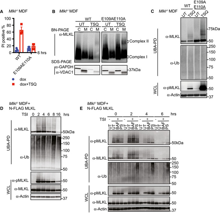
WT and E109AE110A mutant MLKL were inducibly expressed in Mlkl −/− MDFs by doxycycline, and cells were untreated (UT) or treated with TSQ for 6 h (please note that the same WT control was used in Fig EV3A). Cell death was measured by PI staining based on flow cytometry. Data are plotted as mean ± SEM of three independent experiments.
Cytosol (C) and crude membrane (M) cellular fractions from (A) were analysed by Western blot from BN‐PAGE or SDS–PAGE using antibodies as indicated. The same WT control was used in Fig 4A. Representative of three independent experiments.
Cell lysates from (A) were subjected to UBA pulldown and analysed as described above.
N‐FLAG MLKL were inducibly expressed in Mlkl −/− MDFs by doxycycline overnight, and cells were treated with TSI for indicated time, followed by UBA pulldown. Representative of three independent experiments.
N‐FLAG MLKL were inducibly expressed in Mlkl −/− MDFs by doxycycline for 16 h. Cells were stimulated ± TSI for 3 h after withdrawal of doxycycline. Then, TSI medium was removed and replaced to medium containing inhibitors Bafilomycin A1 (BAF), PS341 (PS) or left untreated (UT). IDN‐6556 was added to all conditions to block apoptosis. Cells were collected 0, 2, 4 and 6 h after medium replacement, followed by UBA pulldown. Representative of three independent experiments.

WT MLKL and N‐FLAG MLKL were inducibly expressed in Mlkl −/− MDFs by doxycycline for 12 h, and cells were treated with TSI or TSQ. TS treatment controlled that the response to TS was normal. Cell death was measured by PI staining based on flow cytometry. Data are plotted as mean ± SEM of three independent experiments.
WT MLKL and N‐FLAG MLKL were inducibly expressed in Mlkl −/− MDFs by doxycycline for 6 h, and cells were untreated (UT) or treated with TSQ. Crude membrane (M) and cytosolic (C) cellular fractions were analysed by Western blot from BN‐PAGE or SDS–PAGE using antibodies as indicated. Representative of three independent experiments.
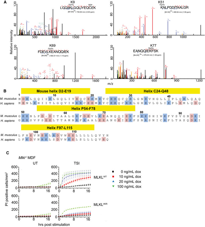
MS spectra were manually validated to confirm the identification of four Gly‐Gly sites on activated MLKL.
Alignment of mouse and human MLKL N‐terminal domain. Positively charged residues are labelled in blue, and negatively charged residues are labelled in pink.
WT and 4KR mutant MLKL were inducibly expressed in Mlkl −/− MDFs by doxycycline (at the indicated concentrations), and cells were treated ± TSI (added simultaneously) for 4 h. Sytox Green‐positive cells were quantified in real time by IncuCyte S3 live cell imaging. Data are plotted as mean ± SEM of three independent experiments.
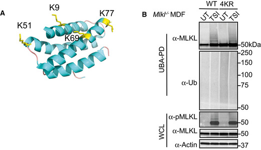
Cartoon of the N‐terminal region (residues 1–180) of mouse MLKL (PDB accession 4BTF [Murphy et al, 2013]) showing the four lysine residues identified from MS analysis as yellow sticks.
WT and 4KR mutant MLKL were inducibly expressed in Mlkl −/− MDFs by doxycycline for 6 h and cells were untreated (UT) or treated with TSI, followed by UBA pulldown. Representative of three independent experiments.
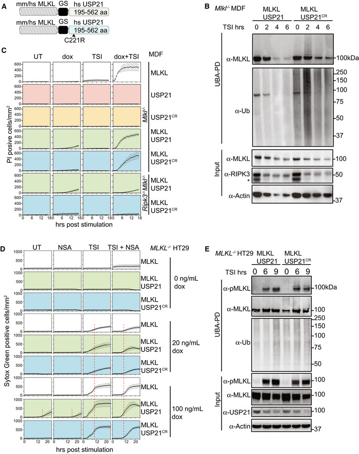
Schematic of the MLKL‐USP21 and USP21 catalytic dead mutant proteins.
Mouse MLKL‐USP21 and MLKL‐USP21C221R were inducibly expressed in Mlkl −/− MDFs by doxycycline (10 ng/ml) for 6 h with addition of a necroptotic stimulus (TSI) for the indicated time, followed by UBA pulldown. Non‐specific band is indicated with an asterisk in the blot of RIPK3. Representative of three independent experiments.
Mlkl −/− MDFs and Ripk3 −/− Mlkl −/− MDFs stably transfected with constructs encoding MLKL, USP21, USP21C221R, MLKL‐USP21 and MLKL‐USP21C221R were treated with doxycycline, TSI or in combination. Propidium iodide‐positive cells were quantified in real time by IncuCyte live cell imaging. Data are plotted as mean ± SEM of three independent experiments.
MLKL −/− HT29 cells stably transfected with constructs encoding human MLKL‐USP21 and MLKL‐USP21C221R, were treated with doxycycline, NSA (1 μM), TSI or combinations thereof (added simultaneously). Sytox Green‐positive cells were quantified in real time by live cell imaging. Data are plotted as mean ± SEM of six independent experiments (A red dashed line is shown to highlight the delay in death kinetics upon treatment with NSA).
Human MLKL‐USP21 and MLKL‐USP21C221R were inducibly expressed in MLKL −/− HT29 cells by doxycycline (10 ng/ml) for 16 h, and TSI was added for the indicated times but all samples were collected 25 h post‐induction, followed by UBA pulldown. Representative of three independent experiments.
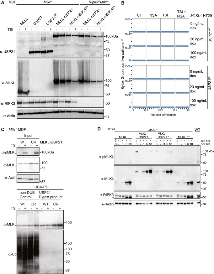
Mouse MLKL, USP21, USP21C221R, MLKL‐USP21 and MLKL‐USP21C221R fusions were inducibly expressed in Mlkl −/− MDFs and Ripk3 −/− Mlkl −/− MDFs as indicated, following doxycycline addition (20 ng/ml) for 6 h ± TSI. Representative of two independent experiments.
MLKL −/− HT29 cells stably transfected with doxycycline‐inducible constructs encoding human USP21 and human USP21C221R were treated with doxycycline, NSA (1 μM), TSI or combinations thereof (added simultaneously). Sytox Green‐positive cells were quantified in real time by live cell imaging. Representative of two independent experiments.
Mouse MLKL‐USP21 and MLKL‐USP21C221R fusions were inducibly expressed in Mlkl −/− MDFs by doxycycline (10 ng/ml) for 8 h with addition of a necroptotic stimulus (TSI) for 3 h, followed by UBA pulldown and USP21 digestion. Antibody (D6E3G, Cell Signaling Technology) was used here to detect MLKL phosphorylation. Representative of two independent experiments.
MLKL −/− HT29 cells were stably transfected with indicated doxycycline‐inducible MLKL alleles (phospho‐mimetic human MLKL mutant T357E/S358E indicated as MLKLTSEE) and treated with doxycycline (100 ng/ml) ± TSI (added simultaneously). A residue band from MLKL blot is indicated by an asterisk in RIPK3 blot due to reprobing. Representative of three independent experiments.
References
-
- Bertrand MJ, Milutinovic S, Dickson KM, Ho WC, Boudreault A, Durkin J, Gillard JW, Jaquith JB, Morris SJ, Barker PA (2008) cIAP1 and cIAP2 facilitate cancer cell survival by functioning as E3 ligases that promote RIP1 ubiquitination. Mol Cell 30: 689–700 - PubMed
-
- Cox J, Neuhauser N, Michalski A, Scheltema RA, Olsen JV, Mann M (2011) Andromeda: a peptide search engine integrated into the MaxQuant environment. J Proteome Res 10: 1794–1805 - PubMed
Publication types
MeSH terms
Substances
LinkOut - more resources
Full Text Sources
Miscellaneous
