Review
. 2022 Apr 1;102(2):893-992.
doi: 10.1152/physrev.00041.2020. Epub 2021 Oct 26.

Mitochondrial calcium exchange in physiology and disease

Affiliations
Review

Mitochondrial calcium exchange in physiology and disease

Joanne F Garbincius et al. Physiol Rev. .

Abstract

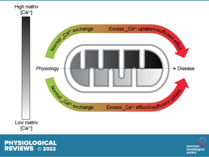
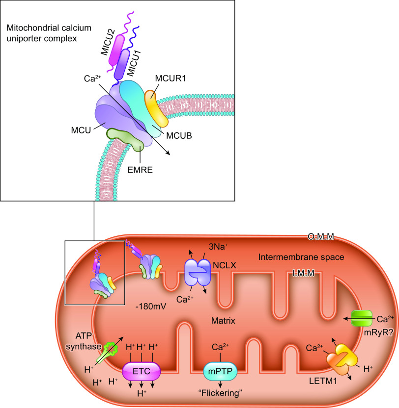
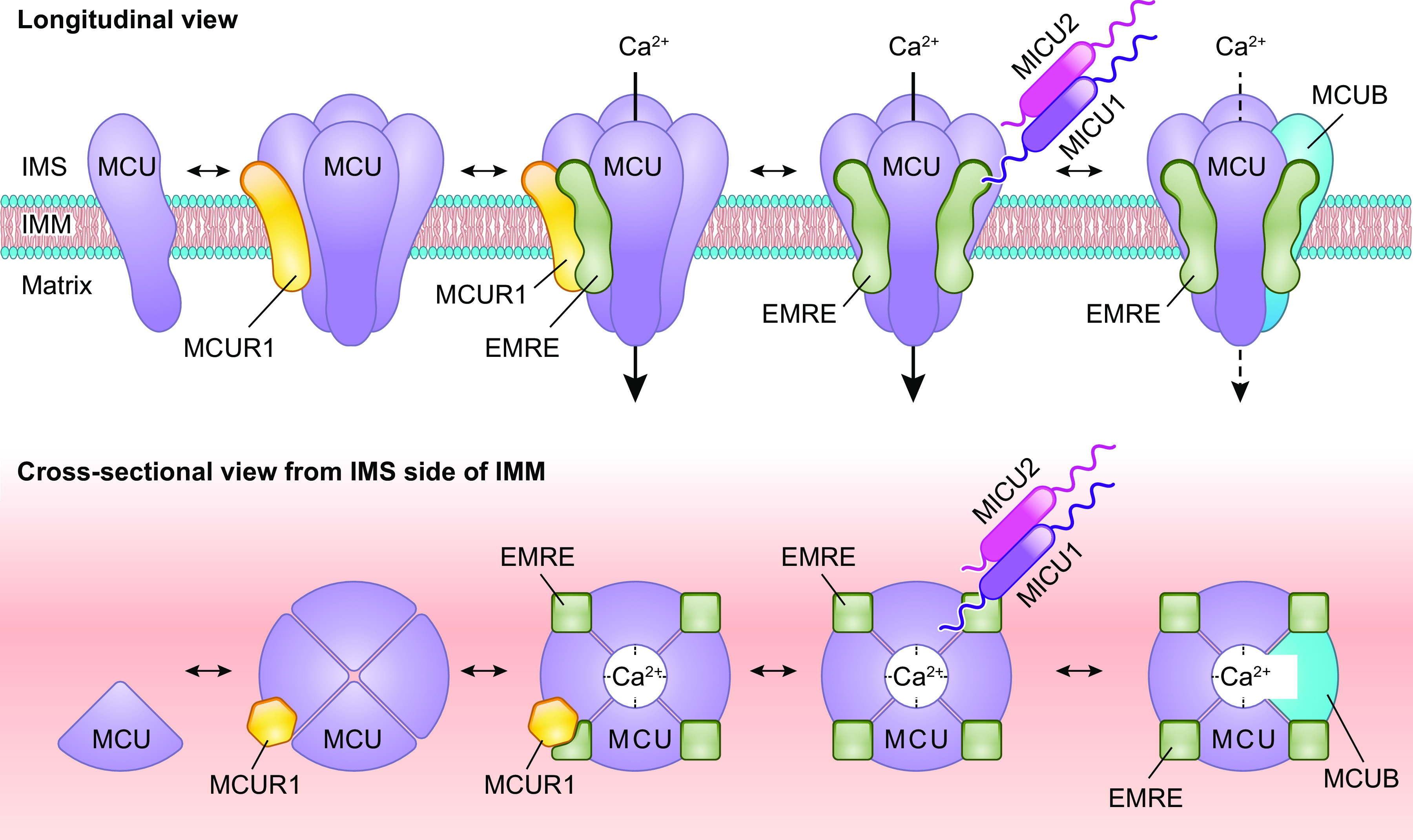
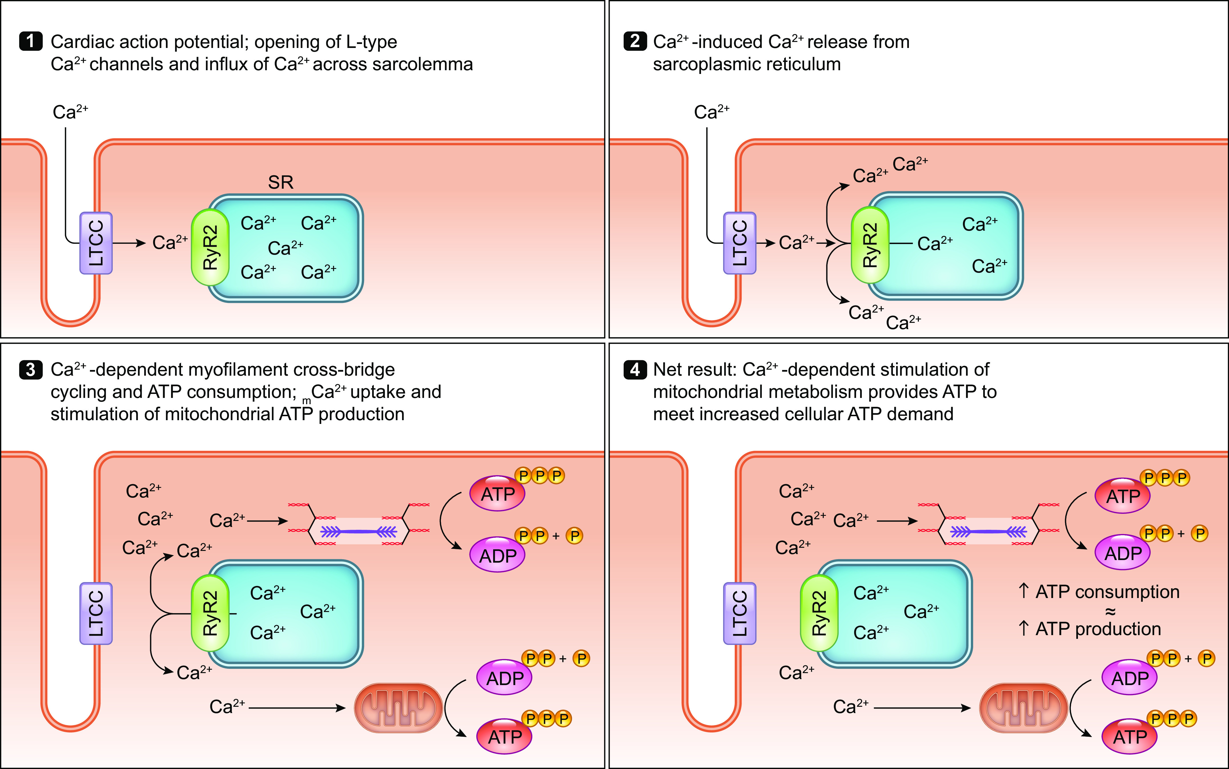
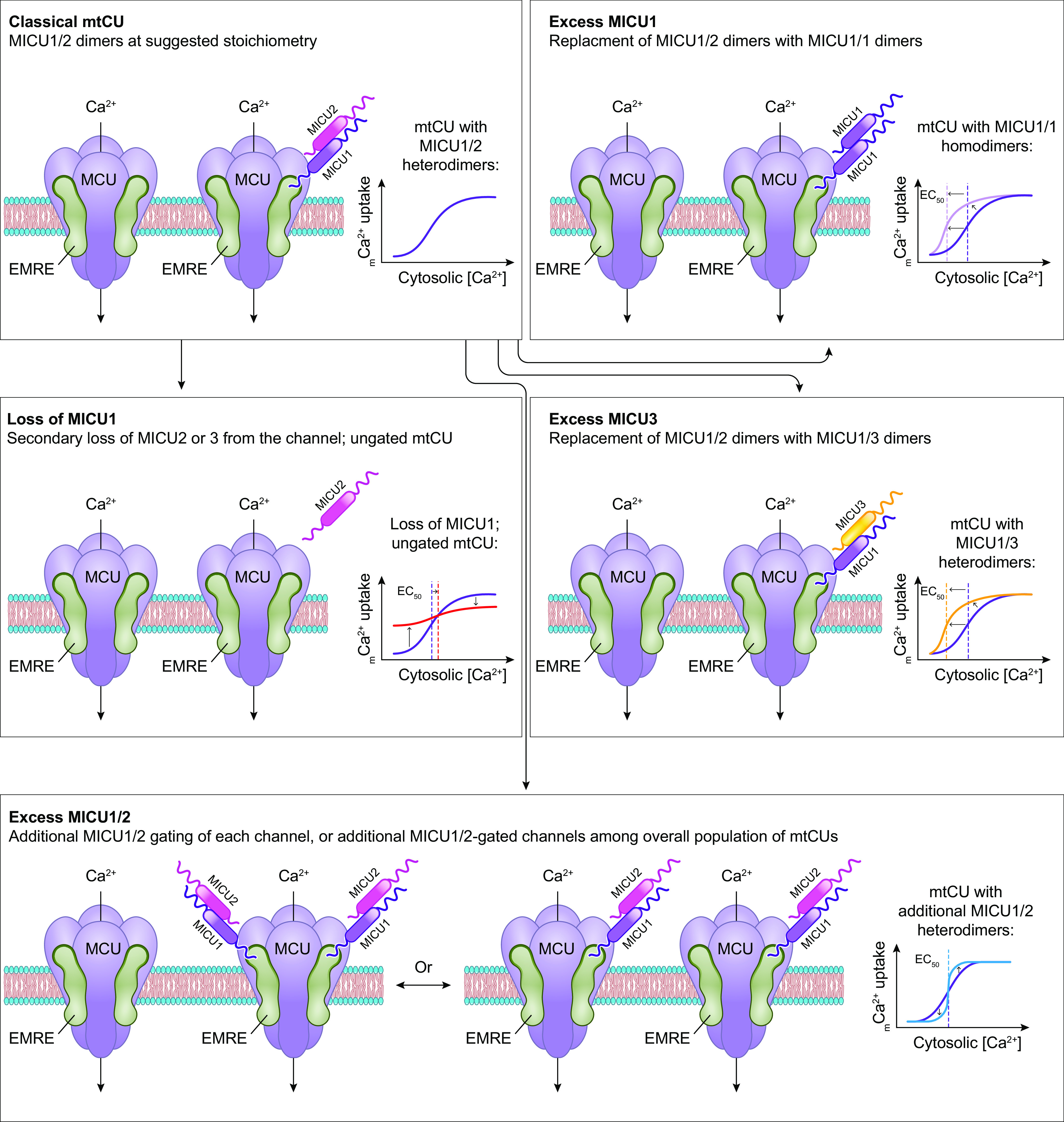
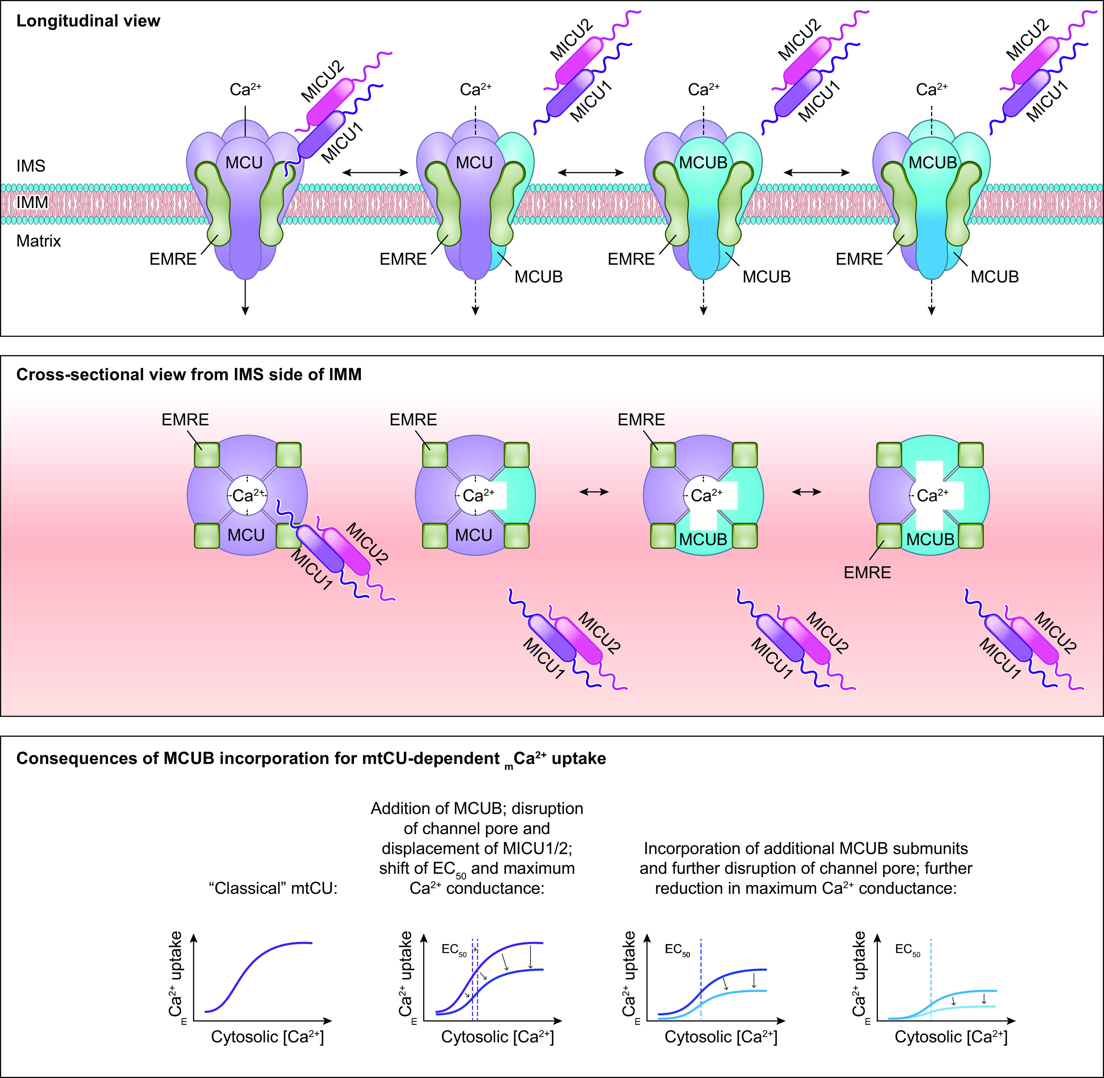
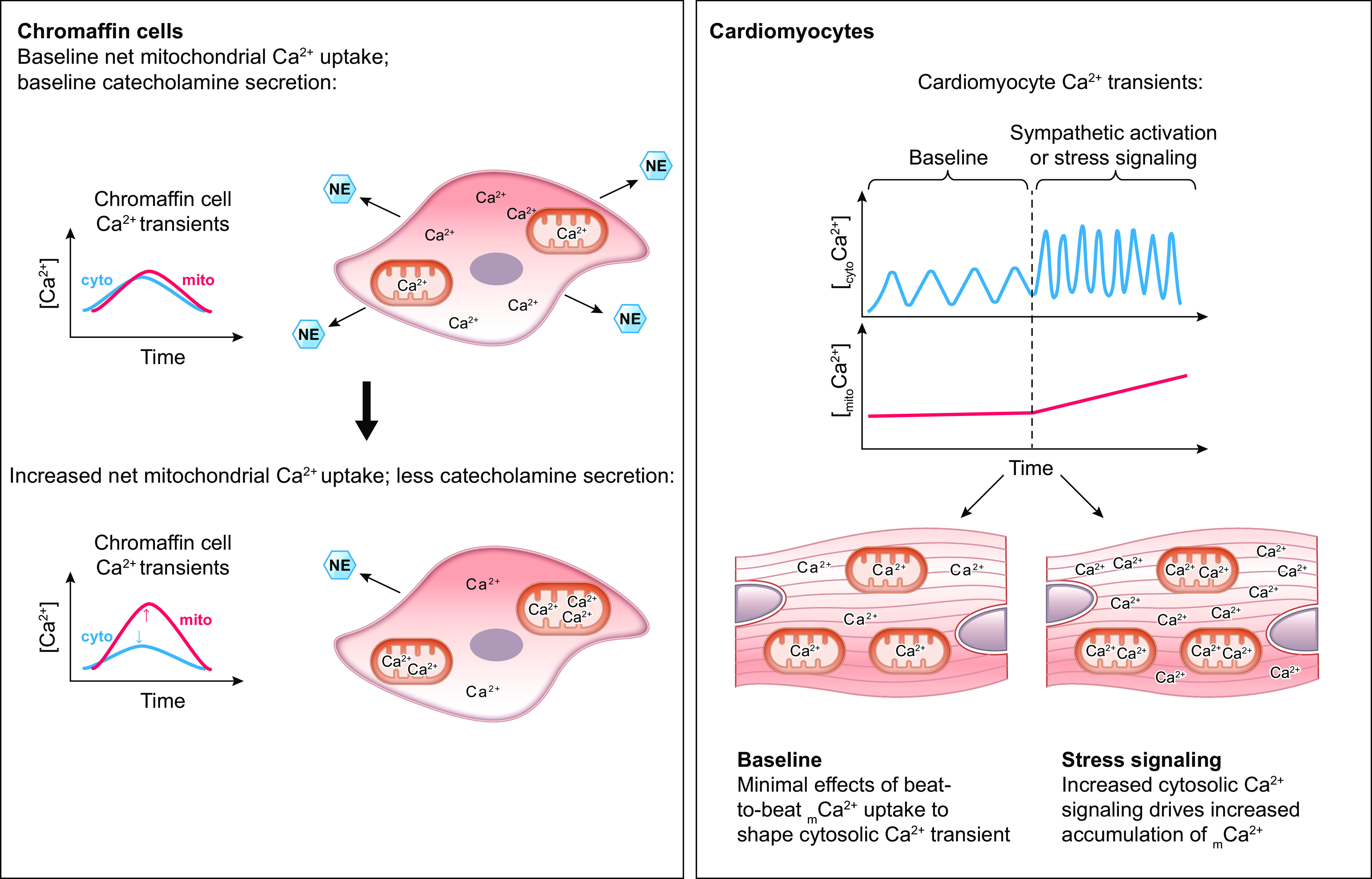
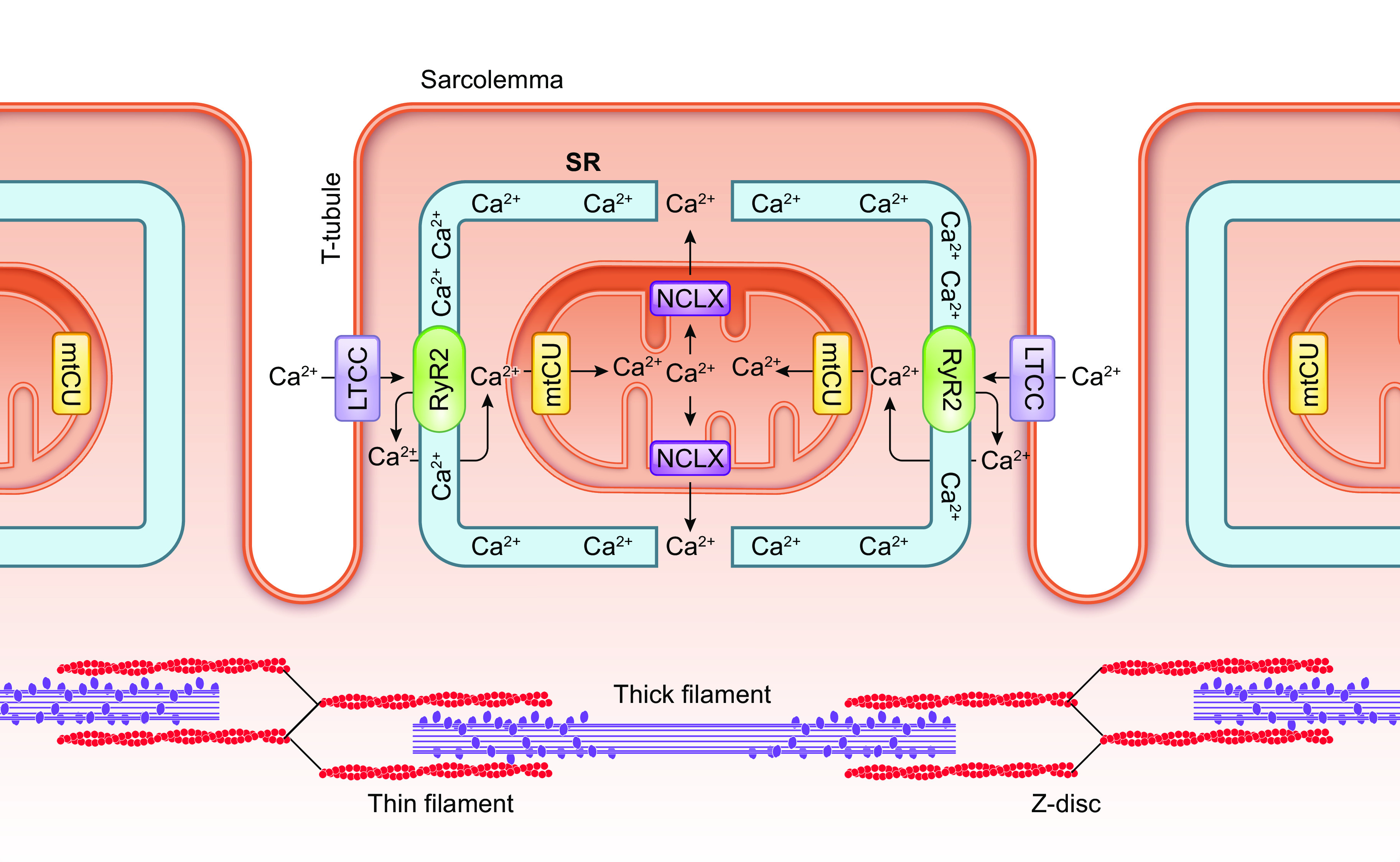
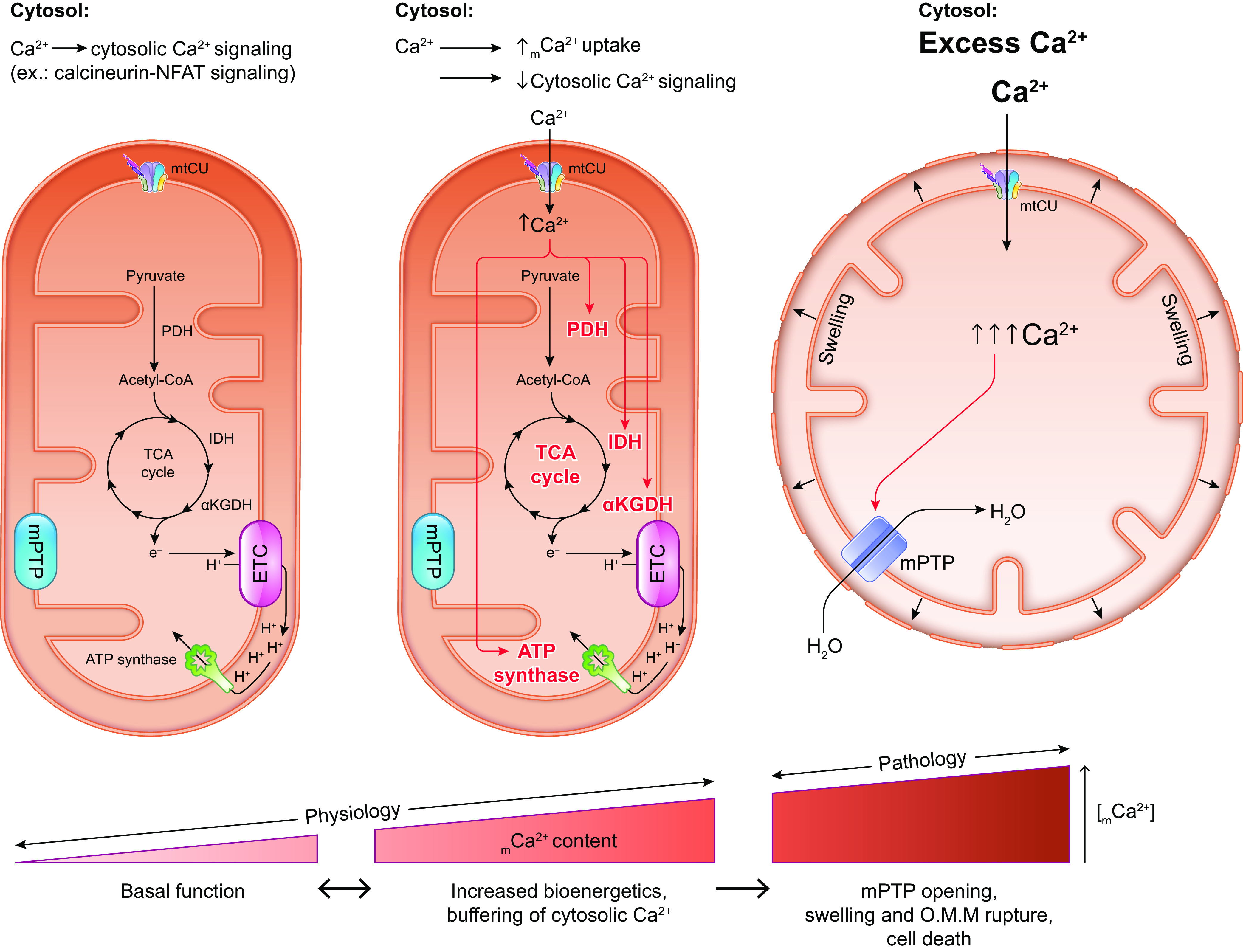
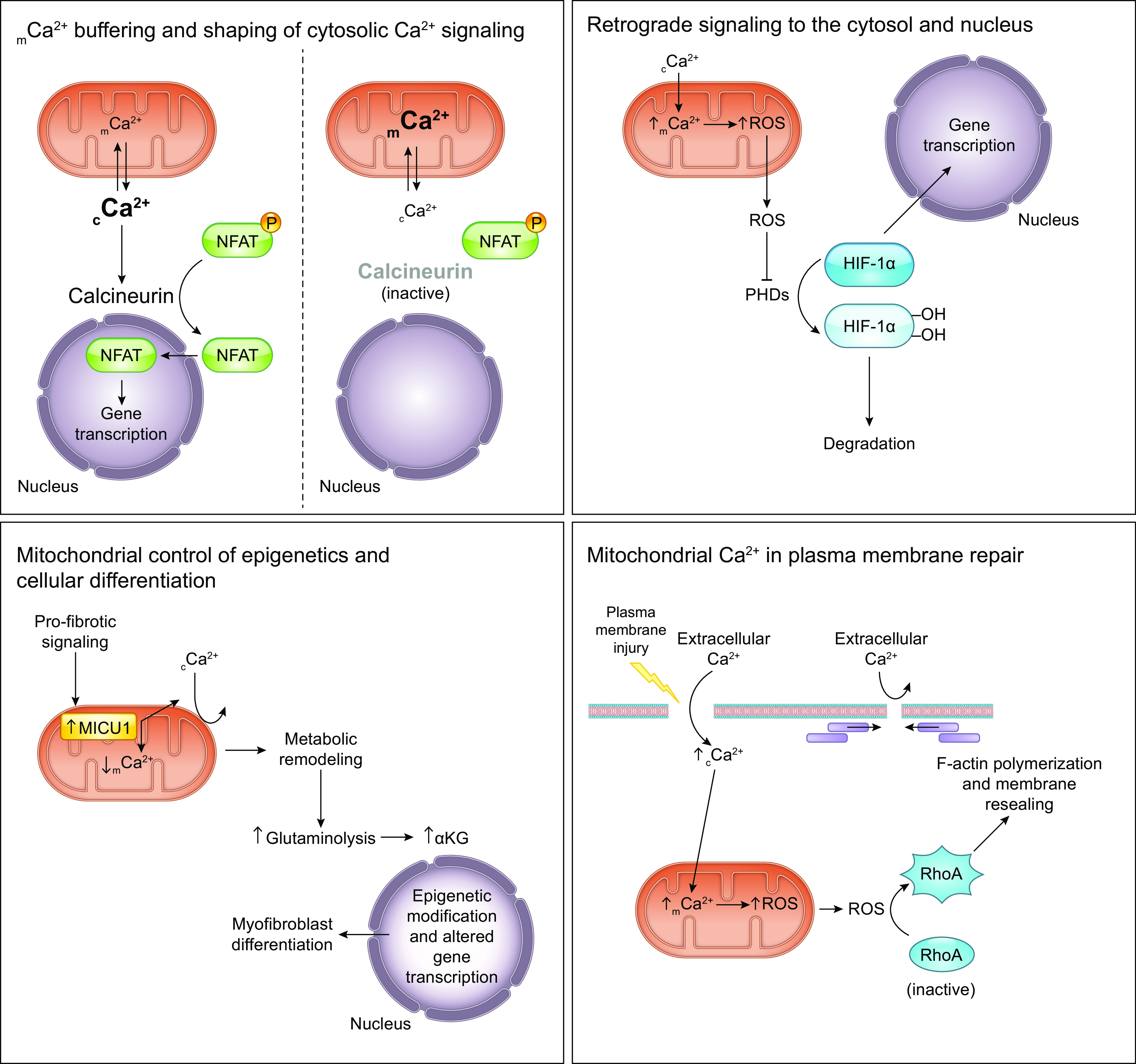
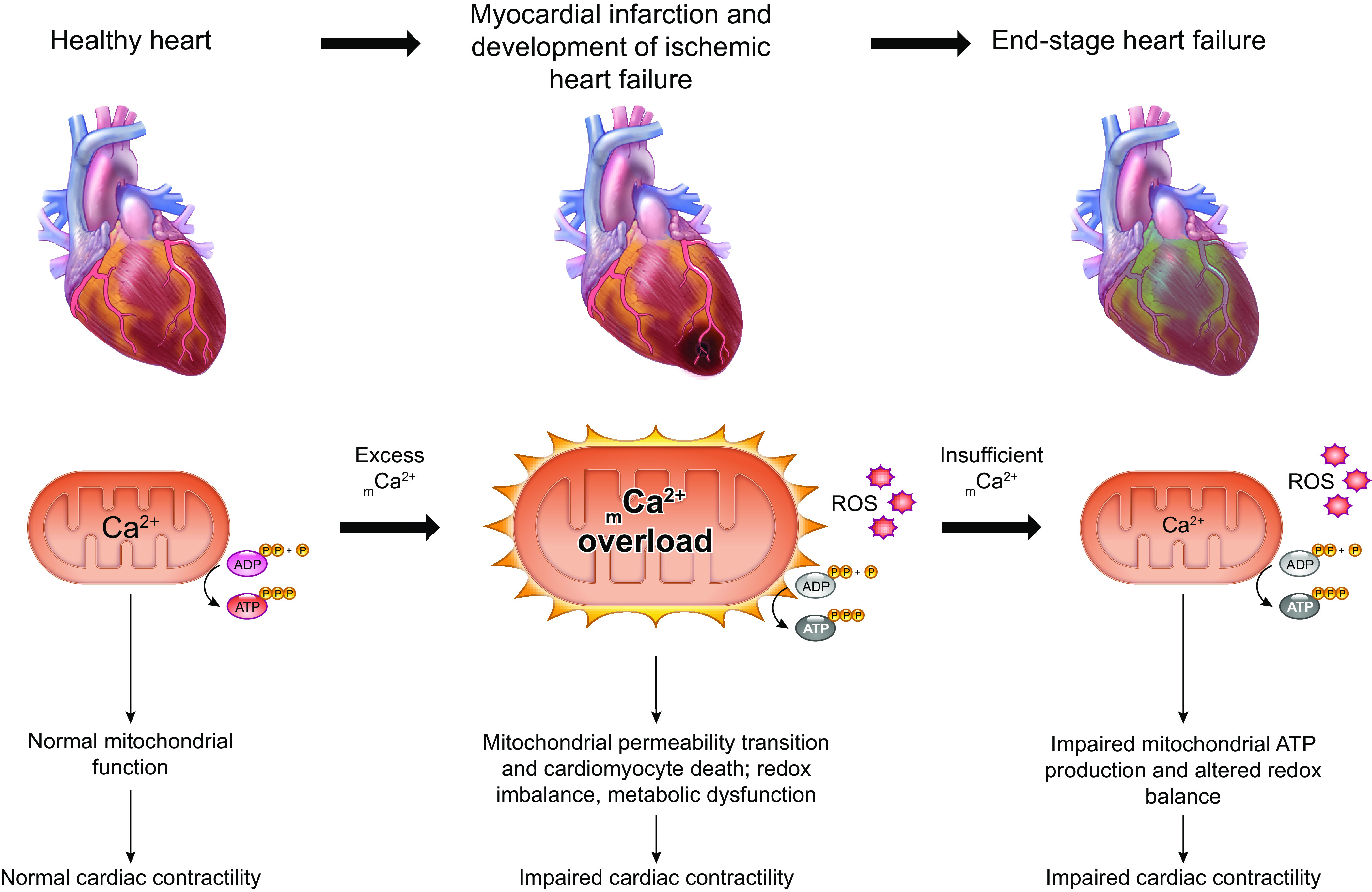
The uptake of calcium into and extrusion of calcium from the mitochondrial matrix is a fundamental biological process that has critical effects on cellular metabolism, signaling, and survival. Disruption of mitochondrial calcium (mCa2+) cycling is implicated in numerous acquired diseases such as heart failure, stroke, neurodegeneration, diabetes, and cancer and is genetically linked to several inherited neuromuscular disorders. Understanding the mechanisms responsible for mCa2+ exchange therefore holds great promise for the treatment of these diseases. The past decade has seen the genetic identification of many of the key proteins that mediate mitochondrial calcium uptake and efflux. Here, we present an overview of the phenomenon of mCa2+ transport and a comprehensive examination of the molecular machinery that mediates calcium flux across the inner mitochondrial membrane: the mitochondrial uniporter complex (consisting of MCU, EMRE, MICU1, MICU2, MICU3, MCUB, and MCUR1), NCLX, LETM1, the mitochondrial ryanodine receptor, and the mitochondrial permeability transition pore. We then consider the physiological implications of mCa2+ flux and evaluate how alterations in mCa2+ homeostasis contribute to human disease. This review concludes by highlighting opportunities and challenges for therapeutic intervention in pathologies characterized by aberrant mCa2+ handling and by summarizing critical unanswered questions regarding the biology of mCa2+ flux.

Keywords: MCU; NCLX; calcium; disease; mitochondria.


Conflict of interest statement

J.W.E. is a paid consultant for Mitobridge and Janssen. J.F.G. has no conflicts of interest, financial or otherwise, to disclose.

Figures

None
Graphical abstract
FIGURE 1.
FIGURE 1.
Ca2+ influx and efflux pathways of the inner mitochondrial membrane. The mitochondrial calcium uniporter channel (mtCU) exists as a channel composed of a total of 4 MCU subunits and up to 4 EMRE subunits. For simplicity, only a single EMRE subunit is shown here. In some cases, the dominant-negative MCU paralog, MCUB, replaces MCU subunits within the complex. The channel is gated by dimers of MICU1/2 or, in some tissues, dimers of MICU1/3. The accessory subunit MCUR1 also binds to and regulates the channel. Two uniporter channels can dimerize, and the function of these dimers is regulated by interactions between the MICU2 subunit associated with each channel. The driving force for Ca2+ entry through the mtCU is the highly negative (approximately −180 mV) potential established across the inner mitochondrial membrane (IMM) by the electron transport chain. Other proposed routes of mitochondrial Ca2+ (mCa2+) uptake may include the mitochondrial ryanodine receptor (mRyR) or reverse-mode operation of NCLX and LETM1. The Na+/Ca2+ exchanger NCLX is a major route for mCa2+ efflux, and the Ca2+/H+ exchanger LETM1 may contribute to mCa2+ efflux in some tissues. Transient opening of the mitochondrial permeability transition pore (mPTP), termed “flickering,” is proposed as another mechanism by which Ca2+ leaves the mitochondrial matrix under physiological conditions. See glossary for abbreviations.
FIGURE 2.
FIGURE 2.
Assembly and stoichiometry of the mitochondrial calcium uniporter channel. Single MCU subunits assemble into tetramers with the assistance of the accessory mtCU subunit, MCUR1. In the absence of EMRE, these MCU tetramers are not capable of conducting Ca2+ across the inner mitochondrial membrane (IMM). EMRE associates with MCU at a likely 1-to-1 ratio in functional Ca2+ channels. The regulatory protein MICU1 binds to MCU and EMRE within the intermembrane space (IMS). In conjunction with its binding partners MICU2 or MICU3, MICU1 acts as a gatekeeper for the mtCU at low cytosolic Ca2+ concentrations and facilitates cooperative activation of the mtCU as cytosolic Ca2+ concentration rises. Current models suggest a stoichiometry of 1 MICU dimer (MICU1/1, MICU1/2, or MICU1/3):4 MCU:up to 4 EMRE. Binding of MICU1 to the channel is proposed to displace the scaffolding factor MCUR1. MCUB, a dominant-negative paralog of MCU, can replace MCU subunits within the channel pore. Incorporation of MCUB into the mtCU impairs Ca2+ conductance through the channel by disrupting the structure of channel pore and by displacing MICU dimers from the channel. See glossary for abbreviations.
FIGURE 3.
FIGURE 3.
Ca2+ signaling in the parallel activation of ATP-consuming processes and ATP production. Ca2+ serves as a second messenger that coordinates the activation of both ATP-consuming and ATP-generating processes within the cell. This process is particularly important for tissues with a high energy demand such as the heart. The parallel activation of ATP consumption and ATP generation within cardiomyocytes is illustrated stepwise as follows: 1) Extracellular Ca2+ enters the cardiomyocyte through the L-type calcium channel (LTCC) that opens with each action potential. 2) Ca2+ that entered the cardiomyocyte through the LTCC activates the ryanodine receptor (RyR2), triggering calcium-induced calcium release from the sarcoplasmic reticulum (SR). 3) Increased cytosolic Ca2+ stimulates the ATP-consuming process of myofilament cross-bridge cycling and cardiac contraction. At the same time, increased cytosolic Ca2+ concentration drives increased mitochondrial Ca2+ uptake that stimulates mitochondrial ATP production. 4) This mechanism of Ca2+-dependent parallel activation allows the heart to respond to an increased workload with a sustained increase in mitochondrial ATP production that matches the increase in ATP consumption by the contractile apparatus. See glossary for abbreviations.
FIGURE 4.
FIGURE 4.
Gating of the mitochondrial calcium uniporter channel and consequences of changes in MICU composition. Clockwise from top left: Classical mtCU: MICU1/2 dimers bind to the mtCU and regulate its activation by Ca2+ within the intermembrane space (IMS). The stoichiometry suggested by structural studies of the mtCU is 4 MCU:3 or 4 EMRE:1 MICU1:1 MICU2, although the proportion of the total mtCUs that are regulated by MICUs is determined by the relative MICU expression within each tissue. At low cytosolic/IMS Ca2+ concentration ([Ca2+]), the MICU1/2 dimer acts as a gatekeeper to keep the channel impermeable to Ca2+. As cytosolic/IMS [Ca2+] rises, the MICU1/2 dimer facilitates the cooperative activation of the channel, resulting in a sigmoidal relationship between mtCU-dependent mCa2+ uptake and cytosolic/IMS [Ca2+]. Excess MICU1: Increased expression of MICU1 may displace MICU2 from existing MICU1/2 dimers, resulting in MICU1 homodimers. Displacement of MICU2 results in loss of its ability to tune MICU1-dependent mtCU gating and cooperative activation, shifting the relationship between mtCU-dependent mCa2+ uptake and cytosolic/IMS Ca2+ concentration to the left. Excess MICU3: Increased expression of MICU3 may displace MICU2 from existing MICU1/2 dimers, resulting in MICU1/3 dimers. The properties of MICU3-dependent regulation of MICU1 differ somewhat from those of MICU2 (see text for details); thus, replacement of MICU2 with MICU3 also shifts the relationship between mtCU-dependent mCa2+ uptake and cytosolic/IMS Ca2+ concentration to the left. Excess MICU1/2: Increased expression of MICU1/2 results in more stringent regulation of Ca2+ flux through the mtCU, with enhanced channel gatekeeping at low cytosolic/IMS [Ca2+], but greater activation of the channel at higher [Ca2+]. This behavior may result from a greater number of MICU1/2 dimers binding to each mtCU, or from an increase in the number of mtCUs within the tissue being gated by MICU1/2. Loss of MICU1: MICU2 and MICU3 are incapable of binding to and regulating the mtCU in the absence of MICU1. Loss of MICU1 therefore results in loss of all MICU-dependent mtCU regulation. The absence of MICU-dependent channel gatekeeping results in increased mCa2+ uptake at low cytosolic/IMS [Ca2+], while the absence of MICU-dependent cooperative activation makes further mtCU activation less responsive to increases in cytosolic/IMS [Ca2+]. See glossary for abbreviations.
FIGURE 5.
FIGURE 5.
Consequences of MCUB incorporation on mitochondrial calcium uniporter channel function. Replacement of MCU with its dominant-negative paralog, MCUB, impairs Ca2+ uptake through the mtCU. At least 2 mechanisms are responsible for this phenomenon. Replacement of MCU with MCUB can disrupt binding between MICU1 and the mtCU, resulting in loss of MICU-dependent regulation of the channel. Here, cooperative activation of the mtCU is impaired, shifting the relationship between mtCU-dependent mCa2+ uptake and cytosolic/IMS Ca2+ concentration to the right. However, the consequences of the loss of MICU-dependent gatekeeping function at low cytosolic/IMS Ca2+ concentration are mitigated by MCUB’s additional effect to disrupt Ca2+ flux through the channel. As a result, mCa2+ uptake through the channel is reduced compared with the classical mtCU. Replacement of additional MCU subunits with MCUB further attenuates Ca2+ flux through the channel. Structural modeling suggests that this effect results from a widening of the channel pore in the presence of MCUB, which may disrupt Ca2+ coordination within the channel. See glossary for abbreviations.
FIGURE 6.
FIGURE 6.
Cell-type dependent effects of mCa2+ uptake on cytosolic Ca2+ signals. Stimulation of secretory cells elicits cytosolic Ca2+ transients that are rapidly transmitted to the mitochondria, resulting in large mCa2+ transients. Modulation of net mCa2+ uptake affects the magnitude of both the cytosolic Ca2+ transient and the mitochondrial Ca2+ transient, and impacts physiologically relevant cellular functions. For example, increased net mCa2+ uptake in adrenal chromaffin cells can limit the magnitude of the cytosolic Ca2+ transient and thereby limit the amount of cytosolic Ca2+ available to stimulate secretion of catecholamines such as norepinephrine (NE). In other cell types, however, mitochondrial Ca2+ signals are not coupled as directly to each cytosolic Ca2+ transient. For example, cardiomyocytes do not generally appear to have beat-to-beat oscillations in mCa2+ content that mirror cytosolic Ca2+ transients. Rather, alterations in the amplitude and/or frequency of cytosolic Ca2+ transients drive integrated changes in mCa2+ concentration. For instance, sympathetic stimulation of cardiomyocytes increases the amplitude and frequency of cytosolic Ca2+ transients, causing a progressive increase in mCa2+ concentration over time. [Ca2+], Ca2+ concentration; [cytoCa2+], cytosolic [Ca2+]; [mitoCa2+], mitochondrial [Ca2+]; see glossary for other abbreviations.
FIGURE 7.
FIGURE 7.
Spatial segregation of mitochondrial Ca2+ influx and efflux pathways in cardiomyocytes. In cardiac muscle, the mitochondrial calcium uniporter complex (mtCU) is found at regions of the inner mitochondrial membrane (IMM) near contacts between the outer mitochondrial membrane and Ca2+ release sites on the sarcoplasmic reticulum (SR). This arrangement enables Ca2+ released from the SR via the ryanodine receptor (RyR2) to be taken up efficiently into the mitochondrial matrix. NCLX, the principal route for mitochondrial Ca2+ efflux in cardiomyocytes, is instead distributed at regions of the IMM that are more distant from the sites of SR-mitochondria contact. This spatial separation of mitochondrial Ca2+ uptake and efflux is thought to minimize futile cycling of Ca2+ flux through the mtCU and NCLX that would otherwise depolarize mitochondrial membrane potential. See glossary for abbreviations.
FIGURE 8.
FIGURE 8.
Physiological and pathological functions of acute mCa2+ uptake. Physiological cellular Ca2+ signaling simulates signal transduction pathways in the cytosol and, if cytosolic Ca2+ concentration reaches a high enough threshold, can trigger mCa2+ uptake through the mitochondrial calcium uniporter complex (mtCU). In some cell types, mitochondrial Ca2+ uptake can buffer further changes in cytosolic Ca2+ concentration and so affect Ca2+-dependent signaling throughout the cell. Increasing matrix Ca2+ concentration stimulates the activity of pyruvate dehydrogenase (PDH), isocitrate dehydrogenase (IDH), and α-ketoglutarate dehydrogenase (αKGDH) to increase TCA cycle flux and delivery of electrons to the electron transport chain to stimulate ATP production. Matrix Ca2+ may also stimulate ATP synthase directly, although this concept is controversial. Rapid mCa2+ uptake through the mtCU allows cells to couple Ca2+-dependent, energy-consuming processes in the cytosol, such as muscle contraction, with a coordinated increase in mitochondrial ATP production in order to supply the cell’s increased energetic demand. A pathological increase in cytosolic Ca2+ concentration, as occurs during ischemia-reperfusion, can drive excessive Ca2+ uptake through the mtCU, leading to activation of the mitochondrial permeability transition pore, collapse of mitochondrial membrane potential, influx of water and swelling of the matrix, and rupture of the outer mitochondrial membrane (OMM), culminating in cell death. See glossary for abbreviations.
FIGURE 9.
FIGURE 9.
Mitochondrial Ca2+ handling as a master regulator of cellular signaling pathways. Clockwise from top left: mCa2+ buffering and shaping of cytosolic Ca2+ signaling: Net mCa2+ flux influences the amount of cytosolic Ca2+ available to participate in cytosolic signaling pathways. For instance, changes in net mCa2+ uptake can influence NFAT signaling and affect gene expression by influencing amount of cytosolic Ca2+ available to activate the Ca2+-sensitive phosphatase calcineurin. Retrograde signaling to the cytosol and nucleus: Prolyl hydroxylases (PHDs) hydroxylate the transcription factor hypoxia-inducible factor (HIF-1α) and target it for degradation. Increased net mCa2+ accumulation triggers the mitochondrial production of reactive oxygen species (ROS), which inhibit PHDs, thereby favoring HIF-1α accumulation and the transcription of its target genes. Mitochondrial Ca2+ in plasma membrane repair: Lesion of the plasma membrane allows extracellular Ca2+ to flow into the cell, elevating cytosolic Ca2+ concentration. Subsequent mCa2+ uptake drives increased mitochondrial production of ROS. ROS activates the GTPase RhoA, which then promotes F-actin polymerization to facilitate plasma membrane repair. Effective resealing of the plasma membrane completes this feedback loop by preventing further influx of extracellular Ca2+. Mitochondrial control of epigenetics and cellular differentiation: Profibrotic stimulation of fibroblasts upregulates the mtCU gatekeeper MICU1, thereby attenuating mCa2+ uptake. Altered mCa2+ handling remodels fibroblast metabolism and increases glutaminolysis to increase the cellular supply of α-ketoglutarate (αKG). αKG serves as a cofactor for histone demethylases and so modulates gene expression that ultimately causes differentiation of the fibroblasts into myofibroblasts. See glossary for abbreviations.
FIGURE 10.
FIGURE 10.
Either too much or too little mCa2+ can contribute to disease. mCa2+ has pleiotropic roles in diseases such as cancer, neurodegeneration, and heart failure that relate to its fundamental functions in supporting mitochondrial bioenergetics, influencing mitochondrial signaling, and cell death. For example, acute mCa2+ overload during cardiac ischemia-reperfusion injury causes excess reactive oxygen species (ROS) production and elicits mitochondrial permeability transition, which compromises mitochondrial ATP production and triggers cardiomyocyte death. These immediate effects of mCa2+ overload impair the heart’s mechanical pump function and over time cause the heart to fail. In end-stage heart failure, however, mCa2+ content may be reduced below normal levels because of excessive mitochondrial Na+/Ca2+ exchange. Here, diminished mCa2+-dependent stimulation of TCA cycle flux can disrupt mitochondrial redox balance, favor ROS production, and impair mitochondrial ATP production. The diminished ATP supply and excessive oxidative stress drive further impairment of the heart’s contractile function. See glossary for abbreviations.

References

    1. Slater EC, Cleland KW. The effect of calcium on the respiratory and phosphorylative activities of heart-muscle sarcosomes. Biochem J 55: 566–590, 1953. doi: 10.1042/bj0550566. - DOI - PMC - PubMed
    1. Lehninger AL. Enzymes and Enzyme Systems: Their State in Nature. Cambridge, MA: Harvard University Press, 1951.
    1. DeLuca HF, Engstrom GW. Calcium uptake by rat kidney mitochondria. Proc Natl Acad Sci USA 47: 1744–1750, 1961. doi: 10.1073/pnas.47.11.1744. - DOI - PMC - PubMed
    1. Vasington FD, Murphy JV. Ca ion uptake by rat kidney mitochondria and its dependence on respiration and phosphorylation. J Biol Chem 237: 2670–2677, 1962. doi: 10.1016/S0021-9258(19)73805-8. - DOI - PubMed
    1. Mitchell P. Coupling of phosphorylation to electron and hydrogen transfer by a chemi-osmotic type of mechanism. Nature 191: 144–148, 1961. doi: 10.1038/191144a0. - DOI - PubMed

Publication types

LinkOut - more resources