Review
. 2022 Apr 1;102(2):815-857.
doi: 10.1152/physrev.00013.2021. Epub 2021 Oct 26.

Physiological roles of mammalian transmembrane adenylyl cyclase isoforms

Affiliations
Review

Physiological roles of mammalian transmembrane adenylyl cyclase isoforms

Katrina F Ostrom et al. Physiol Rev. .

Abstract

Adenylyl cyclases (ACs) catalyze the conversion of ATP to the ubiquitous second messenger cAMP. Mammals possess nine isoforms of transmembrane ACs, dubbed AC1-9, that serve as major effector enzymes of G protein-coupled receptors (GPCRs). The transmembrane ACs display varying expression patterns across tissues, giving the potential for them to have a wide array of physiological roles. Cells express multiple AC isoforms, implying that ACs have redundant functions. Furthermore, all transmembrane ACs are activated by Gαs, so it was long assumed that all ACs are activated by Gαs-coupled GPCRs. AC isoforms partition to different microdomains of the plasma membrane and form prearranged signaling complexes with specific GPCRs that contribute to cAMP signaling compartments. This compartmentation allows for a diversity of cellular and physiological responses by enabling unique signaling events to be triggered by different pools of cAMP. Isoform-specific pharmacological activators or inhibitors are lacking for most ACs, making knockdown and overexpression the primary tools for examining the physiological roles of a given isoform. Much progress has been made in understanding the physiological effects mediated through individual transmembrane ACs. GPCR-AC-cAMP signaling pathways play significant roles in regulating functions of every cell and tissue, so understanding each AC isoform's role holds potential for uncovering new approaches for treating a vast array of pathophysiological conditions.

Keywords: G protein-coupled receptors; adenylyl cyclase; cAMP.


Conflict of interest statement

No conflicts of interest, financial or otherwise, are declared by the authors.

Figures

None
Graphical abstract
FIGURE 1.
FIGURE 1.
General structure of mammalian transmembrane adenylyl cyclases (ACs). TM1 and TM2 are transmembrane domains each consisting of 6 transmembrane α-helices. The second extracellular loop of TM2 contains N-linked glycosylation sites that target some isoforms to lipid rafts (12). C1 and C2 are intracellular cytoplasmic loops, with C1a and C2a forming the catalytic domain and forskolin binding site (in the case of AC1–8). The C1a domain is the site of Gαi binding, whereas the C2a domain interacts with Gαs. C1b and C2b are regulatory subdomains, and the NH2-terminal domain is involved in many protein-protein interactions. The C1 and C2 domains are separated from each transmembrane domain by 2 coiled-coil helices (shown in yellow). These 2 helices and the C1b domain of AC5 contain the indicated mutations associated with dyskinesias (–16). This structural model is based largely on the recent cryo-EM structure of AC9 (10).
FIGURE 2.
FIGURE 2.
Schematic diagram of signaling via transmembrane adenylyl cyclases (ACs). Once activated by an agonist, a G protein-coupled receptor (GPCR) will activate coupled G proteins. Gαs activates AC activity, which catalyzes the conversion of ATP to cAMP, whereas Gαi inhibits AC activity. AC isoforms are also regulated by various other signals, as described in TABLE 1. Phosphodiesterases (PDEs) terminate the signal by decyclizing cAMP to 5′-AMP. cAMP activates several effector proteins, including PKA, Epac, Popeye Domain Containing (POPDC), and cyclic nucleotide-gated channels. PKA is anchored by A kinase anchoring proteins (AKAPs) to specific phosphorylation targets. PKA, Epac, cyclic nucleotide-gated channels, and POPDCs elicit various downstream physiological responses depending on the cell type.
FIGURE 3.
FIGURE 3.
Tissue distribution of transmembrane adenylyl cyclases (ACs): the predominant AC isoforms expressed by major organs and systems in the human body. AC isoforms are color coded by group (group I in blue, group II in green, group III in red, group IV in brown). The digestive tract expresses all AC isoforms except AC2 (52). The vasculature expresses primarily AC3, AC5, and AC6 (–55).
FIGURE 4.
FIGURE 4.
Regulation of mammalian transmembrane adenylyl cyclases (ACs). Numerous signals directly regulate enzymatic activity of specific AC isoforms. AC isoforms are classified into 4 groups (and color coded) based on these regulatory properties. Green arrows denote stimulatory effects, red arrows denote inhibitory effects, and dashed arrows show conditional effects that are dependent upon other, coincident stimuli. NO, nitric oxide.
FIGURE 5.
FIGURE 5.
Representation of cAMP signaling compartmentation. Membrane microdomains and prearranged complexes form in most cells that couple specific G protein-coupled receptors (GPCRs) to unique subsets of physiological responses. Transmembrane adenylyl cyclase (AC) isoforms characteristically localize in either lipid raft (AC1, 3, 5, 6, and 8) or nonraft (AC2, 4, 7, and 9) domains, where they form complexes with unique signaling partners [GPCRs, A kinase anchoring proteins (AKAPs), phosphodiesterases (PDEs), and others] to create distinct cAMP signaling compartments. There are 43 AKAP isoforms and 24 PDE genes (plus many splice variants), making the combinatorial possibilities for cAMP signaling complex formation enormous. Diffusion of cAMP is limited by PKA buffering, colocalized PDE isoforms, and cellular barriers to diffusion (represented as a brown matrix). Effector proteins downstream of PKA can also be localized in specific domains such that each compartment can give rise to a unique pattern of cellular and physiological responses. AC isoforms are color coded by group (group I in blue, group II in green, group II in red, group IV in brown).
FIGURE 6.
FIGURE 6.
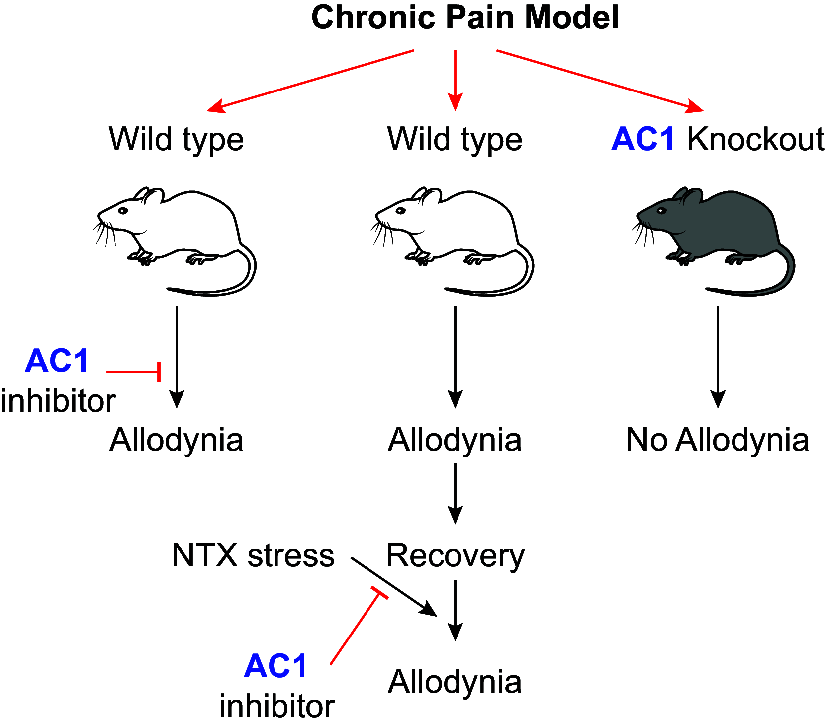
The role of adenylyl cyclase (AC)1 in chronic pain-induced allodynia in vivo. In wild-type mice subjected to a chronic pain model, AC1 inhibitors can block allodynia. In wild-type mice that have recovered from a chronic pain model, AC1 inhibitors can block naltrexone (NTX)-induced allodynia (latent sensitization model). It is likely that stress-induced allodynia in chronic pain models will also be blocked by AC1 inhibitors. In AC1 knockout mice, chronic pain models do not produce allodynia.
FIGURE 7.
FIGURE 7.
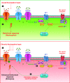
Cellular adaptations to chronic nociceptive input promote allodynia through adenylyl cyclase (AC)1. Top: sufficient acute nociceptive input results in activation of voltage-gated calcium (CaV) channels, α-amino-3-hydroxy-5-methyl-4-isoxazolepropionic acid (AMPA) receptors (AMPARs), and N-methyl-d-aspartate (NMDA) receptors (NMDARs). This activation at varying levels of the pain pathways (i.e., spinal cord, anterior cingulate cortex, insular cortex) can produce an acute behavioral response in rodents, typically a response to a noxious mechanical/thermal stimulus. Although calcium influx through these channels will activate AC1, AC1 does not modulate acute nociceptive responses. Bottom: chronic nociceptive input produces sustained calcium influx and AC1 activation. This promotes a positive feedback loop whereby AC1 activation results in phosphorylation of AMPARs and increased expression of GluN2B-containing NMDARs, both of which further promote increased AC1 signaling. This ultimately results in a sensitized system where nonnoxious stimuli produce a behavioral response (allodynia). Both panels are generalized schemes and likely occur at multiple levels of the pain pathways. Mu opioid receptor (MOR) agonists can inhibit AC1, among other functions. AC1 regulation by MORs in the acute nociceptive setting (top) is likely different from that in the chronic setting (bottom). In the chronic setting there is an interplay between NMDARs, MORs, and AC1 that are involved in latent sensitization (see text).
FIGURE 8.
FIGURE 8.
Adenylyl cyclase (AC)-A kinase anchoring protein (AKAP) signaling complexes regulate specific cardiac functions. AC isoforms associate with specific AKAPs to form signaling complexes. These complexes are localized in specific structures and spaces within the cardiac myocyte, likely based on localization of AC. AKAP interactions with downstream effector and regulator proteins allow a complex to regulate unique physiological functions. Cardiac repolarization is facilitated by an AKAP9 (Yotiao) complex that includes KCNQ1, PKA, protein phosphatase 1 (PP1), and phosphodiesterase 4DE3 (PDE4D3). Disruption of the complex prolongs the action potential. Calcium-stimulated calcium release is regulated by a macromolecular complex consisting of AC5/6 and AKAP5 (AKAP79/150). AKAP5 anchors a cAMP regulatory unit in the transverse tubule, where it is in register with phospholamban (PLN), ryanodine receptors (RyRs), and the sarco(endo)plasmic reticulum calcium ATPase (SERCA). Molecules involved in cardiac hypertrophy, including protein phosphatase 2B (PP2B), PDE4D3, and phospholipase Cε (PLCε), are anchored by AKAP6 (mAKAP) to both AC5 in the transverse tubule and the nuclear membrane via nesprin. The close proximity of the transverse tubules and the nucleus allows functional coupling of the AC5 complex to nuclear calcium signaling. The cardiac stress response is further regulated by binding of AC9 to heat shock protein (HSP)20, facilitating PKA regulation of HSP20 phosphorylation and promoting cardioprotection. LTCC, L-type Ca2+ channel. Figure adapted from Ref. , with permission from Journal of Cardiovascular Development and Disease.
FIGURE 9.
FIGURE 9.
Internalization and endosomal signaling by adenylyl cyclase (AC)9 but not AC1. β2 adrenergic receptors (β2ARs) activated by agonist stimulate cAMP at the plasma membrane but then internalize via arrestin-mediated endocytosis into endosomes. When complexed with AC1, the receptor ceases cAMP signaling once internalized, creating a short-duration cAMP signal delimited to the plasma membrane. When complexed with AC9, the receptor internalizes with the AC and continues to generate cAMP in the endosomal compartment (131, 460). AC9 internalization occurs independently of the receptor and does not require arrestins or AC activity (460).

References

    1. Cooper DM. Regulation and organization of adenylyl cyclases and cAMP. Biochem J 375: 517–529, 2003. doi:10.1042/bj20031061. - DOI - PMC - PubMed
    1. Tang WJ, Yan S, Drum CL. Class III adenylyl cyclases: regulation and underlying mechanisms. Adv Second Messenger Phosphoprotein Res 32: 137–151, 1998. doi:10.1016/s1040-7952(98)80009-8. - DOI - PubMed
    1. Sunahara RK, Dessauer CW, Gilman AG. Complexity and diversity of mammalian adenylyl cyclases. Annu Rev Pharmacol Toxicol 36: 461–480, 1996. doi:10.1146/annurev.pa.36.040196.002333. - DOI - PubMed
    1. Khannpnavar B, Mehta V, Qi C, Korkhov V. Structure and function of adenylyl cyclases, key enzymes in cellular signaling. Curr Opin Struct Biol 63: 34–41, 2020. doi:10.1016/j.sbi.2020.03.003. - DOI - PubMed
    1. Linder JU, Schultz JE. The class III adenylyl cyclases: multi-purpose signalling modules. Cell Signal 15: 1081–1089, 2003. doi:10.1016/S0898-6568(03)00130-X. - DOI - PubMed

Publication types