Review
. 2022 May;32(3):e2305.
doi: 10.1002/rmv.2305. Epub 2021 Oct 26.

Safety and potency of BIV1-CovIran inactivated vaccine candidate for SARS-CoV-2: A preclinical study

Affiliations
Review

Safety and potency of BIV1-CovIran inactivated vaccine candidate for SARS-CoV-2: A preclinical study

Asghar Abdoli et al. Rev Med Virol. 2022 May.

Abstract

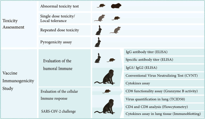
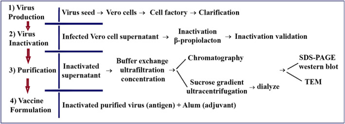
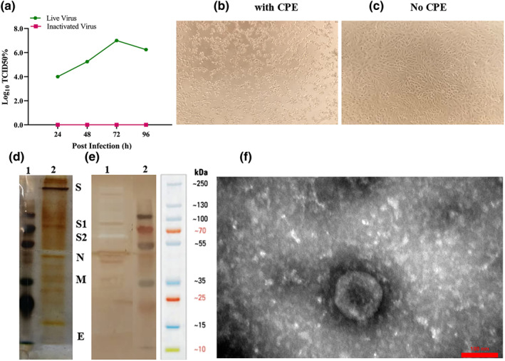
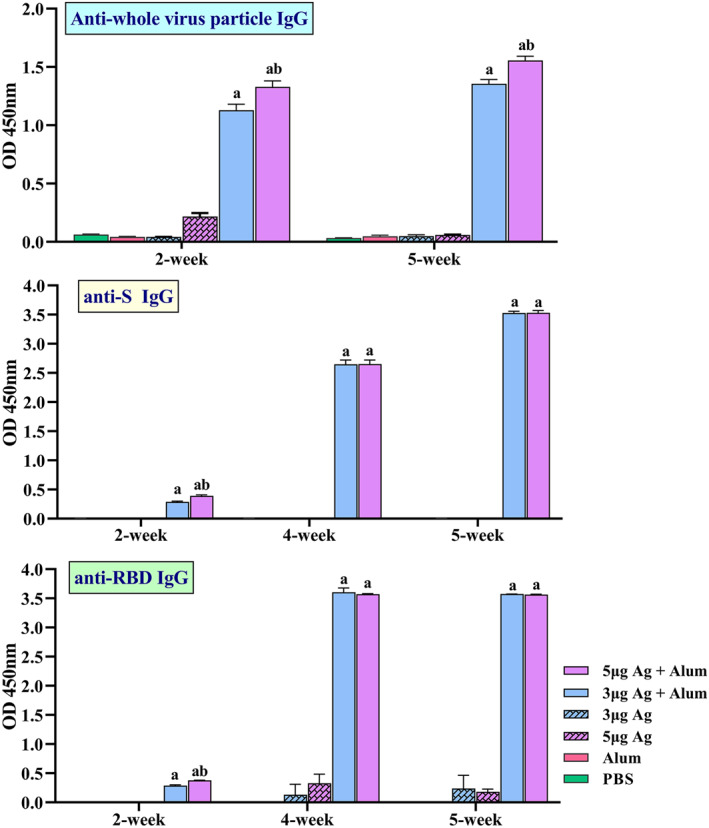
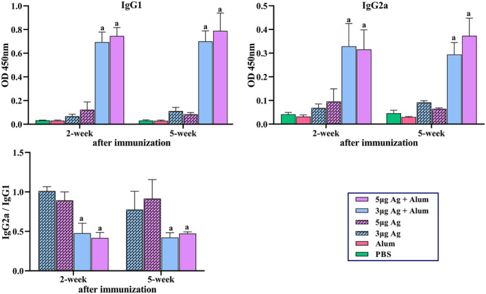
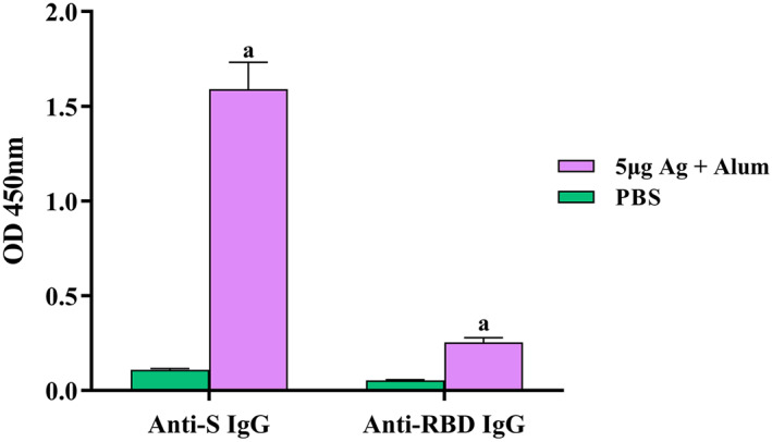
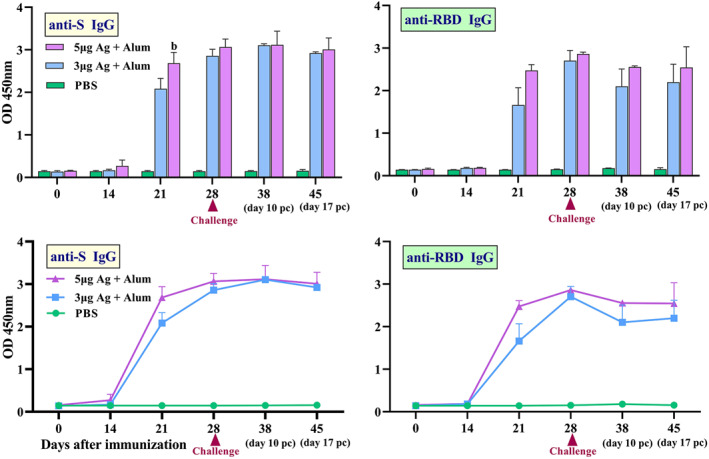
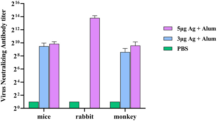
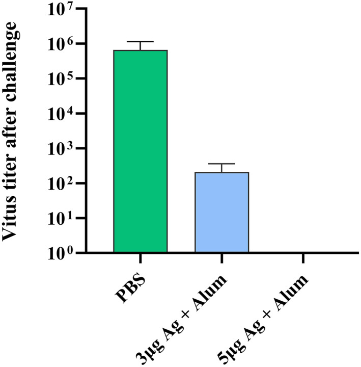
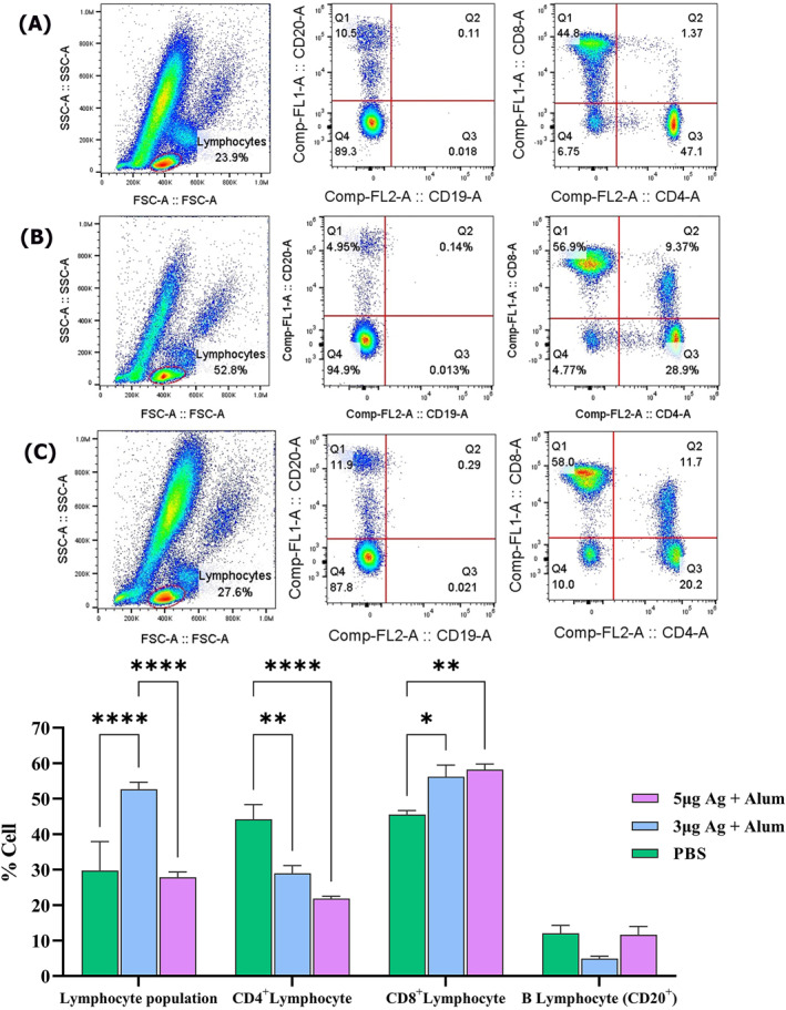
The development of effective and safe COVID-19 vaccines is a major move forward in our global effort to control the SARS-CoV-2 pandemic. The aims of this study were (1) to develop an inactivated whole-virus SARS-CoV-2 candidate vaccine named BIV1-CovIran and (2) to determine the safety and potency of BIV1-CovIran inactivated vaccine candidate against SARS-CoV-2. Infectious virus was isolated from nasopharyngeal swab specimen and propagated in Vero cells with clear cytopathic effects in a biosafety level-3 facility using the World Health Organization's laboratory biosafety guidance related to COVID-19. After characterisation of viral seed stocks, the virus working seed was scaled-up in Vero cells. After chemical inactivation and purification, it was formulated with alum adjuvant. Finally, different animal species were used to determine the toxicity and immunogenicity of the vaccine candidate. The study showed the safety profile in studied animals including guinea pig, rabbit, mice and monkeys. Immunisation at two different doses (3 or 5 μg per dose) elicited a high level of SARS-CoV-2 specific and neutralising antibodies in mice, rabbits and nonhuman primates. Rhesus macaques were immunised with the two-dose schedule of 5 or 3 μg of the BIV1-CovIran vaccine and showed highly efficient protection against 104 TCID50 of SARS-CoV-2 intratracheal challenge compared with the control group. These results highlight the BIV1-CovIran vaccine as a potential candidate to induce a strong and potent immune response that may be a promising and feasible vaccine to protect against SARS-CoV-2 infection.

Keywords: BIV1-CovIran; COVID-19; SARS-CoV-2; immunisation; inactivated vaccine.


Conflict of interest statement

Mohammad Taqavian and Mohammadreza Hosseinpour are employees of Shifa Pharmed, with no stock options or incentives. Hamidreza Jamshidi and Hasan Jalili are the chairman and managing director of the vaccine research and development unit in Shifa Pharmed, respectively. Dr. Asghar Abdoli is the founder of Amirabad Virology Lab and the only shareholder of this laboratory; Dr Abdoli is also the scientific director of Amirabad Virology Lab. Further, Dr. Abdoli is a faculty member of the Pasteur Institute of Iran and a project consultant on the PastoCoAd vaccine project, which was initiated after the BIV1‐CovIran vaccine project.

All other authors declare no competing interests.

Figures

FIGURE 1
FIGURE 1
Animal studies for evaluating the safety and efficacy of the BIV1‐CovIran vaccine
FIGURE 2
FIGURE 2
Flowchart of BIV1‐CovIran vaccine preparation
FIGURE 3
FIGURE 3
Analysis of the purified inactivated viral particles. (a) Cytopathic effect (CPE) of SARS‐CoV‐2 virus before and after inactivation. Virus titre (104–107) measured by CCID50 at three‐time points: 24, 48 and 72 h, respectively. (b) Image of Vero cell monolayer with CPE before inactivation. (c) No CPE after inactivation. (d) Samples of the purified inactivated viral particles were separated on 10% SDS‐PAGE and stained with silver nitrate: 1, molecular size markers; 2 purified viral particles. (e) Western blot: proteins on SDS‐PAGE gel were transferred onto PVDF membrane and SARS‐CoV‐2 proteins were detected using the anti‐rabbit polyclonal antibody: 1, purified viral particles and 2, molecular size markers. SDS‐PAGE and Western blot patterns show the major structural proteins: spike protein (S), nucleocapsid protein (N), membrane protein (M), and envelope protein (E). (f) Electron micrographs of negatively stained purified viral particles under TEM. Purified viral particles were negatively stained with 1% uranyl acetate and observed under TEM (TEM, EM208S, Philips, 100 kV) at 90,000 × magnification
FIGURE 4
FIGURE 4
Induction of specific IgG antibodies by different vaccine preparation in mice groups. Immunisations were carried out two times during 2 weeks with 100 μL intramuscular injection of different vaccine preparations according to Table 1. A PBS solution containing three or 5 μg of purified inactivated viruses as antigens, administered alone or in combination with alum. The control groups received alum or PBS. Anti‐whole virus particle IgG, Anti‐spike glycoprotein (S) IgG and Anti‐receptor binding domain (RBD) IgG antibody titres in different dilutions of serum were assayed by ELISA at 2, 4 and 5 weeks post‐immunisation. Data are mean ± SD. The levels of statistical significance for differences between test groups were determined using two‐way ANOVA followed by Tukey’s post hoc test. a: Statistical significance (P < 0.0001) in comparison with groups that received 3 or 5 μg antigen alone and b: Statistical significance (P < 0.0001) between groups that received 3 μg antigens + alum and groups immunised with 5 μg antigens + alum
FIGURE 5
FIGURE 5
Measurement of specific IgG1, IgG2a antibodies and IgG2a/IgG1 ratio in mice groups that received different vaccine preparations. Immunisations were carried out two times during 2 weeks with 100 μL intramuscular injection of different vaccine preparations according to Table 1. A PBS solution containing 3 or 5 μg of purified inactivated viruses as antigens, administered alone or in combination with alum. The control groups received alum or PBS. The mice were bled at 2 and 5 weeks post‐immunisation, and individual sera were assayed by ELISA. (a) Anti‐COV IgG1. (b) IgG2a antibody titres in 1/1000 dilutions of serum at 2 and 5 weeks post‐immunisation. (c) IgG2a/IgG1 ratio. Data are mean ± SD. The levels of statistical significance for differences between test groups were determined using two‐way ANOVA followed by Tukey’s post hoc test. (a) Statistical significance (P < 0.01) in comparison with groups that received 3 or 5 μg antigen alone
FIGURE 6
FIGURE 6
Induction of specific IgG antibodies by candidate vaccine in rabbit groups. Immunisations were carried out three times (on days 0, 14 and 28) using 5 μg antigens in combination with alum. The control groups received PBS. After the third injections, the rabbits were bled at 28 days after last vaccination. S‐specific and RBD‐specific IgG antibody titre in sera were assayed by ELISA kits. Data are reported as mean ± SD. (a) Statistical significance (P < 0.0001) in comparison with groups
FIGURE 7
FIGURE 7
Induction of specific IgG antibodies by candidate vaccine in monkey groups. Rhesus macaques were intramuscularly immunised two times (at days 0 and 14) with 3 or 5 μg doses of candidate vaccine (antigen + alum). The control group was injected with PBS. The challenge study was carried out 14 days after the second immunisation. Anti‐spike glycoprotein (S) and anti‐receptor binding domain (RBD) IgG antibody titre in serum samples of monkey groups was determined by ELISA over time and data are reported as mean ± SD. The statistically significant differences between groups were determined using two‐way ANOVA followed by Tukey’s post hoc test. (b) Statistical significance (P < 0.01) between vaccinated groups
FIGURE 8
FIGURE 8
Conventional virus neutralisation test (cVNT) in animal groups that received different vaccine preparations
FIGURE 9
FIGURE 9
Measurement of cytokine levels secreted by splenocytes of immunised mice. BALB/c mice were immunised two times biweekly, and 2‐week post‐immunisation, spleens were harvested. Splenocytes were isolated and stimulated ex vivo in the presence of the purified inactivated virus 60 h. Cytokines released into culture media were determined by ELISA kit. Data are mean ± SD. The levels of statistical significance for differences between test groups were determined using ANOVA followed by Tukey’s post hoc test. *Statistical significance (P < 0.05)
FIGURE 10
FIGURE 10
Cytokine level in serum samples of monkeys. Macaques were immunised two times on days 0 and 14 through the intramuscular route with 3 or 5 μg antigen + alum. The control group was injected with PBS. A number of key cytokines in serum samples were measured over times after immunisation by ELISA kits. Data presented as mean ± SD. The statistically significant differences between groups were determined using two‐way ANOVA followed by Tukey’s post hoc test
FIGURE 11
FIGURE 11
Cytokine level in homogenised lung tissue samples. Macaques were immunised two times on days 0 and 14 through the intramuscular route with 3 or 5 μg antigen + alum. The control group was injected with PBS. A number of key cytokines in lung tissues were measured on day 45 after immunisation by western blot analysis. The lung tissue samples were used for protein extraction. The total protein concentration was determined by the Bradford assay. Assessment of IL‐6, IL‐4, IL‐10 and IFN‐γ in lung extracted proteins was performed by SDS‐PAGE followed by western blot analysis. Data presented as mean ± SD. The statistically significant differences between groups were determined using two‐way ANOVA followed by Tukey’s post hoc test. Asterisks indicate significance: *P < 0.05, **P < 0.001, ***P < 0.0001 and ****P < 0.00001 in comparison with the control group
FIGURE 12
FIGURE 12
Measurement of Gzm B activity in mice groups that received different vaccine preparations. Immunisations were carried out two times over 2 weeks with 100 μL intramuscular injection of different vaccine preparations. The control groups received alum or PBS. At 2 weeks after the last immunisation, spleens were isolated from immunised mice and Gzm B activity was measured. Data are reported as mean ± SD. The levels of statistical significance for differences between test groups were determined using one‐way ANOVA followed by Tukey’s post hoc test. (a) Statistical significance (P < 0.0001) in comparison with groups that received 3 and 5 μg antigen alone. (b) Statistical significance (P < 0.0001) between groups that received 3 μg antigens with the groups immunised with 5 μg antigens
FIGURE 13
FIGURE 13
Viral clearance from the lungs of immunised rhesus macaques after challenge
FIGURE 14
FIGURE 14
The flow cytometric evaluation of immune cell subsets (CD4+, CD8+ and CD20+) in the peripheral blood of rhesus macaques after the post‐vaccination viral challenge. (a) Control group. (b) Animals vaccinated with 3 µg Ag + alum. (c) Animals vaccinated with 5 µg Ag + alum. (D) Comparison of data in all groups
FIGURE 15
FIGURE 15
Monkeys’ lung tissue sections stained with haematoxylin and eosin. (a and b) Different magnifications of cranial and middle lung lobes in the control group, indicating severe interstitial pneumonia with marked thickening of alveolar septa and mononuclear inflammatory cell infiltration. (c) Bronchus section from the cranial lung lobe in the animal vaccinated with 5 µg Ag + alum which shows epithelial cell necrosis and sloughing due to SARS‐Cov‐2 infection. (d) Mild interstitial inflammation of only cranial lung lobe tissue in animals vaccinated with 3 µg Ag + alum. (e and f) Show some areas of the cranial and the whole middle and caudal lung lobes in monkeys vaccinated with 3 and 5 µg Ag + alum, respectively
FIGURE 16
FIGURE 16
Immunohistochemical staining of immune cells within the lung tissue sections

References

    1. Plotkin S. History of vaccination. Proc Natl Acad Sci. 2014;111(34):12283‐12287. - PMC - PubMed
    1. Jeyanathan M, Afkhami S, Smaill F, Miller MS, Lichty BD, Xing Z. Immunological considerations for COVID‐19 vaccine strategies. Nat Rev Immunol. 2020;20(10):615‐632. - PMC - PubMed
    1. Zakarya K, Kutumbetov L, Orynbayev M, et al. Safety and immunogenicity of a QazCovid‐in® inactivated whole‐virion vaccine against COVID‐19 in healthy adults: a randomized, single‐blind, placebo‐controlled phase 1 clinical trial with a 6 months follow‐up and an open‐label phase 2 clinical trial in Kazakhstan. Lancet Infect Dis. 2021;21(7):950‐961. - PMC - PubMed
    1. Crooke SN, Ovsyannikova IG, Kennedy RB, Poland GA. Immunoinformatic identification of B cell and T cell epitopes in the SARS‐CoV‐2 proteome. Sci Rep. 2020;10(1):1‐15. - PMC - PubMed
    1. Nunnally BK, Turula VE, Sitrin RD. Vaccine Analysis: Strategies, Principles, and Control. Verlag Berlin Heidelberg: Springer; 2015. doi: 10.1007/978-3-662-45024-6 - DOI

Publication types