. 2022 Jan:154:116234.
doi: 10.1016/j.bone.2021.116234. Epub 2021 Oct 23.

Brd4 is required for chondrocyte differentiation and endochondral ossification

Affiliations

Brd4 is required for chondrocyte differentiation and endochondral ossification

Christopher R Paradise et al. Bone. 2022 Jan.

Abstract

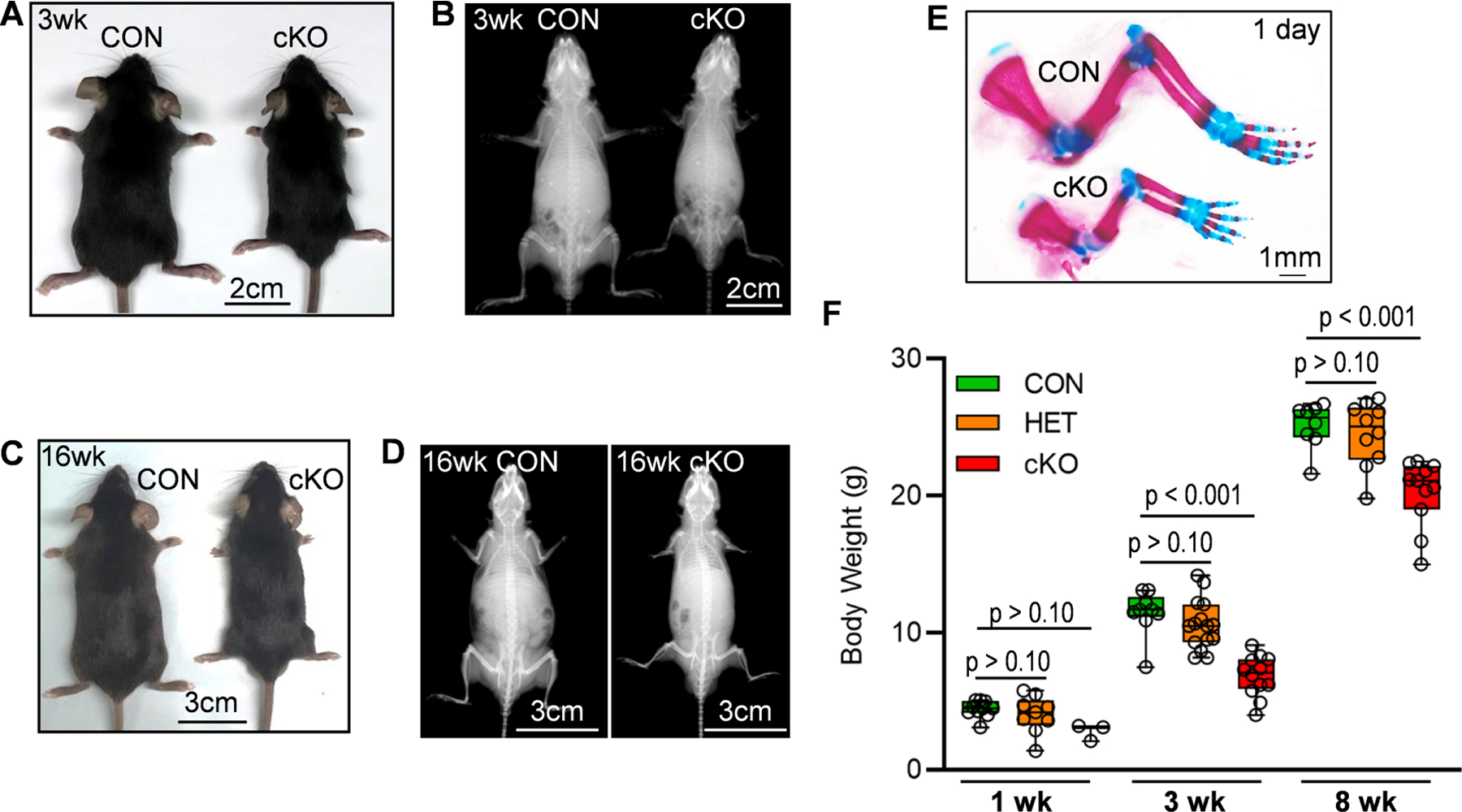
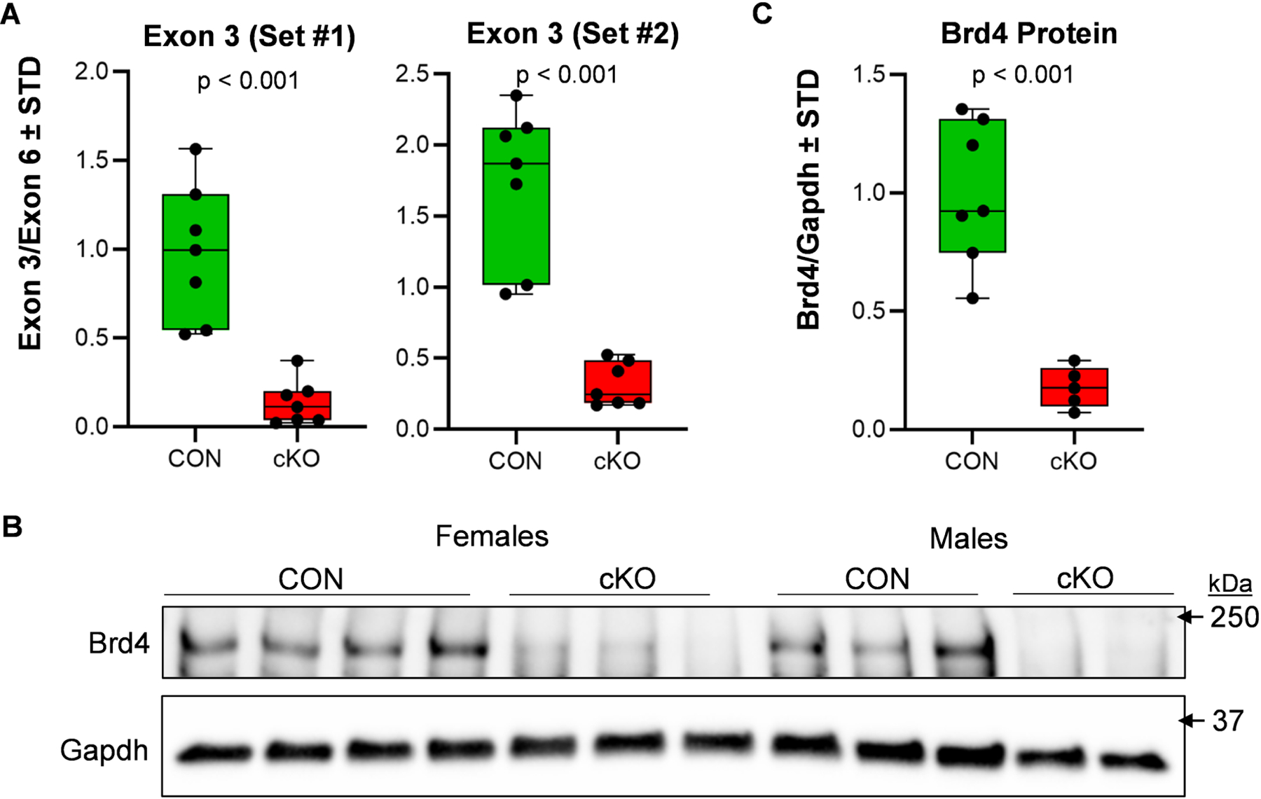
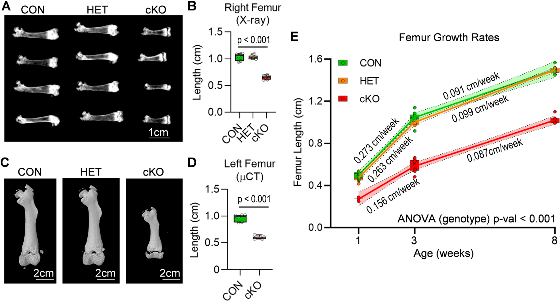
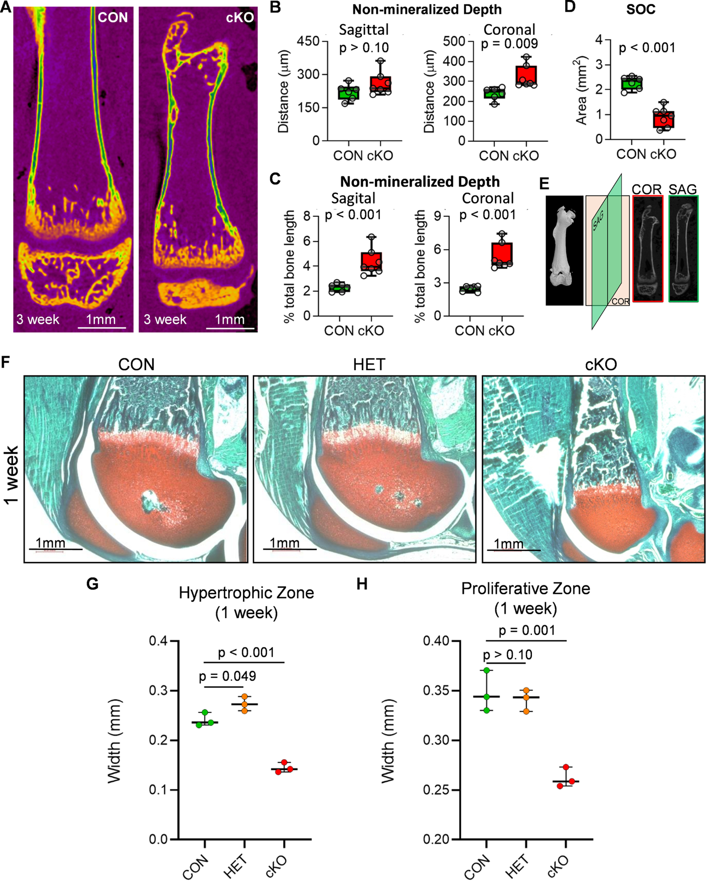
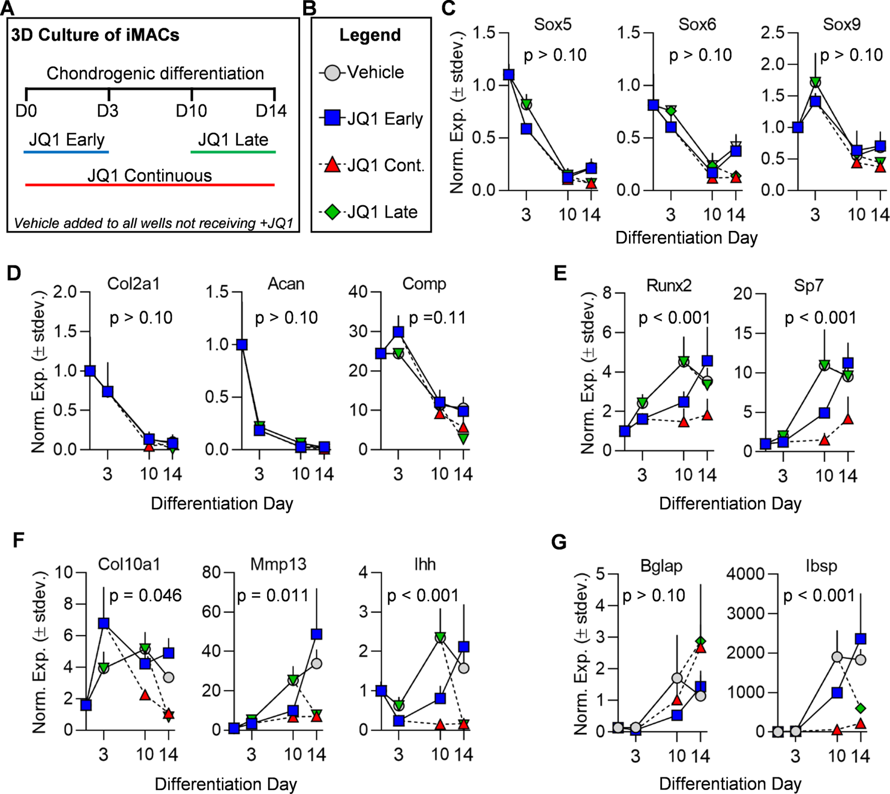
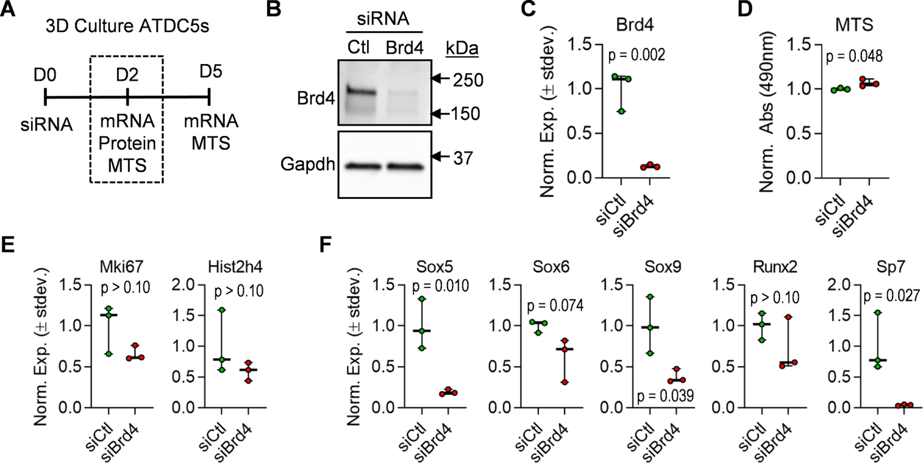
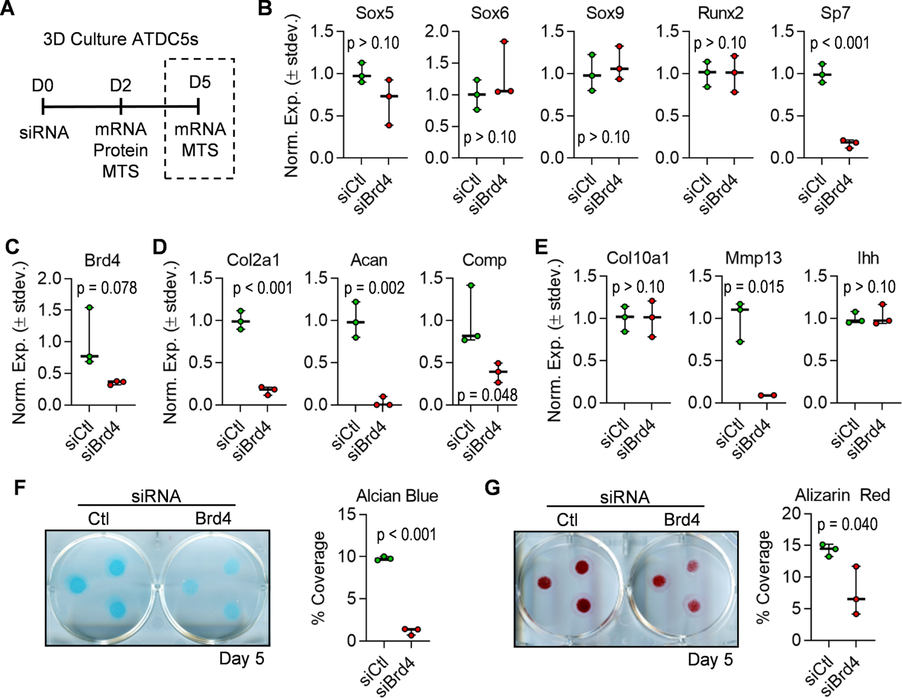
Differentiation of multi-potent mesenchymal stromal cells (MSCs) is directed by the activities of lineage-specific transcription factors and co-factors. A subset of these proteins controls the accessibility of chromatin by recruiting histone acetyl transferases or deacetylases that regulate acetylation of the N-termini of H3 and H4 histone proteins. Bromodomain (BRD) proteins recognize these acetylation marks and recruit the RNA pol II containing transcriptional machinery. Our previous studies have shown that Brd4 is required for osteoblast differentiation in vitro. Here, we investigated the role of Brd4 on endochondral ossification in C57BL/6 mice and chondrogenic differentiation in cell culture models. Conditional loss of Brd4 in the mesenchyme (Brd4 cKO, Brd4fl/fl: Prrx1-Cre) yields smaller mice that exhibit alteration in endochondral ossification. Importantly, abnormal growth plate morphology and delayed long bone formation is observed in juvenile Brd4 cKO mice. One week old Brd4 cKO mice have reduced proliferative and hypertrophic zones within the physis and exhibit a delay in the formation of the secondary ossification center. At the cellular level, Brd4 function is required for chondrogenic differentiation and maturation of both ATDC5 cells and immature mouse articular chondrocytes. Mechanistically, Brd4 loss suppresses Sox9 levels and reduces expression of Sox9 and Runx2 responsive endochondral genes (e.g., Col2a1, Acan, Mmp13 and Sp7/Osx). Collectively, our results indicate that Brd4 is a key epigenetic regulator required for normal chondrogenesis and endochondral ossification.

Keywords: Brd4; Epigenetics; Genetic animal model; Growth plate; Histone; Limb patterning.


Figures

Figure 1.
Figure 1.. Mesenchymal loss of Brd4 results in smaller mice.
Brd4 CON, (Brd4wt/wt: Prrx1-Cre), HET (Brd4wt/f l: Prrx1-Cre), and cKO (Brd4f l/f l: Prrx1-Cre) mice were assessed at several postnatal time points. Photograph (A) and X-ray radiograph (B) of three week old male mice. Photograph (C) and X-ray (D) of sixteen week old male mice. Whole mount staining (front legs) of one day old male pups (E). Total body weight of male mice at one, three, and eight weeks (F). Boxplots indicate the median, interquartile range, and the minimum and maximum value in each dataset. Individual mice are represented by a single point on the graph (1 Week: CON (n = 10), HET (9), cKO (n = 3); 3 Weeks: CON (n = 9), HET (15), cKO (n = 12); 8 Weeks: CON (n = 8), HET (n = 10), cKO (n = 11). A one-way ANOVA was performed across all groups in panel F and demonstrated significant variation within the dataset (p < 0.001). A follow-up multiple comparisons test was performed using the Tukey method (correct for multiple comparisons) to compare the means of each group one-to-one. P-values shown in panel E represent the results from the comparison between the indicated group and the CON group at the same age.
Figure 2.
Figure 2.. Efficient deletion of Brd4 by Prrx1-Cre recombinase.
Limb buds were collected from CON (Brd4f l/wt and Brd4f l/f l: Prrx1-Cre negative) and cKO (Brd4f l/f l: Prrx1-Cre positive) 14.5 dpc embryos. Quantitative PCR analysis was conducted on DNA isolated from the limb buds. Both male and female embryos were included in the analysis. Deletion of exon three was assessed using two distinct primer pairs targeting the floxed region within exon 3 and evaluated in relation to non-targeted exon six (n = 7) (A). Western blotting for Brd4 in limb bud tissues of 14.5 dpc embryos (B) and quantification when normalized to Gapdh (n = 7 and 5) (C). Boxplots indicate the median, interquartile range, and the minimum and maximum value in each dataset. Individual mice are represented by a single point on the graph. P-values shown on the graphs represent the results of an unpaired, two-tailed t-test between CON and cKO.
Figure 3.
Figure 3.. Long bone alterations in mice lacking mesenchymal Brd4 expression.
CON (Brd4wt/wt: Prrx1-Cre), HET (Brd4wt/f l: Prrx1-Cre), and cKO (Brd4f l/f l: Prrx1-Cre) female mice were assessed by X-ray and µCT analysis. X-ray radiographs (A) and length quantification (n = 6) (B) of femora collected from three week old female mice. Micro-computed tomography (µCT) reconstructions (C) and length quantification (n = 6 and 7) (D) of femora collected from three week old female mice. Measurements of femora lengths obtained from X-ray images from one, three, and eight week old male mice (1 Week: CON (n = 7), HET (n = 7), cKO (n = 3); 3 Weeks: CON (n = 9), HET (n = 7), cKO (n = 9); 8 Weeks: CON (n = 3), HET (7), cKO (n = 7). (E). Linear regression analysis was performed for each individual line segment (1 to 3 weeks and 3 to 8 weeks). The slope of the line (growth rate) is indicated on the graph, and the 95% confidence interval is indicated by the shaded region. Boxplots indicate the median, interquartile range, and the minimum and maximum value in each dataset. Individual mice are represented by a single point on the graph. P-value shown in panel B represents the results of a one-way ANOVA followed by a multiple comparisons test that was performed using the Tukey method (correct for multiple comparisons) to compare the means of the cKO and CON groups. P-value shown in panel D represent the results from an unpaired, two-tailed t-test between CON and cKO. For panel E, a two-way ANOVA was performed across the three groups. The time variable accounted for 73.36% of the variation observed (p < 0.001). The genotype variable (reported on the graph) accounted for 18.08% of the variation observed (p < 0.001).
Figure 4.
Figure 4.. Growth plate alterations in mice lacking mesenchymal Brd4 expression.
Distal femora of three week old CON (Brd4wt/wt: Prrx1-Cre) and cKO (Brd4f l/f l: Prrx1-Cre) female mice were assessed by µCT analysis (n = 6 and 7) (A-E). µCT reconstructions of femora collected from three week old female mice (A). Non-mineralized surface within the growth plate area measured throughout the sagittal (left) and coronal (right) planes (B). Each point represents an average of fifteen measurements taken for each mouse femur. Quantification of non-mineralized surface within growth plate area as a percentage of total femur length obtained from µCT images (C). Area of secondary ossification center (SOC) determined from medial slice of µCT images (D). Schematic depicting orientation of cross sections used to measure non-mineralized surface within the growth plate area of distal femora (E). Trichrome staining of distal femora derived from one week old CON (Brd4wt/wt: Prrx1-Cre), HET (Brd4wt/f l: Prrx1-Cre), and cKO (Brd4f l/f l: Prrx1-Cre) male mice (F). Red = cartilage, blue/green = bone, brown = nuclei. Measurements of hypertrophic (G) and proliferative (H) growth plate zones from trichrome stained sections of one week old male femora (n = 3). Individual measurements are represented by a single point on the graph for each mouse. Boxplots indicate the median, interquartile range, and the minimum and maximum value in each dataset. P-values shown in panels B-D represent the results from unpaired, two-tailed Student’s t-test between CON and cKO groups on each graph. P-values shown in panels G and H represent the results of a one-way ANOVA followed by a multiple comparisons test performed using the Tukey method (correct for multiple comparisons) to compare the means of the indicated groups.
Figure 5.
Figure 5.. +JQ1 treatment prevents maturation of iMACs.
iMACs were collected from five day-old wild-type C57BL/6J mice and cultured in three-dimensional micromass (3D-µmass) conditions in chondrogenic differentiation media. Schematic of treatment regimen with vehicle (DMSO) control or 100nm +JQ1 (A). Legend indicating treatment groups in subsequent graphs (B). RT-qPCR analysis of chondrogenic transcription factors (C), early chondrogenic markers (D), hypertrophic/osteogenic transcription factors (E), hypertrophic markers (F), and hypertrophic/osteogenic markers (G). Experiments were performed in n = 3 biological replicates and the mean is indicated by each point on the graph. Error bars represent standard deviation and statistical significance is indicated in each panel. A mixed-effects model (REML) analysis was performed for each graph (alpha = 0.05). The p-value reported for the fixed effects was then assessed. For all datasets, the time variable had a statistically significant impact with a p-value < 0.001. The p-value calculated for the “treatment group” variable is reported on each graph.
Figure 6.
Figure 6.. Depletion of Brd4 prevents early transcription factor expression in ATDC5 cells.
ATDC5 cells were transfected with non-targeting siRNA (siCtl) or Brd4-targeting siRNA (siBrd4) and cultured in three-dimensional micromass cultures in chondrogenic differentiation media. Schematic of siRNA transfection experiment and data collection points (A). Western blot analysis performed on protein lysates collected two days after transfection (B). RT-qPCR analysis on mRNA isolated two days post-transfection (C). MTS assay conducted two days after transfection (D). RT-qPCR analysis of cell cycle genes (E) and transcription factors performed on mRNA isolated two days post-transfection. Boxplots indicate the median, interquartile range, and the minimum and maximum value in each dataset (n = 3). Individual biological replicates are represented by a single point on the graph. P-values indicate results from unpaired, two-tailed Student’s t-test between siCtl and siBrd4 groups.
Figure 7.
Figure 7.. Brd4 knock-down prevents maturation of ATDC5 cells.
ATDC5 cells were transfected with non-targeting siRNA (siCtl) or Brd4-targeting siRNA (siBrd4) and cultured in three-dimensional micromass cultures in chondrogenic differentiation media. Schematic of siRNA transfection experiment and data collection points (A). RT-qPCR analysis of transcription factors (B), Brd4 (C), early chondrogenic genes (D), and hypertrophic genes (E) performed on mRNA isolated five days post-transfection. Alcian Blue staining and quantification performed five days after induction of chondrogenic differentiation (F). Alizarin red staining and quantification performed five days after induction of chondrogenic differentiation (G). For quantification of staining, percent coverage of a 1.13cm2 circle was measured for each micromass (n = 3 micromasses per well). Boxplots indicate the median, interquartile range, and the minimum and maximum value in each dataset (n = 3). Individual biological replicates are represented by a single point on the graph. P-values indicate results from unpaired, two-tailed Student’s t-test between siCtl and siBrd4 groups.

References

    1. Kobayashi T, Kronenberg HM, Overview of skeletal development, Methods Mol Biol 1130 (2014) 3–12. - PubMed
    1. Breeland G, Menezes RG, Embryology, Bone Ossification, StatPearls Publishing, Available from: https://www.ncbi.nlm.nih.gov/books/NBK539718/, 2019. - PubMed
    1. Hall BK, Miyake T, The membranous skeleton: the role of cell condensations in vertebrate skeletogenesis, Anat Embryol (Berl) 186(2) (1992) 107–24. - PubMed
    1. Berendsen AD, Olsen BR, Bone development, Bone 80 (2015) 14–18. - PMC - PubMed
    1. Long F, Building strong bones: molecular regulation of the osteoblast lineage, Nat Rev Mol Cell Biol 13(1) (2011) 27–38. - PubMed

Publication types