. 2021 Oct 26;7(1):64.
doi: 10.1186/s40942-021-00339-z.

Spontaneous closure of degenerative lamellar macular hole with epiretinal membrane proliferation

Affiliations

Spontaneous closure of degenerative lamellar macular hole with epiretinal membrane proliferation

Rony C Preti et al. Int J Retina Vitreous. .

Abstract

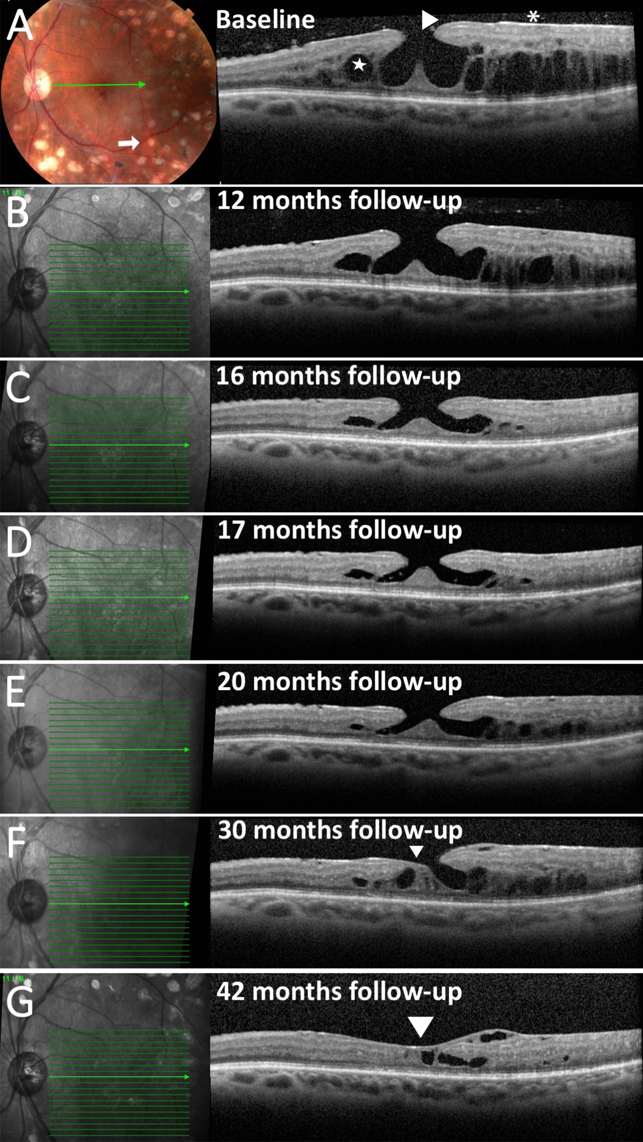
Background: To describe the spontaneous closure of a degenerative lamellar macular hole with epiretinal proliferation (LHEP) as documented with tracked spectral domain optical coherence tomography (SD-OCT).

Case presentation: A 54-years-old diabetic female patient presented with progressive vision loss in the left eye. SD-OCT illustrated LHEP associated with cystic fluid in the outer nuclear layer. Sequentially tracked SD-OCT showed progressive closure of the degenerative lamellar macular hole and resolution of the CME over almost 4 years, in the absence of any surgical intervention.

Discussion/conclusion: LHEP may represent a specialized form of degenerative epiretinal membrane associated with Muller cell activation. Spontaneous degenerative LMH closure may rarely occur with these lesion types, in the absence of surgical intervention, possibly due to Muller cell proliferation preceded by PVD.

Keywords: Epiretinal membrane; Epiretinal proliferation; Lamellar hole–associated epiretinal proliferation; Lamellar macular hole; Spectral domain optical coherence tomography.


Conflict of interest statement

The following authors have no financial disclosures: RCP, LCZ, LPC, MRLM.

Figures

Fig. 1
Fig. 1
A Left. Fundus photography shows multiple panretinal photocoagulation laser scars consistent with regressed proliferative diabetic retinopathy. Right, SD-OCT B-scan illustrates cysts in the outer nuclear layer (star), lamellar hole with epiretinal proliferation (LHEP) (arrow-heads), and epiretinal proliferation (asterisk). Note the grade 0 posterior vitreous detachment. B Left. Registered near infrared reflectance with level of OCT section Right. BE A partial grade 3 PVD is noted. While LHEP is unchanged, CME is improved. EG Progressive closure of the LMH (arrowheads) and resolution of the CME is illustrated with sequentially tracked SD-OCT B scans. Complete grade 4 PVD is noted

References

    1. Pang CE, Maberley DA, Freund KB, White VA, Rasmussen S, To E, et al. Lamellar hole-associated epiretinal proliferation: a clinicopathologic correlation. Retina. 2016;36(7):1408–1412. doi: 10.1097/IAE.0000000000001069. - DOI - PubMed
    1. Choi WS, Merlau DJ, Chang S. Vitrectomy for macular disorders associated with lamellar macular hole epiretinal proliferation. Retina. 2018;38(4):664–669. doi: 10.1097/IAE.0000000000001591. - DOI - PubMed
    1. Govetto A, Dacquay Y, Farajzadeh M, Platner E, Hirabayashi K, Hosseini H, et al. Lamellar macular hole: two distinct clinical entities? Am J Ophthalmol. 2016;164:99–109. doi: 10.1016/j.ajo.2016.02.008. - DOI - PubMed
    1. Ghadiali Q, Zahid S, Dolz-Marco R, Tan A, Engelbert M. An Assessment of vitreous degeneration in eyes with vitreomacular traction and macular holes. J Ophthalmol. 2017;2017:6834692. doi: 10.1155/2017/6834692. - DOI - PMC - PubMed
    1. Uchino E, Uemura A, Ohba N. Initial stages of posterior vitreous detachment in healthy eyes of older persons evaluated by optical coherence tomography. Arch Ophthalmol. 2001;119(10):1475–1479. doi: 10.1001/archopht.119.10.1475. - DOI - PubMed

LinkOut - more resources