Review
. 2021 Nov:130:112466.
doi: 10.1016/j.msec.2021.112466. Epub 2021 Sep 29.

FDA-approved bone grafts and bone graft substitute devices in bone regeneration

Affiliations
Review

FDA-approved bone grafts and bone graft substitute devices in bone regeneration

Cassidy E Gillman et al. Mater Sci Eng C Mater Biol Appl. 2021 Nov.

Abstract

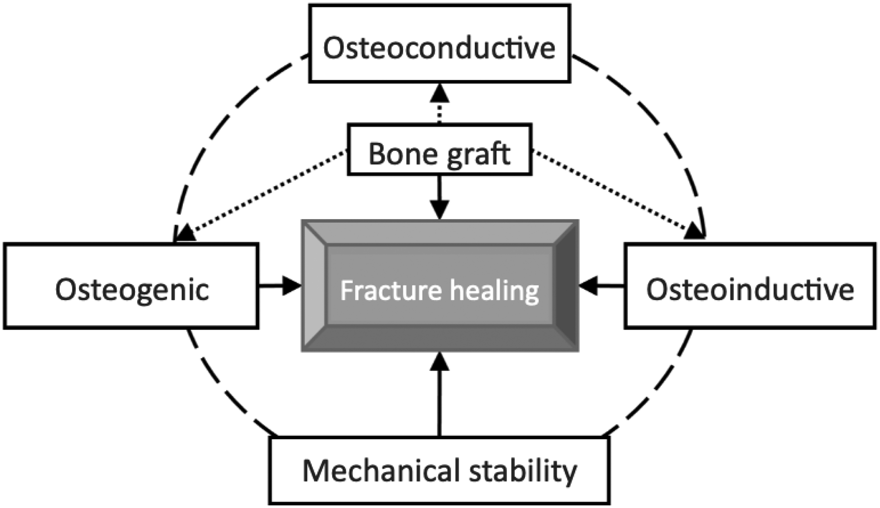
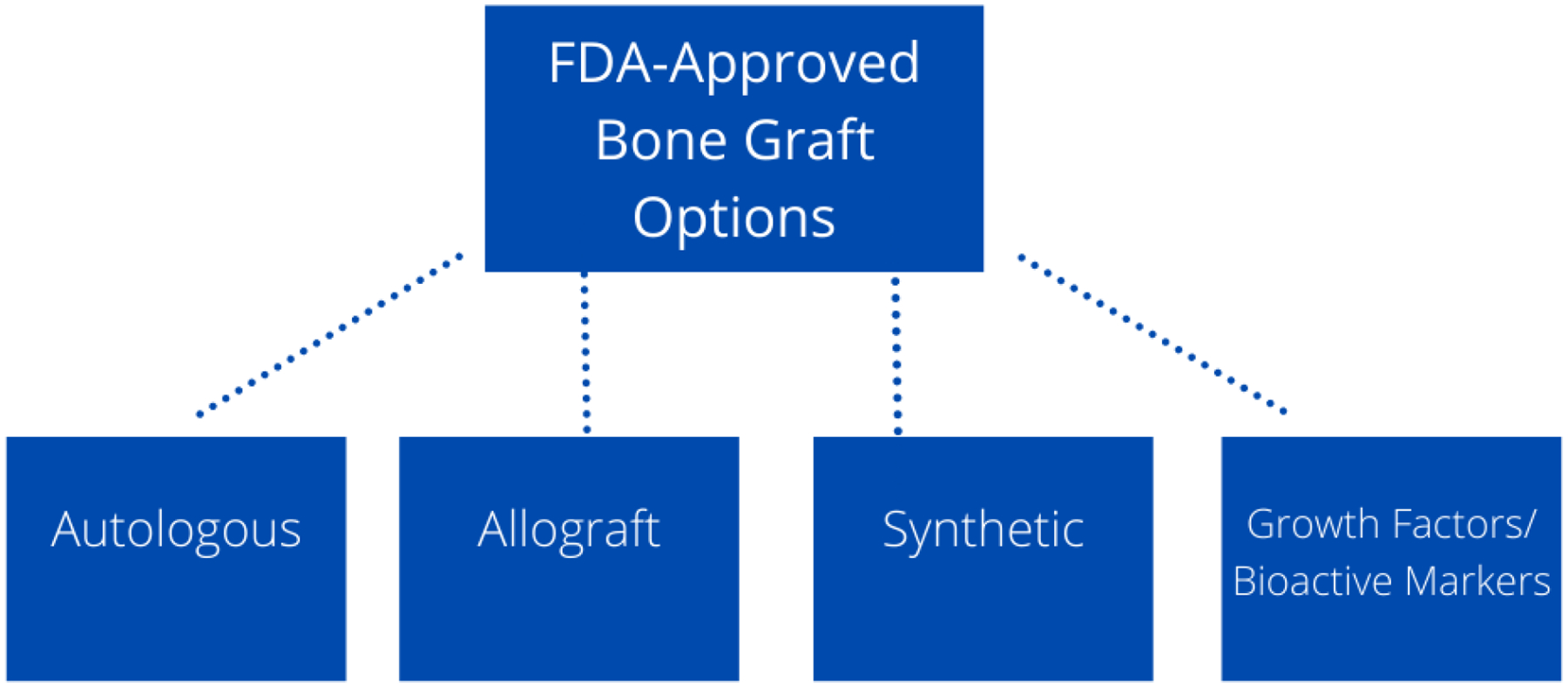
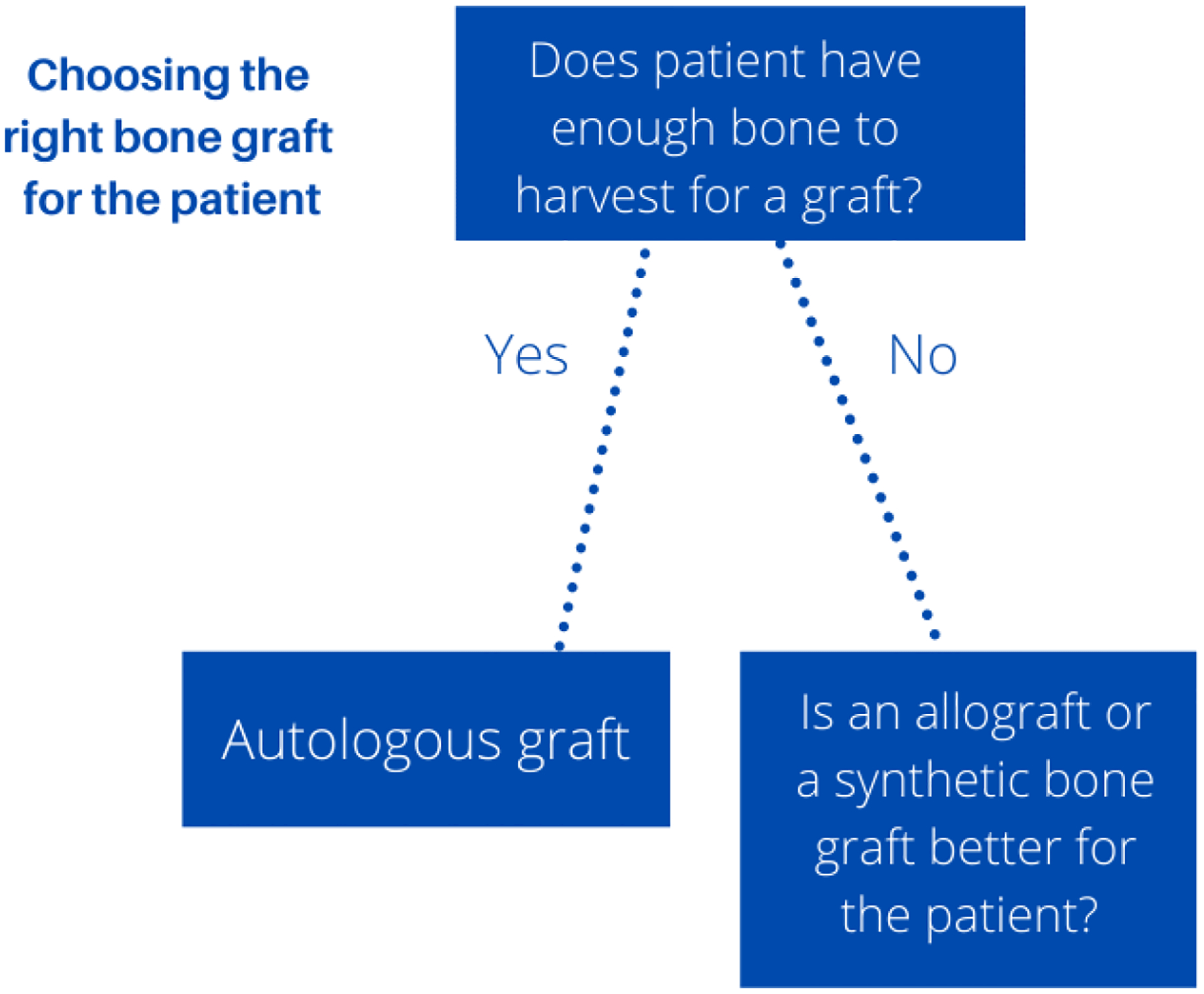
To induce bone regeneration there is a complex cascade of growth factors. Growth factors such as recombinant BMP-2, BMP-7, and PDGF are FDA-approved therapies in bone regeneration. Although, BMP shows promising results as being an alternative to autograft, it also has its own downfalls. BMP-2 has many adverse effects such as inflammatory complications such as massive soft-tissue swelling that can compromise a patient's airway, ectopic bone formation, and tumor formation. BMP-2 may also be advantageous for patients not willing to give up smoking as it shows bone regeneration success with smokers. BMP-7 is no longer an option for bone regeneration as it has withdrawn off the market. PDGF-BB grafts in studies have shown PDGF had similar fusion rates to autologous grafts and fewer adverse effects. There is also an FDA-approved bioactive molecule for bone regeneration, a peptide P-15. P-15 was found to be effective, safe, and have similar outcomes to autograft at 2 years post-op for cervical radiculopathy due to cervical degenerative disc disease. Growth factors and bioactive molecules show some promising results in bone regeneration, although more research is needed to avoid their adverse effects and learn about the long-term effects of these therapies. There is a need of a bone regeneration method of similar quality of an autograft that is osteoconductive, osteoinductive, and osteogenic. This review covers all FDA-approved bone regeneration therapies such as the "gold standard" autografts, allografts, synthetic bone grafts, and the newer growth factors/bioactive molecules. It also covers international bone grafts not yet approved in the United States and upcoming technologies in bone grafts.

Keywords: Bone cement; Bone grafts; Bone morphogenetic proteins; Clinical indications; Food and drug administration; Platelet derived growth factor.


Figures

Figure 1:
Figure 1:
Radiographic image of atrophic nonunion of fracture of the humerus [24]. Reprinted/adapted by permission from [CopyRight Clearance Center]: [Springer Nature]
Figure 2:
Figure 2:
Radiographic image of malunion of the proximal femur [24]. Reprinted/adapted by permission from [CopyRight Clearance Center]: [Springer Nature]
Figure 3:
Figure 3:
Radiographic image of bone cyst in the calcaneus. Reprinted from [29] with permission from the Creative Commons Attribution 4.0 International License (http://creativecommons.org/licenses/by/4.0/).
Figure 4:
Figure 4:
Radiographic image of avascular necrosis of the femoral head. Reprinted from [30] with permission from The Radiological Society of North America.
Figure 5:
Figure 5:
Typical fracture healing process shown at different phases of bone healing with primal cell types found at each stage and time of each stage. Below the fracture healing illustration, metabolic phases (blue bars) and biological stages (brown bars) are shown. The metabolic stages are anabolic and catabolic. The three major biological stages of healing are inflammatory, endochondral bone formation, and coupled remodeling. The time scale of healing is equivalent to a mouse closed femur fracture fixed with an intramedullary rod. Reprinted from [38] with permission from Journal of Bioactive Materials under the Creative Commons Attribution-NonCommercial License (https://creativecommons.org/licenses/by-nc-nd/4.0/).
Figure 6:
Figure 6:
The process of a fractured bone healing is controlled by properties of osteoconduction, osteoinduction, and osteogenesis. Bone graft materials are osteoconductive to allow angiogenesis and cell growth to occur. Then by osteoinduction, recruitment of mesenchymal stem cells occurs. Then osteogenesis releases growth factors to help form new bone. Other important aspects of bone healing include vascularity as adequate blood supply is needed for healing and mechanical stability. Reprinted from [41] with permission from Creative Commons Attribution-NonCommercial License (https://creativecommons.org/licenses/by-nc/4.0/).
Figure 7:
Figure 7:
Cascade of Growth Factors. Illustration show actions of prevalent growth factors (BMP-2, FGF, PDGF, and VEGF) in vascularized bone regeneration. Reprinted by permission from [CopyRight Clearance Center]: [Elsevier] [45].
Figure 8:
Figure 8:
FDA-Approved Bone Graft Options: The four main options of bone grafting include autologous, allograft, synthetic grafts, and growth factors/bioactive markers.
Figure 9:
Figure 9:
Choosing right bone graft for the patient: the “gold” standard for bone grafting is still using autologous grafts, but the limitation of this method is if there is enough graft to harvest. If there is not enough graft to harvest other bone grafting options such as allografts and synthetic bone grafts will be chosen for the patient.

References

    1. Campana V, Milano G, Pagano E, Barba M, Cicione C, Salonna G, Lattanzi W, and Logroscino G, Bone substitutes in orthopaedic surgery: from basic science to clinical practice. J Mater Sci Mater Med, 2014. 25(10): p. 2445–61. - PMC - PubMed
    1. Greenwald AS, Boden SD, Goldberg VM, Khan Y, Laurencin CT, Rosier RN, and American I. Academy of Orthopaedic Surgeons. The Committee on Biological, Bone-graft substitutes: facts, fictions, and applications. J Bone Joint Surg Am, 2001. 83-A Suppl 2 Pt 2: p. 98–103. - PubMed
    1. Finkemeier CG, Bone-grafting and bone-graft substitutes. J Bone Joint Surg Am, 2002. 84(3): p. 454–64. - PubMed
    1. Faour O, Dimitriou R, Cousins CA, and Giannoudis PV, The use of bone graft substitutes in large cancellous voids: any specific needs? Injury, 2011. 42 Suppl 2: p. S87–90. - PubMed
    1. Van Heest A and Swiontkowski M, Bone-graft substitutes. The Lancet, 1999. 353: p. S28–S29. - PubMed

Substances

LinkOut - more resources