. 2021 Oct 26;12(1):6174.
doi: 10.1038/s41467-021-26429-y.

Structural basis for protein glutamylation by the Legionella pseudokinase SidJ

Affiliations

Structural basis for protein glutamylation by the Legionella pseudokinase SidJ

Michael Adams et al. Nat Commun. .

Abstract

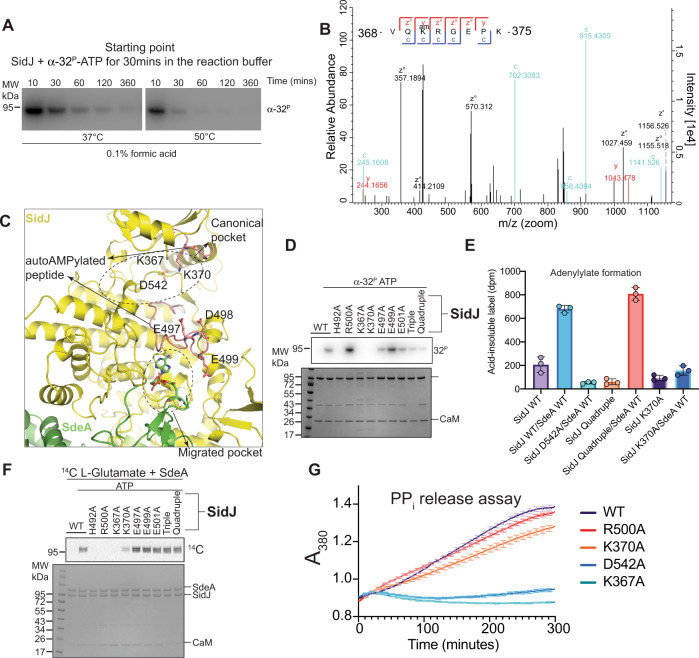
Legionella pneumophila (LP) avoids phagocytosis by secreting nearly 300 effector proteins into the host cytosol. SidE family of effectors (SdeA, SdeB, SdeC and SidE) employ phosphoribosyl ubiquitination to target multiple host Rab GTPases and innate immune factors. To suppress the deleterious toxicity of SidE enzymes in a timely manner, LP employs a metaeffector named SidJ. Upon activation by host Calmodulin (CaM), SidJ executes an ATP-dependent glutamylation to modify the catalytic residue Glu860 in the mono-ADP-ribosyl transferase (mART) domain of SdeA. SidJ is a unique glutamylase that adopts a kinase-like fold but contains two nucleotide-binding pockets. There is a lack of consensus about the substrate recognition and catalytic mechanism of SidJ. Here, we determined the cryo-EM structure of SidJ in complex with its substrate SdeA in two different states of catalysis. Our structures reveal that both phosphodiesterase (PDE) and mART domains of SdeA make extensive contacts with SidJ. In the pre-glutamylation state structure of the SidJ-SdeA complex, adenylylated E860 of SdeA is inserted into the non-canonical (migrated) nucleotide-binding pocket of SidJ. Structure-based mutational analysis indicates that SidJ employs its migrated pocket for the glutamylation of SdeA. Finally, using mass spectrometry, we identified several transient autoAMPylation sites close to both the catalytic pockets of SidJ. Our data provide unique insights into the substrate recognition and the mechanism of protein glutamylation by the pseudokinase SidJ.


Conflict of interest statement

The authors declare no competing interests.

Figures

Fig. 1
Fig. 1. SidJ E565A/CaM and SdeA form a stable reaction intermediate complex.
A Size-exclusion chromatography (SEC) profiles of SidJ/CaM and SdeA in the presence of ATP. The highlighted fraction in the SidJ/CaM E565A + SdeA sample is shown on SDS-PAGE (right). B SEC profiles of SidJ/CaM E565A + SdeA in the presence of various cofactors. C Schematic representation of the reaction scheme of SidJ-mediate glutamylation of SdeA highlighting the trapped adenylylated reaction intermediate between SidJ/CaM and SdeA. D Time course experiment measuring the incorporation of [14C]-Glu into SdeA catalyzed by SidJ using SidJ WT, SidJ E565A. Samples were separated by SDS-PAGE and visualized by Coomassie stain and autoradiography (Top). The experiment was done in triplicates and [14C]-bands were quantified and plotted (Bottom). Purple corresponds to WT, blue corresponds to the E565A mutant. Individual measurements are shown. E Flow chart of the sample preparation process of SidJ E565A/CaM + SdeA for Cryo-EM. From left to right, samples are incubated and then cross-linked via GraFix method. Fractions are analyzed via SDS-PAGE and further purified via SEC.
Fig. 2
Fig. 2. Adenylylated SdeA E860 binds to the migrated pocket of SidJ.
A Cryo-EM map of SidJ E565A (Yellow), Calmodulin (Teal), and SdeA (Green). B Overall structure of the reaction intermediate SidJ E565A/CaM/SdeA heterotrimer. Both N- and C-lobe of SidJ (Yellow), as well as mART and PDE lobes of SdeA (Green) are highlighted, with Calmodulin (Teal) bound to the C-terminus of SidJ. C Closer view of the migrated nucleotide-binding pocket of SidJ (Yellow), and adenylylated SdeA E860 (Stick representation, Green) inserted. The cryo-EM density of E860 of SdeA and AMP is shown in the mesh. SdeA catalytic loop is colored in salmon. E862, another glutamylation target of SidJ is also shown. D Incorporation of [14 C]-Glu into SdeA with and without its PDE domain present. Reaction components were separated by SDS-PAGE and either visualized by Coomassie stain (Top) or autoradiography (Bottom).
Fig. 3
Fig. 3. SidJ and SdeA form an extensive binding interface.
A Overview of SidJ/CaM and SdeA intermediate complex showing the three binding sites between SidJ and SdeA. Site 1 being the loop insertion site (Shown in 3B, C), site 2 being the mART site (Shown in 3E), and site 3 being the migrated pocket site (Shown in 4A). B The insertion loop of site 1 (Red) of SidJ inserting itself into the cleft between SdeA’s PDE and mART lobes (Shown in green, surface representation). SidJ Proline 290 is shown in stick representation. C Detailed view of the SidJ–SdeA interaction site 1—the insertion loop of SidJ (Yellow) and SdeA (green) with key residues shown. D Incorporation of [14C]-Glu into SdeA catalyzed by SidJ, with and without the insertion loop (Left) and using mutations on both SidJ and SdeA from site 1 (Right). Samples were separated by SDS-PAGE and visualized by Coomassie stain (Bottom) or autoradiography (Top). E Detailed view of the SidJ–SdeA interaction site 2—the interactions between SidJ C-lobe (Yellow) and SdeA mART, with key residues shown. F Incorporation of [14C]-Glu into SdeA catalyzed by SidJ using mutations on both SidJ and SdeA from site 2. Samples were separated by SDS-PAGE and visualized by Coomassie stain (Bottom) or autoradiography (Top).
Fig. 4
Fig. 4. SidJ catalyzes glutamylation in the migrated pocket.
A Detailed view of the SidJ–SdeA Interaction site 3- adenylylated E860 of SdeA (Green) inserted into the migrated pocket of SidJ (Yellow), with key residues shown. B Incorporation of [14C]-Glu into SdeA catalyzed by SidJ using mutations on both SidJ and SdeA from site 3. Samples were separated by SDS-PAGE and visualized by Coomassie stain (Bottom) or autoradiography (Top). C Structural comparison between SidJ E565A (Yellow)/CaM + SdeA (Green) in its pre-glutamylation and post-catalytic states. Highlighted is a loop of SidJ (Red) located in the migrated pocket that undergoes structural change post-catalysis. D Overlay of SidJ canonical pocket in pre-glutamylation (Gray) and post-catalytic (Yellow) state, with key residues highlighted.
Fig. 5
Fig. 5. Characterization of SidJ autoAMPylation.
A Time course of SidJ autoAMPylation in an acidic environment at different temperatures. Reactions were performed using SidJ WT + CaM and α-[32 P]-ATP. Samples were separated by SDS-PAGE and visualized by autoradiography. B ETD fragmentation spectrum of SidJ peptide VQKRGEPK, with the AMPylation site K370 highlighted. AutoAMPylation reactions of SidJ WT were analyzed using LC–MS/MS. C Detailed view of SidJ autoAMPylation sites, with peptides identified in mass spectrometry shown in red, and modification sites highlighted. D AutoAMPylation of SidJs. CaM + SidJ WT or indicated mutants were reacted with α-[32 P]-ATP. Samples were separated by SDS-PAGE and visualized by Coomassie stain (Bottom) and autoradiography (Top). E Measuring α-[32P]-AMP incorporation into various SidJ and SdeA mutants. The samples indicated were reacted with α-[32P]-ATP and TCA precipitated followed by scintillation counting to measure the AMPylation levels. Each condition was measured in triplicates. Error bars denote standard deviation. F Incorporation of [14 C]-Glu into SdeA catalyzed by SidJ using SidJ WT and mutants of the autoAMPylation sites. Samples were separated by SDS-PAGE and visualized by Coomassie stain (Bottom) and autoradiography (Top). G Pyrophosphate release assay with SidJ and mutants indicated. The assay was performed using the EnzCheck™ pyrophosphate assay kit (Thermo Fischer Scientific). The kit components (see methods) react with the PPi in solution and release ribose 1-phosphate and 2-amino-6-mercapto-7-methylpurine which shows an absorption peak at 380 nm. The average of triplicate measurements is plotted, with standard deviation bars being shown in 10-min intervals.

References

    1. Grabbe C, Husnjak K, Dikic I. The spatial and temporal organization of ubiquitin networks. Nat. Rev. Mol. Cell Biol. 2011;12:295–307. doi: 10.1038/nrm3099. - DOI - PMC - PubMed
    1. Yau R, Rape M. The increasing complexity of the ubiquitin code. Nat. Cell Biol. 2016;18:579–586. doi: 10.1038/ncb3358. - DOI - PubMed
    1. Maculins T, Fiskin E, Bhogaraju S, Dikic I. Bacteria-host relationship: ubiquitin ligases as weapons of invasion. Cell Res. 2016;26:499–510. doi: 10.1038/cr.2016.30. - DOI - PMC - PubMed
    1. Hubber, A. & Roy, C. R. Modulation of host cell function by Legionella pneumophila type IV effectors. 26, 261–283 (2010). - PubMed
    1. Xu L, Luo Z-Q. Cell biology of infection by Legionella pneumophila. Microbes Infect. 2013;15:157–167. doi: 10.1016/j.micinf.2012.11.001. - DOI - PMC - PubMed

MeSH terms