A novel technique to assess rotational deformities in lower extremities using CT-based motion analysis
- PMID: 34702869
- PMCID: PMC8548303
- DOI: 10.1038/s41598-021-00532-y
A novel technique to assess rotational deformities in lower extremities using CT-based motion analysis
Abstract
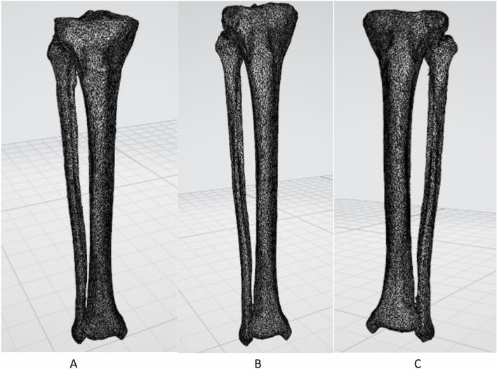
Rotational deformities following intramedullary (IM) nailing of tibia has a reported incidence of as high as 20%. Common techniques to measure deformities following IM nailing of tibia are either based on clinical assessment, plain X-rays or Computed Tomography (CT) comparing the treated leg with the uninjured contralateral side. All these techniques are based on examiners manual calculation inherently subject to bias. Following our previous rigorous motion analysis and symmetry studies on hemi pelvises, femurs and orthopaedic implants, we aimed to introduce a novel fully digital technique to measure rotational deformities in the lower legs. Following formal institutional approval from the Imperial College, CT images of 10 pairs of human lower legs were retrieved. Images were anonymized and uploaded to a research server. Three dimensional CT images of the lower legs were bilaterally reconstructed. CT-based motion analysis (CTMA) was used and the mirrored images of the left side were merged with the right side proximally as stationary and distally as moving objects. Discrepancies in translation and rotation were automatically calculated. Our study population had a mean age of 54 ± 20 years. There were six males and four females. We observed a greater variation in translation (mm) of Centre of Mass (COM) in sagittal plane (95% CI - 2.959-.292) which was also presented as rotational difference alongside the antero-posterior direction or Y axis (95% CI .370-1.035). In other word the right lower legs in our study were more likely to be in varus compared to the left side. However, there were no statistically significant differences in coronal or axial planes. Using our proposed fully digital technique we found that lower legs of the human adults were symmetrical in axial and coronal plane. We found sagittal plane differences which need further addressing in future using bigger sample size. Our novel recommended technique is fully digital and commercially available. This new technique can be useful in clinical practice addressing rotational deformities following orthopaedic surgical intervention. This new technique can substitute the previously introduced techniques.
© 2021. The Author(s).
Conflict of interest statement
The authors declare no competing interests.
Figures