

E3-ligase knock down revealed differential titin degradation by autopagy and the ubiquitin proteasome system
- PMID: 34702928
- PMCID: PMC8548520
- DOI: 10.1038/s41598-021-00618-7
E3-ligase knock down revealed differential titin degradation by autopagy and the ubiquitin proteasome system
Abstract
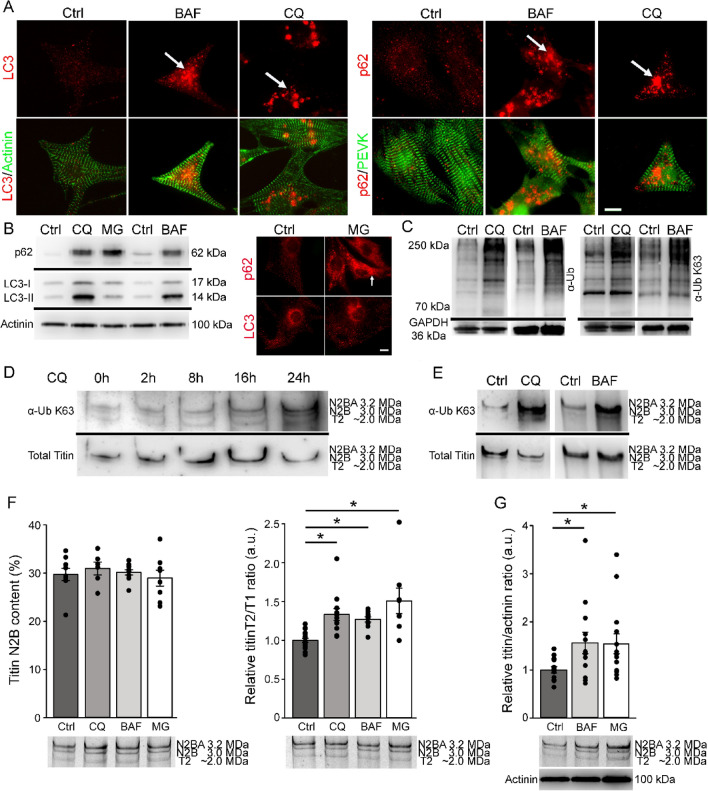
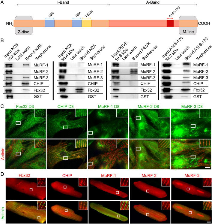
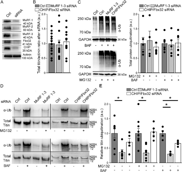
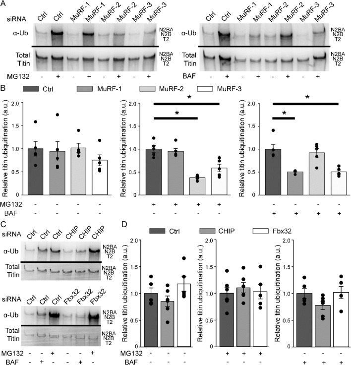
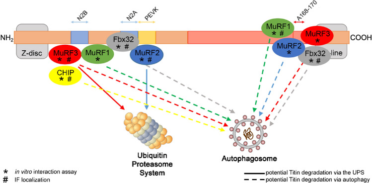
The sarcomere protein titin is a major determinant of cardiomyocyte stiffness and ventricular distensibility. The constant mechanical stress on titin requires well-controlled protein quality control, the exact mechanisms of which have not yet been fully elucidated. Here, we analyzed E3-ligases potentially responsible for cardiac titin ubiquitination and specifically studied the involvement of the autophagosomal system in titin degradation. Pharmacological inhibition of autophagy and the proteasome in cultured primary rat cardiomyocytes significantly elevated titin ubiquitination and increased titin degradation. Using in-vitro pull down assays we identified binding of E3-ligases MuRF1-3, CHIP and Fbx32 to several titin domains. Immunofluorescence analysis showed sarcomeric localization of the E3-ligases. siRNA-mediated knock-down of the E3-ligases MuRF-1, -3 and a combination of CHIP/Fbx32 significantly reduced autophagy-related titin ubiquitination, whereas knock-down of MuRF-2 and -3 reduced proteasome-related titin ubiquitination. We demonstrated that the proteasomal and the autophagosomal-lysosomal system participate in degradation of the titin filament. We found that ubiquitination and degradation of titin are partially regulated by E3-ligases of the MuRF family. We further identified CHIP and Fbx32 as E3-ligases involved in titin ubiquitination.
© 2021. The Author(s).
Conflict of interest statement
The authors declare no competing interests.
Figures
Similar articles
-
Titin kinase ubiquitination aligns autophagy receptors with mechanical signals in the sarcomere.EMBO Rep. 2021 Oct 5;22(10):e48018. doi: 10.15252/embr.201948018. Epub 2021 Aug 17. EMBO Rep. 2021. PMID: 34402565 Free PMC article.
-
Design Principles Involving Protein Disorder Facilitate Specific Substrate Selection and Degradation by the Ubiquitin-Proteasome System.J Biol Chem. 2016 Mar 25;291(13):6723-31. doi: 10.1074/jbc.R115.692665. Epub 2016 Feb 5. J Biol Chem. 2016. PMID: 26851277 Free PMC article. Review.
-
P62 Links the Autophagy Pathway and the Ubiquitin-Proteasome System in Endothelial Cells during Atherosclerosis.Int J Mol Sci. 2021 Jul 21;22(15):7791. doi: 10.3390/ijms22157791. Int J Mol Sci. 2021. PMID: 34360560 Free PMC article.
-
SYVN1, NEDD8, and FBXO2 Proteins Regulate ΔF508 Cystic Fibrosis Transmembrane Conductance Regulator (CFTR) Ubiquitin-mediated Proteasomal Degradation.J Biol Chem. 2016 Dec 2;291(49):25489-25504. doi: 10.1074/jbc.M116.754283. Epub 2016 Oct 18. J Biol Chem. 2016. PMID: 27756846 Free PMC article.
-
[The role of different E3 ubiquitin ligases in regulation of the P53 tumor suppressor protein].Tsitologiia. 2013;55(10):673-87. Tsitologiia. 2013. PMID: 25509121 Review. Russian.
Cited by
-
RING Finger Protein 10 Regulates AP-1/Meox2 to Mediate Pirarubicin-Induced Cardiomyocyte Apoptosis.Oxid Med Cell Longev. 2023 Jan 20;2023:7872193. doi: 10.1155/2023/7872193. eCollection 2023. Oxid Med Cell Longev. 2023. PMID: 36713029 Free PMC article.
-
MyoMed205 Counteracts Titin Hyperphosphorylation and the Expression of Contraction-Regulating Proteins in a Rat Model of HFpEF.J Cachexia Sarcopenia Muscle. 2025 Jun;16(3):e13843. doi: 10.1002/jcsm.13843. J Cachexia Sarcopenia Muscle. 2025. PMID: 40464169 Free PMC article.
-
Oxygen-dependent regulation of E3(SCF)ubiquitin ligases and a Skp1-associated JmjD6 homolog in development of the social amoeba Dictyostelium.J Biol Chem. 2022 Sep;298(9):102305. doi: 10.1016/j.jbc.2022.102305. Epub 2022 Aug 4. J Biol Chem. 2022. PMID: 35933019 Free PMC article.
-
Titin: roles in cardiac function and diseases.Front Physiol. 2024 Apr 10;15:1385821. doi: 10.3389/fphys.2024.1385821. eCollection 2024. Front Physiol. 2024. PMID: 38660537 Free PMC article. Review.
-
Modulation of Titin and Contraction-Regulating Proteins in a Rat Model of Heart Failure with Preserved Ejection Fraction: Limb vs. Diaphragmatic Muscle.Int J Mol Sci. 2024 Jun 16;25(12):6618. doi: 10.3390/ijms25126618. Int J Mol Sci. 2024. PMID: 38928324 Free PMC article.
References
Publication types
MeSH terms
Substances
Grants and funding
LinkOut - more resources
Full Text Sources
