. 2021 Oct 26;11(1):21059.
doi: 10.1038/s41598-021-99965-8.

Preventing adolescent synaptic pruning in mouse prelimbic cortex via local knockdown of α4βδ GABAA receptors increases anxiety response in adulthood

Affiliations

Preventing adolescent synaptic pruning in mouse prelimbic cortex via local knockdown of α4βδ GABAA receptors increases anxiety response in adulthood

Matthew R Evrard et al. Sci Rep. .

Abstract

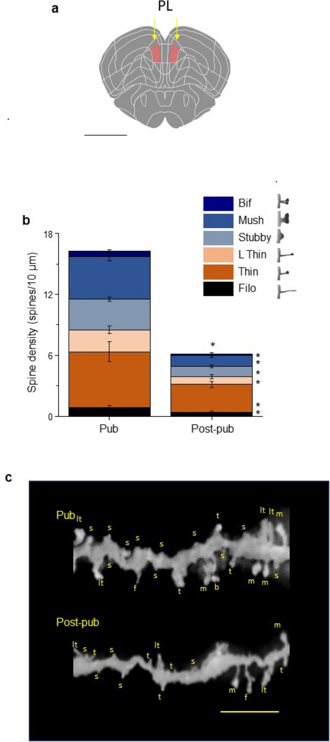
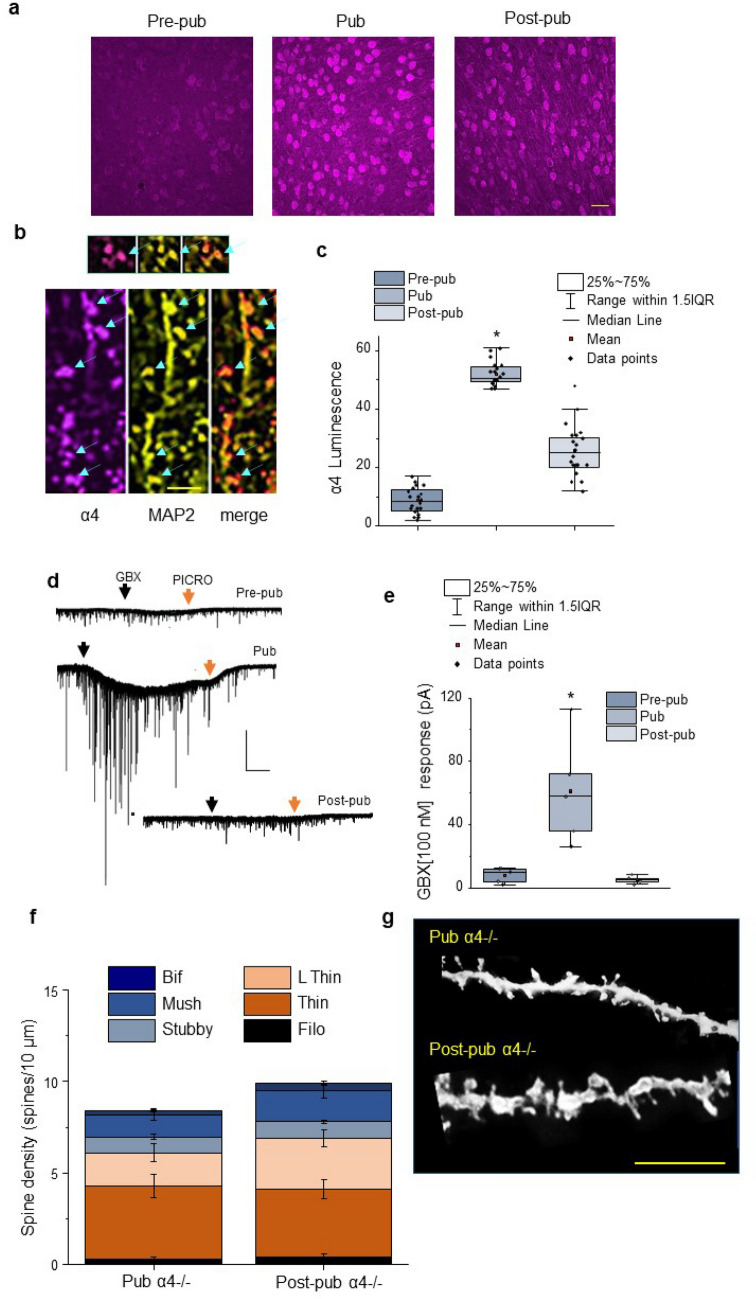
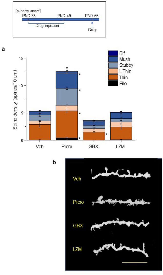
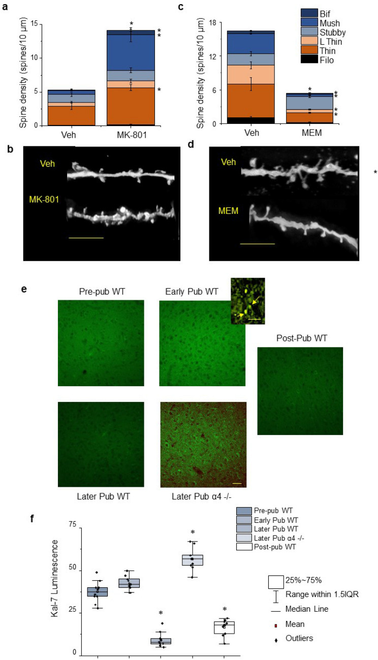
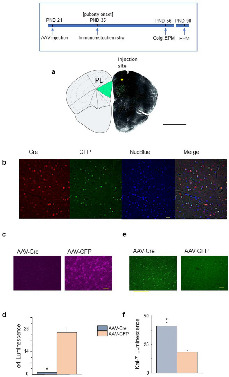
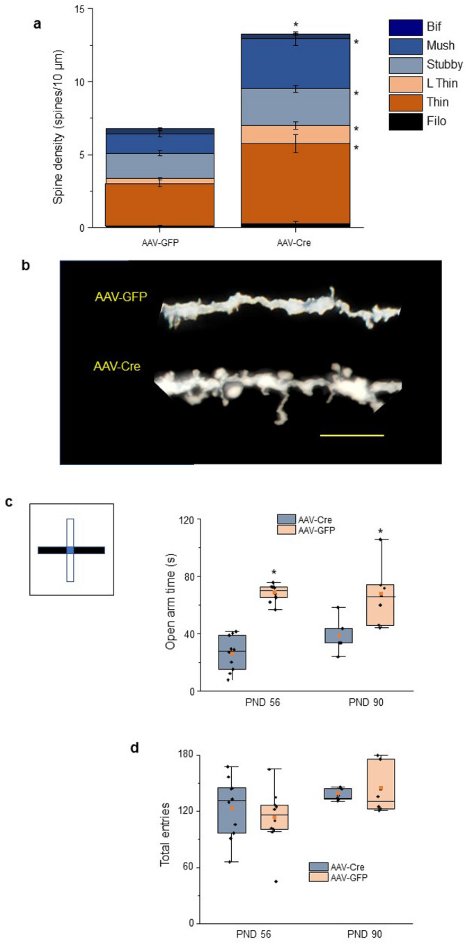
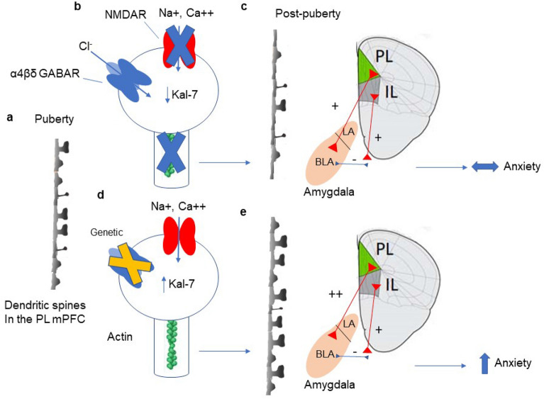
Anxiety is increasingly reported, especially in adolescent females. The etiology is largely unknown, which limits effective treatment. Layer 5 prelimbic cortex (L5PL) increases anxiety responses but undergoes adolescent synaptic pruning, raising the question of the impact of pruning on anxiety. Here we show that preventing L5PL pruning increases anxiety in response to an aversive event in adolescent and adult female mice. Spine density of Golgi-stained neurons decreased ~ 63% from puberty (~ PND35, vaginal opening) to post-puberty (PND56, P < 0.0001). Expression of α4βδ GABAA receptors (GABARs) transiently increased tenfold in L5PL at puberty (P < 0.00001), but decreased post-pubertally. Both global and local knockdown of these receptors during puberty prevented pruning, increasing spine density post-pubertally (P < 0.0001), an effect reversed by blocking NMDA receptors (NMDARs). Pubertal expression of the NMDAR-dependent spine protein kalirin7 decreased (50%, P < 0.0001), an effect prevented by α4 knock-out, suggesting that α4βδ-induced reductions in kalirin7 underlie pruning. Increased spine density due to local α4 knockdown at puberty decreased open arm time on the elevated plus maze post-pubertally (62%, P < 0.0001) in response to an aversive stimulus, suggesting that increases in L5PL synapses increase anxiety responses. These findings suggest that prelimbic synaptic pruning is necessary to limit anxiety in adulthood and may suggest novel therapies.


Conflict of interest statement

The authors declare no competing interests.

Figures

Figure 1
Figure 1
Spine density in layer 5 prelimbic cortex (L5 PL) of the female mouse decreases by half during adolescence: assessment of spine-types. (a) Schematic diagram of prelimbic cortex (PL) localization in mouse brain (pink shading, arrows). Coronal section, 2.09 mm anterior to Bregma. Scale, 1 mm. (b) Averaged data, spine density (#spines/10 μm) of layer 5 (L5) PL for pubertal (Pub, ~ PND 35, assessed by vaginal opening) and post-pubertal (Post-pub, PND 56) female mice. Total spines, t(20) = 6.43, *P < 0.0001; bifurcated, t(20) = 3.71, *P = 0.0014; mushroom, t(20) = 6.2, *P < 0.0001; stubby, t(20) = 6.18, *P < 0.0001; long thin, t(20) = 3.46, *P = 0.0025; thin, t(20) = 2.79, *P = 0.0114; filopodia, t(20) = 2.24, *P = 0.037. (c) Representative images of basal dendrites from Golgi-stained neurons, from Pub and Post-pub female mice. Spine-types: f filopodia, lt long thin, t thin, s stubby, m mushroom. Scale, 5 μm. n = 43–44 neurons, 11 mice/group.
Figure 2
Figure 2
Increases in α4βδ GABAR expression in L5 PL at puberty are necessary for synaptic pruning in female mice. (a) Representative images, α4 immunostaining (magenta), L5 PL pyramidal cells from pre-pubertal (pre-pub, left), pubertal (pub, center), and post-pubertal (post-pub, right) female mice (α4, magenta). Scale, 100 μm. Z-stack sequences used for merged images are presented in Supp. Figure 1. (b) Representative images, α4 (magenta, left), MAP2 (yellow, middle), and merged (orange, right) on the dendrites and spines (arrows) of pyramidal cell in L5 PL. from a pubertal mouse. Inset, Co-localization of α4 and MAP2 on a spine (arrow). Scale, 2 μm. (c) Averaged data, mean, median, and interquartile range (IQR). F(2,57) = 248.9, P < 0.00001, *P < 0.05 vs. other groups. n = 15 neurons, 10 mice/group. (d) Representative whole cell voltage-clamp recordings of L5 PL pyramidal cell response to 100 nM gaboxadol (GBX, black arrow) from Pre-pub, Pub, and Post-pub female mice. PICRO (picrotoxin, 100 μM, red arrow). Scale, 50 s, 200 pA. (e) Averaged data, mean, median, interquartile range, and individual data points for GBX responses. F(2,12) = 12.28, *P = 0.00125. *P < 0.05 vs. other groups. n = 5 mice/group. (f) Averaged data, spine density (#spines/10 μm) of L5 PL from a Pub and Post-pub female α4−/− mouse. (g) Representative images, Golgi-stained dendrites from pub and post-pub female α4−/− mice. Scale, 5 μm. n = 13–31 neurons, 8 mice/group.
Figure 3
Figure 3
Pharmacological manipulation of GABAR-gated current during puberty alters spine density post-pubertally. Inset, timeline of drug administration during the pubertal period of high α4 expression (PND 35–49) using picrotoxin (PICRO, 3 mg/kg, i.p.), gaboxadol (GBX, 0.1 mg/kg,i.p.), lorazepam (LZM, 0.25 mg/kg, i.p.) or vehicle (VEH). Mice were euthanized for Golgi procedures on PND 56. (a) Averaged data, Total spines, F(3,22) = 119.5, P < 0.0001 [Veh vs. Picro, *P < 0.0001]; mushroom, F(3,22) = 25.53, P < 0.0001 [Veh vs. Picro, *P < 0.0001]; stubby, F(3,22) = 43.76, P < 0.0001 [Veh vs. Picro, *P < 0.0001]; thin, F(3,22) = 15.45, P < 0.0001 [Veh vs. Picro, *P = 0.0019; Veh vs. GBX, *P = 0.0445]; filopodia, F(3,22) = 4.71, P = 0.011 [Veh vs. Picro, *P < 0.02]. (b) Representative images of basal dendrites from Golgi-stained neurons from Post-pub female WT mice treated with the indicated drugs during puberty. Scale, 10 μm. n = 14–23 neurons, 7 mice/group.
Figure 4
Figure 4
Pubertal state and α4 knock-out: Effects on NMDA receptor-regulated pruning and expression of the spine protein kalirin-7. (a) Averaged data, spine density (#spines/10 μm) of post-pubertal (PND 56) female WT mice treated during the pubertal period (PND 35–49, inset) with MK-801 (0.1 mg/kg, i.p.) which paradoxically increases NMDA receptor expression in mPFC (Supp. Figure 3). Total spines, t(12) = 5.32, *P = 0.0002; bifurcated, t(12) = 4.16, *P = 0.0002; mushroom, t(12) = 4.53, *P = 0.0007; stubby, t(12) = 1.1, P = 0.206; thin, t(12) = 3.76, *P = 0.0027; long thin, t(12) = 1.25, *P = 0.0027. (b) Representative images of basal dendrites from Golgi-stained neurons, from the indicated groups. Scale, 5 μm. n = 18–21 neurons, 7 mice/group. (c) Averaged data, spine density of post-pubertal female α4−/− mice treated during the pubertal period (PND 35–49) with memantine (0.1 mg/kg, i.p.) an NMDA receptor antagonist. Total spines, t(18) = 4.54, *P = 0.0003; bifurcated, t(18) = 2.6, *P = 0.018; mushroom, t(18) = 3.87, *P = 0.0011; long thin, t(18) = 4.53, *P = 0.0003; thin, t(18) = 3.69, *P = 0.0017; filopodia, t(18) = 5.46, *P < 0.0001. (d) Representative images of basal dendrites from Golgi-stained neurons, from the indicated groups. Scale, 5 μm. n = 29–41 neurons, 10 mice/group. (e) Representative images, kalirin-7 (Kal-7, green) immunostaining of L5 PL from pre-pubertal (pre-pub), early pubertal (early pub, day of vaginal opening, later pubertal (later pub) and post-pubertal (post-pub) WT female mice as well as from a pubertal α4−/− mouse (Later Pub α4−/−). Scale, 100 μm. Inset, spine localization of Kal-7. Scale, 4 μm. (Z-stack sequences used for merged images are presented in Supp. Figure 4). (f) Averaged data, mean, median and interquartile range (IQR) from the indicated groups. F(4,45) = 139, P < 0.0001; *P < 0.05 vs. other unstarred groups. n = 20 neurons, 9 mice/group.
Figure 5
Figure 5
Local infusion of AAV-Cre into PL on PND 21 results in Cre expression and α4 knockdown in PND 35 female mice. Inset, Timeline showing the day of AAV injection (PND 21) when transgenic female mice expressing LoxP sites flanking the GABRA4 gene were injected bilaterally into the PL with adeno-associated virus (AAV)-Cre recombinase or AAV-green fluorescent protein (GFP) and testing (PND 35, α4 immunohistochemistry; PND 56, Golgi spine protocol; PND 56 and 90, anxiety tested using the EPM). (a) Left, schematic showing PL (green). Right, Representative image at PND 35 of GFP staining restricted to the PL (green) boundaries following injection of AAV-Cre/GFP at PND 21. Scale, 2067 μm. This coronal slice was taken 1.767 μm anterior to Bregma. (b) Representative images (×40) at PND 35 following injection of AAV-Cre/GFP at PND 21 of Cre recombinase (Cre) immunostaining (left to right, Cre, GFP, nuclear blue (Nuc blue), merged); Scale, 50 μm. (c) Representative images of α4 immunostaining at PND 35 after infusion of AAV-Cre or AAV-GFP on PND 21 . Scale, 100 μm. (Z-stack sequence, Supp. Figure 5). (d) Averaged data, t(18) = 7.27, *P < 0.00001. n = 10 neurons, 5 mice/group. (e) Representative images of Kal-7 immunostaining after infusion of AAV-Cre or AAV-GFP on PND 21. Scale, 100 μm. (Z-stack sequence, Supp. Figure 6). (f) Averaged data, t(18) = 6.36, *P < 0.00001. n = 10 neurons, 4 mice/group.
Figure 6
Figure 6
Local α4 knockdown at puberty increases spine density of L5 PL pyramidal cells at PND 56 and increases the anxiety response to an aversive stimulus. (a) Averaged data, spine density (#spines/10 μm) in post-pubertal (PND 56) female L5 PL following local α4 knockdown with AAV-Cre (right) compared to the AAV-GFP control (left). Total spines, t(16) = 4.94, *P = 0.0001; mushroom, t(16) = 3.89, *P = 0.0013; stubby, t(16) = 2.38, *P = 0.03; thin, t(16) = 3.63, *P = 0.0023; long thin, t(16) = 3.13, *P = 0.0065. (b) Representative images of basal dendrites from Golgi-stained neurons from the indicated groups. Scale, 5 μm. n = 36 neurons, 9 mice/group. (c) Inset, schematic drawing of elevated plus maze (EPM; arms, 30 cm long, 5 cm wide, elevated 40 cm): white, open arms, half enclosed by walls 7 cm high, extending 15 cm from the center; black, closed arms enclosed by walls 15.25 cm high for the entire length of the arm. (c) Mean (red square), median line, 25–75% interquartile range (IQR), SEM, and individual data points for open arm time in the EPM for PND 56 or PND 90 female mice injected with AAV-GFP or AAV-Cre in the PL on PND 21. PND 56, t(18) = 9.97, *P < 0.00001; PND 90, t(9) = 2.45, *P = 0.0366. A decrease in open arm time is an increase in avoidance behavior which reflects anxiety. (d) Mean (red square), median line, 25–75% interquartile range (IQR), SEM, and individual data points for total crossings in the EPM, measuring locomotor activity. n = 10 mice/group (PND 56); n = 5–6 mice/group (PND 90).
Figure 7
Figure 7
Summary figure. (a) Schematic diagram showing representative dendritic spines in L5 PL at puberty. (b) Normally, expression of α4βδ GABARs increases on the spine at puberty where they gate a Cl current that is inhibitory and impairs activation of NMDARs, which gate Na+ and Ca++ conductances. This decreases Kal-7 expression, resulting in actin de-stabilization, which causes synaptic pruning of L5 PL pyramidal cells. (c) The decrease in spine density represents a loss of synaptic input to L5 PL post-pubertally, which reduces excitatory output to the basolateral amygdala. Because this is offset by inhibitory input from the IL, anxiety behavior is unchanged in late adolescence. (d) When α4βδ GABAR expression is reduced at puberty (genetically or pharmacologically), NMDARs can generate Kal-7 expression, which stabilizes the actin cytoskeleton, preventing pruning. (e) This increased spine density of L5 PL would increase activation of the basolateral amygdala, thereby increasing anxiety in late adolescence. PL prelimbic mPFC, IL infralimbic mPFC, LA lateral amygdala, BLA basolateral amygdala.

Similar articles

Cited by

References

    1. Kessler RC, et al. Lifetime prevalence and age-of-onset distributions of DSM-IV disorders in the National Comorbidity Survey Replication. Arch. Gen. Psychiatry. 2005;62:593–602. doi: 10.1001/archpsyc.62.6.593. - DOI - PubMed
    1. Hantsoo L, Epperson CN. Anxiety disorders among women: A female lifespan approach. Focus. 2017;15:162–172. doi: 10.1176/appi.focus.20160042. - DOI - PMC - PubMed
    1. Goodwin RD, Weinberger AH, Kim JH, Wu M, Galea S. Trends in anxiety among adults in the United States, 2008–2018: Rapid increases among young adults. J. Psychiatr. Res. 2020;130:441–446. doi: 10.1016/j.jpsychires.2020.08.014. - DOI - PMC - PubMed
    1. National College Health Assessment (2008–2019). https://www.acha.org/NCHA/ACHA-NCHA_Data/Publications_and_Reports. Accessed Mar 2021
    1. Hayward C, Sanborn K. Puberty and the emergence of gender differences in psychopathology. J. Adolesc. Health. 2002;30:49–58. doi: 10.1016/S1054-139X(02)00336-1. - DOI - PubMed

Publication types

Substances