Review
. 2022 Jan;18(1):23-42.
doi: 10.1038/s41574-021-00575-1. Epub 2021 Oct 26.

Adaptive immune cells shape obesity-associated type 2 diabetes mellitus and less prominent comorbidities

Affiliations
Review

Adaptive immune cells shape obesity-associated type 2 diabetes mellitus and less prominent comorbidities

Sara SantaCruz-Calvo et al. Nat Rev Endocrinol. 2022 Jan.

Abstract

Obesity and type 2 diabetes mellitus (T2DM) are increasing in prevalence owing to decreases in physical activity levels and a shift to diets that include addictive and/or high-calorie foods. These changes are associated with the adoption of modern lifestyles and the presence of an obesogenic environment, which have resulted in alterations to metabolism, adaptive immunity and endocrine regulation. The size and quality of adipose tissue depots in obesity, including the adipose tissue immune compartment, are critical determinants of overall health. In obesity, chronic low-grade inflammation can occur in adipose tissue that can progress to systemic inflammation; this inflammation contributes to the development of insulin resistance, T2DM and other comorbidities. An improved understanding of adaptive immune cell dysregulation that occurs during obesity and its associated metabolic comorbidities, with an appreciation of sex differences, will be critical for repurposing or developing immunomodulatory therapies to treat obesity and/or T2DM-associated inflammation. This Review critically discusses how activation and metabolic reprogramming of lymphocytes, that is, T cells and B cells, triggers the onset, development and progression of obesity and T2DM. We also consider the role of immunity in under-appreciated comorbidities of obesity and/or T2DM, such as oral cavity inflammation, neuroinflammation in Alzheimer disease and gut microbiome dysbiosis. Finally, we discuss previous clinical trials of anti-inflammatory medications in T2DM and consider the path forward.


Conflict of interest statement

Competing interests

The authors declare no competing interests

Figures
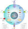
Figure 1:
Figure 1:. Insulin signalling in activated T cells favors the expansion of effector T cells over TReg.
Insulin binding activates the insulin receptor by autophosphorylation of tyrosine residues on the intracellular tail that allows binding and phosphorylation of the adaptor protein IRS1. IRS1 activates PI3K, which in turn catalyzes conversion of plasma membrane PIP2 to PIP3. PIP3 subsequently activates AKT, which ultimately stimulates glucose internalization by GLUT1 and downstream glycolysis. Insulin stimulation upon TCR activation favors the expansion of effector T cells (left side, teal) by promoting IFNγ expression and AKT–mTOR-mediated expression of MYC. Later, MYC mediates the transcription of several transporters (including GLUT1 and the glutamine transporter) that regulate glycolysis, mitochondrial function glutamine translocation and metabolism. Furthermore, TReg insulin receptor stimulation will limit TReg (right side, light blue) clonal expansion by reducing glycolysis and IL-10 expression, and instead promoting IFNγ expression. Text boxes summarize the main steps driving effector T cell proliferation in lean conditions and the downregulation of TReg in conditions of obesity upon TCR stimulation and insulin receptor activation. AKT, protein kinase B; GLUT1, glucose transporter 1; IFNγ, interferon gamma; IRS1, insulin receptor substrate 1; PI3K, phosphoinositide 3 kinase; PIP2, phosphatidyl-inositol-4,5-biphosphate; PIP3, phosphatidyl-inositol-4,5-triphosphate; TNF, tumour necrosis factor; TCR: T cell receptor; TReg, regulatory T cell.
Figure 2:
Figure 2:. Adipocyte leptin promotes lymphocyte activation and TH17 differentiation.
In mouse effector T cells (left side, teal), leptin signalling promotes CD4+ T cell differentiation to TH17 by promoting STAT3-activated expression of the genes encoding the transcription factor RORγt (RORC in human) and the cytokine IL-17, which are both characteristic factors expressed by TH17. In effector B cells (right side, purple) leptin receptor stimulation by leptin activates JAK2–STAT3-dependent expression of cytokines from B cells, including IL-10, IFNγ and TNF. Leptin signalling in both B and T cells additionally activates the PI3K–Akt signalling pathway outlined in Figure 1 and shown again here, which ultimately favours glycolysis to increase effector cell expansion in response to BCR (in B cells) or TCR (in T cells) engagement. AKT, protein kinase B; BCR, B cell receptor; GLUT1, glucose transporter 1; IFNγ: interferon gamma; r; PI3K: phosphoinositide 3 -kinase;; PIP2: phosphatidyl-inositol-4,5-biphosphate; PIP3: phosphatidyl-inositol-4,5-triphosphate; TCR, T cell receptor; TH17, T helper 17 cells; TNF, tumor necrosis factor.
Figure 3.
Figure 3.. Calcium signalling upon TCR or BCR activation.
T cells and B cells rely on sustained intracellular concentration of calcium (i[Ca2+]) to activate development, proliferation, and differentiation. TCR activation in T cells and BCR engagement in B cells triggers several outcomes. The cytoskeleton reorganizes to establish the immunological synapse between lymphocytes and antigen presenting cells. Lymphocytes activate PLCγ to trigger the breakdown of PIP2 into DAG and IP3, which binds to its receptor stimulate Ca2+-pool depletion, coupled with inward-Na+ voltage-gated channels. Sustained i[Ca2+] is facilitated by extracellular calcium influx mediated by CRAC in the plasma membrane and STIM1 and STIM2 in the ER through the process termed store-operated Ca2+ entry. Ca2+ stored in the ER is shuttled from the mitochondria that previously buffered cytosolic Ca2+ through the MCU; Mitochondria–ER Ca2+ shuttling is facilitated by contact points (MAMs), formed by IP3R in the ER and VDAC in the mitochondria. Finally, sustained i[Ca2+] promotes calcineurin–camodulin-mediated activation of the nuclear factor of activated T cells that: A | activates the transcription of genes encoding IL-17 and RORγt to activate STAT3 and favour TH17 differentiation; B | activates the expression of the genes encoding the transcription factor FOXP3 and the cytokine IL-2 to activate STAT5 and downstream IL-2 receptor to favour regulatory T cell differentiation and proliferation; C | promotes B cell development together with the NFκB-mediated expression of the anti-apoptotic factor BCL-xL. CRAC, calcium release activated calcium channels; DAG, diacylglycerol; IP3: inositol-1,4,5-triphosphate; IP3R: inositol-1,4,5-triphosphate receptor; STIM1: sensor stromal interaction molecule 1; MCU: mitochondrial calcium uniporter channel; MAMs: mitochondria-associated membranes; PIP2, phosphatidyl-inositol-4,5-biphosphate; STIM1, sensor stromal interaction molecule 1; TGFβ: transforming growth factor β; VDAC, voltage-dependent channels.
Figure 4.
Figure 4.. Insulin resistance in the brain during neuroinflammation.
(A) Insulin resistance in a neuron during neuroinflammation. TNF binding to TNF receptor activates JNK, which phosphorylates multiple serine residues in IRS1. This event limits IRS1 phosphorylation at Tyr-465 and reduces downstream signalling through PI3K and AKT, thereby promoting insulin resistance. Disrupted insulin signalling favours assembly of Aβ-plaques from AβO, which are formed (along with sAPPβ) from cleavage of the precursor molecule APP. Aβ plaques further favour IRS1 phosphorylation on Ser-616, which worsens insulin resistance. The T2DM drugs exendin-4 and liraglutide (both GLP1R agonists) trigger GLP1R to prevent IRS1 Ser-616 phosphorylation and improve insulin signalling. (B) Molecules and immune cell trafficking from circulation across the BBB during neuroinflammation. During T2DM–Alzheimer disease-mediated insulin resistance, the permeability of the BBB (formed by endothelial cells, pericytes, astrocytes and neurons), is altered causing increased trafficking of insulin, TH17, IL-17 and IL-22, B cells along with the accumulation of Aβ-plaques, which ultimately promotes neurodegeneration. Aβ, amyloid beta; AβO, amyloid beta oligomers; AKT, protein kinase B; BBB, blood–brain barrier; GLP1R, glucagon-like peptide 1 receptor; IRS1, insulin receptor substrate 1; PI3K, phosphoinositide 3-kinase; APP, amyloid precursor protein; sAPP, secreted amyloid precursor protein; ; TH17: T helper 17 cells.
Figure 5:
Figure 5:. Taste perception in taste bud cells is subject to immunoregulation.
(A) Type I taste bud cells (left) are glial-like cells with a structural role; they express ENac channels to trigger the response to salts. Type II cells (middle; receptor cells) express G-coupled protein receptors that detect sweet (via TAS1R2 and TAS1R3), umami (via TAS1R1 and TAS1R3) or bitter (TAS2R) and the lipid translocator CD36. Type III cells (right; presynaptic) express PKD2L1 and PKD1L3 (detecting sour) and convey sensory information through the afferent nerve fibers. Taste receptors on type II cells trigger a signalling cascade that starts with the activation the G-protein α-gustducin, which ultimately results in cell depolarization and the release of non-vesicular ATP. Upon ATP-mediated engagement of PX2R in afferent nerves and type III cells, Ca2+-gated channels promote the release of vesicular neurotransmitters (i.e. 5-HT and GABA) to convey sensory information to the brain through the sensory afferent nerves. (B) Crosstalk amongst taste bud cells is driven by regulators of inflammation in obesity-associated T2DM. For example, TNF is secreted by sweet and umami-sensing type II cells. This local TNF triggers aversion to bitter compounds by interacting with bitter-sensing type II cells, and also activates type I and type III cells. IL-10 is secreted by bitter-sensing type II cells and is essential to maintain taste bud structure, perhaps in part by regulating the IL-10 receptor-expressing subset of type II cells (sweet–umami). IFNγ and IFNGR are upregulated by inflammatory stimulation of some type II cells and most type III cells. TLR4 is expressed in all taste bud cell subsets and is also involved in modulation of taste preferences. Additionally, TLR4 interacts with CD36 to facilitate fatty acid uptake and signalling. IFNγ, interferon-γ; IFNGR, interferon-γ receptor; PX2R, purinergic ionotropic receptors; TLR4, toll-like 4 receptor. TNF, tumor necrosis factor.

References

    1. Obesity and overweight. Available at: https://www.who.int/news-room/fact-sheets/detail/obesity-and-overweight. (Accessed: 27th June 2020)
    1. Diabetes. Available at: https://www.who.int/news-room/fact-sheets/detail/diabetes. (Accessed: 27th June 2020)
    1. Singh-Manoux A et al. Obesity trajectories and risk of dementia: 28 years of follow-up in the Whitehall II Study. Alzheimer’s Dement. 14, 178–186 (2018). - PMC - PubMed
    1. Kearns CE & Bero LA Conflicts of interest between the sugary food and beverage industry and dental research organisations: time for reform. The Lancet 394, 194–196 (2019). - PubMed
    1. Wernstedt Asterholm I et al. Adipocyte inflammation is essential for healthy adipose tissue expansion and remodeling. Cell Metab. 20, 103–118 (2014). - PMC - PubMed

Publication types