

Odontogenic myxoma with pain and uncommon histological feature in the mandible: A case report and review the literature
- PMID: 34703133
- PMCID: PMC8491336
- DOI: 10.4103/0973-029X.325240
Odontogenic myxoma with pain and uncommon histological feature in the mandible: A case report and review the literature
Abstract
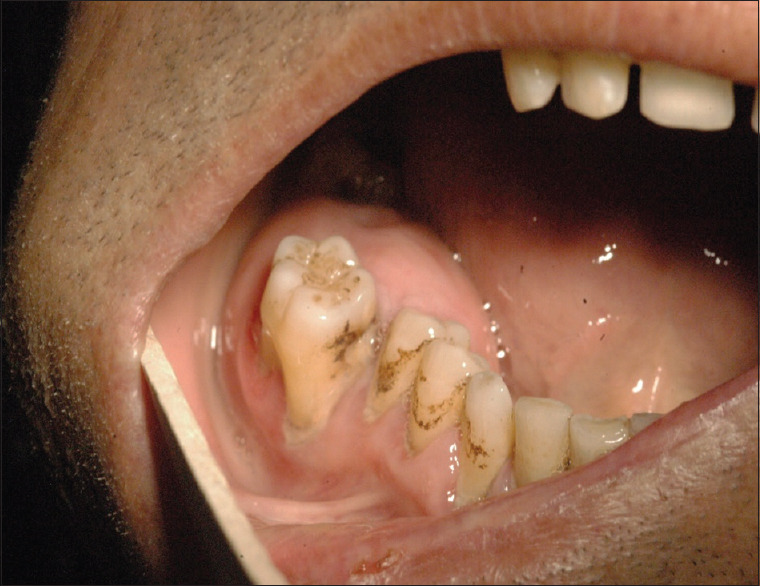
Odontogenic myxoma (OM) is a rare benign painless, slow-growing lesion with local aggressive behavior. Pain and sensory disturbance and fibro-osseous appearance in histopathology have been rarely reported in OM. The authors reported a 52-year-old male case presented with a large gingival mass around a mobile mandibular right first molar extended to the distal aspect of the third molar. Microscopic examination of the incisional and excisional biopsy revealed an OM with numerous newly formed bone or cementum-like material present throughout the specimen like those seen in fibro-osseous lesions. For avoiding to recurrence, a segmental mandibulectomy was performed and a metal plate was inserted to the right mandible defect under general anesthesia. Rehabilitation was completed with the placement of implants. We review and discuss about this variety.
Keywords: Bone disease; case report; odontogenic tumor.
Copyright: © 2021 Journal of Oral and Maxillofacial Pathology.
Conflict of interest statement
There are no conflicts of interest.
Figures


References
-
- Ansari A, Thomas A, Bohra R, Vare R, Rathod J, Dongardive SJOCR. Recurrent myxoma of the maxilla: A rare case. J Otolaryngology Case Reports. 2019;10:1–4.
-
- Thabusum DA, Rajesh N, Reddy RS, Ravikanth M, Raju US. Odontogenic myxoma of maxilla–A rare case report. WJPMR. 2017;3:282–5.
-
- Thoma KH, Goldman HM. Central myxoma of the jaw. Oral Surg Oral Med Oral Pathol. 1947;33:B532–40. - PubMed
Publication types
LinkOut - more resources
Full Text Sources