. 2021 Aug 20;20(3):215-221.
doi: 10.4103/wjnm.WJNM_116_20. eCollection 2021 Jul-Sep.

Diagnostic performance of 18F-fluorodeoxyglucose positron emission tomography/computed tomography in anorectal melanoma

Affiliations

Diagnostic performance of 18F-fluorodeoxyglucose positron emission tomography/computed tomography in anorectal melanoma

Ajinkya N Bakare et al. World J Nucl Med. .

Abstract

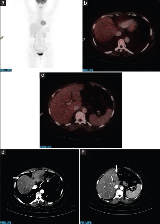
To evaluate the diagnostic role of 18F-fluorodeoxyglucose positron emission tomography/computed tomography (PET/CT) in initial staging and restaging of anorectal melanoma. This was a single-institution, retrospective observational study; patients for initial staging and with clinical or radiological suspicion of disease recurrence referred for PET/CT between January 2006 and December 2015 were included in the study. Diagnostic performance of PET/CT was evaluated for baseline staging and disease recurrence. A total of 61 patients who were referred for initial staging were included. PET/CT correctly detected primary lesion in 57 (93.44%) cases, regional nodes in 46 (75.4%) cases, nonregional nodes in 22 (36%) cases, and distant metastases in 25 (41%) cases. The sensitivity (SN); specificity (SP); positive predictive value (PPV); negative predictive value (NPV); and accuracy for primary lesion, regional nodes, nonregional nodes, and distant metastases were 96.6%, 100%, 100%, 50%, and 96.7%; 97.9%, 100%, 100%, 93.3%, and 98.4%; 100%, 100%, 100%, 100%, and 100%; and 100%, 100%, 100%, 100%, and 100%, respectively. A total of 24 patients were included for suspected recurrence/restaging. All the patients were treated previously by surgery, radiotherapy, or chemotherapy. PET/CT detected disease recurrence in 20 (83.3%) patients. Ten patients had recurrence at the primary site, 8 of whom also had distant metastases and 2 had only locoregional metastatic nodes. In the remaining 10 patients, there was no primary site recurrence; however, 2 patients had locoregional nodal and distant metastases and 8 patients had only distant metastases. PET/CT was false negative in 1 patient, which missed liver metastasis. SN, SP, PPV, and NPV of PET/CT was found to be 95%, 100%, 100%, and 75%, respectively, with accuracy of 96%. PET/CT demonstrates overall high diagnostic accuracy in the initial staging and detection of recurrent disease in cases of anorectal melanoma.

Keywords: Accuracy; anorectal; diagnostic; melanoma; positron emission tomography/computed tomography.


Conflict of interest statement

There are no conflicts of interest.

Figures

Figure 1
Figure 1
A 52-year-old patient presented with bleeding per rectum. Biopsy revealed features consistent with malignant melanoma. FDG PET/CT was done as baseline imaging. MIP image (a) shows no abnormal focal increased tracer uptake in the pelvic region. No focus of hypermetabolism or soft tissue lesion is seen in the anal canal region. Postsurgery histopathology results revealed submucosal malignant melanoma of rectum with reactive regional nodes. Thus, PET/CT showed false-negative results at the primary tumor site. MIP (b) and fused (c) images show focal uptake in the reactive right hilar node. FDG: Fluorodeoxyglucose; PET/CT: Positron emission tomography/computed tomography; MIP: Maximum Intensity Projection
Figure 2
Figure 2
A 41-year-old patient presented with blood in stools, pain in perianal area, and weight loss. Biopsy revealed malignant tumor which had epithelioid and slightly spindle-shaped appearance. Immunohistochemistry was positive for HMB 45 and S100 proteins. MIP image (a) shows increased FDG tracer uptake in the pelvis and below it. Fused transaxial PET/CT images reveal FDG-avid growth in the anal canal (b) with metastatic right internal iliac node (c) and perirectal nodule (d). FDG: Fluorodeoxyglucose; PET/CT: Positron emission tomography/computed tomography; MIP: Maximum Intensity Projection
Figure 3
Figure 3
A 33-year-old patient presented with bleeding per rectum. Rectal growth biopsy is suggestive of malignant melanoma. Immunohistochemistry revealed tumor positive for HMB 45 and S100 protein. FDG PET/CT scan was done for initial staging. MIP images (a) show increased tracer uptake at multiple sites in the pelvis, liver, and multiple vertebrae. Fused transaxial PET/CT images show large anorectal mass (b), metastatic left internal iliac node (c), bilateral inguinal nodes (d), metastatic liver lesions (e), skeletal lesions (f), and lung nodules(g). The patient was considered for palliative chemoradiotherapy. FDG: Fluorodeoxyglucose; PET/CT: Positron emission tomography/computed tomography; MIP: Maximum Intensity Projection
Figure 4
Figure 4
A 89-year-old patient presented with biopsy proven anal malignant melanoma. PET/CT scan was for restaging in the view of growth detected on per rectal examination. MIP image (a) shows focal increased tracer uptake in the pelvis, liver, right lung, and dorsal vertebra. Fused transaxial FDG PET/CT images show anorectal soft tissue mass (short arrow) and left inguinal node (long arrow) (b); pararectal nodule (c); liver metastases (d), metastatic dorsal vertebral lesion (e); and lung nodules (f). The patient was considered for palliative chemotherapy. FDG: Fluorodeoxyglucose; PET/CT: Positron emission tomography/computed tomography; MIP: Maximum Intensity Projection
Figure 5
Figure 5
A 53-year-old patient initially presented with pain in abdomen and underwent anal polypectomy. Biopsy report revealed anorectal melanoma with immunohistochemistry results positive for HMB 45 and S100 protein. Follow-up USG done revealed left liver lobe hypoechoic suspicious lesion. USG done six months later showed increase in the size of suspicious left liver lobe lesion. FNAC revealed metastatic malignant melanoma consistent with known primary in the anorectum. FDG PET/CT done for restaging showed no abnormal focus of increased tracer uptake in the scan as seen in MIP (a) and fused transaxial images (b and c); triphasic diagnostic CT scan done a week after shows subcentimeter size enhancing liver lesions in segment VIII and III (d and e), best appreciated in portovenous phase. Thus, PET/CT did not detect the subcentimeter size metastatic liver lesions. FDG: Fluorodeoxyglucose; PET/CT: Positron emission tomography/computed tomography; USG: Ultrasonography; FNAC: Fine-needle aspiration cytology

References

    1. Stefanou A, Nalamati SP. Anorectal Melanoma. Clin Colon Rectal Surg. 2011;24:171–6. - PMC - PubMed
    1. Row D, Weiser MR. Anorectal melanoma. Clin Colon Rectal Surg. 2009;22:120–6. - PMC - PubMed
    1. Singer M, Mutch MG. Anal melanoma. Clin Colon Rectal Surg. 2006;19:78–87. - PMC - PubMed
    1. Meguerditchian AN, Meterissian SH, Dunn KB. Anorectal melanoma: Diagnosis and treatment. Dis Colon Rectum. 2011;54:638–44. - PubMed
    1. Gilchrest BA, Eller MS, Geller AC, Yaar M. The pathogenesis of melanoma induced by ultraviolet radiation. N Engl J Med. 1999;340:1341–8. - PubMed