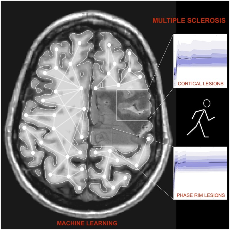
Cortical and phase rim lesions on 7 T MRI as markers of multiple sclerosis disease progression
- PMID: 34704024
- PMCID: PMC8361394
- DOI: 10.1093/braincomms/fcab134
Cortical and phase rim lesions on 7 T MRI as markers of multiple sclerosis disease progression
Abstract
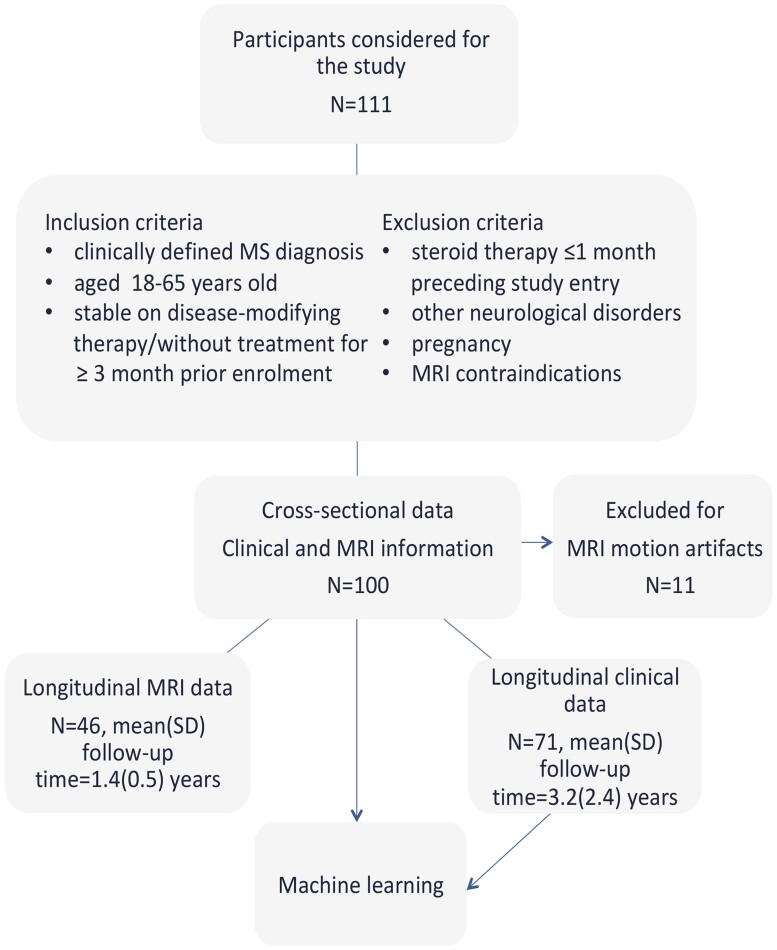
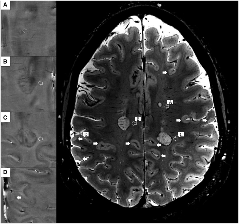
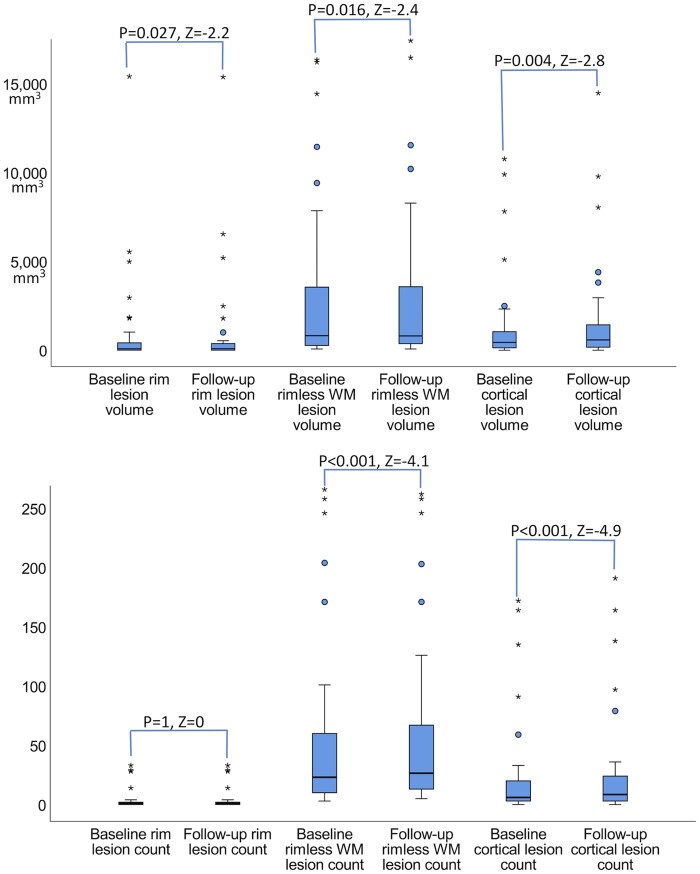
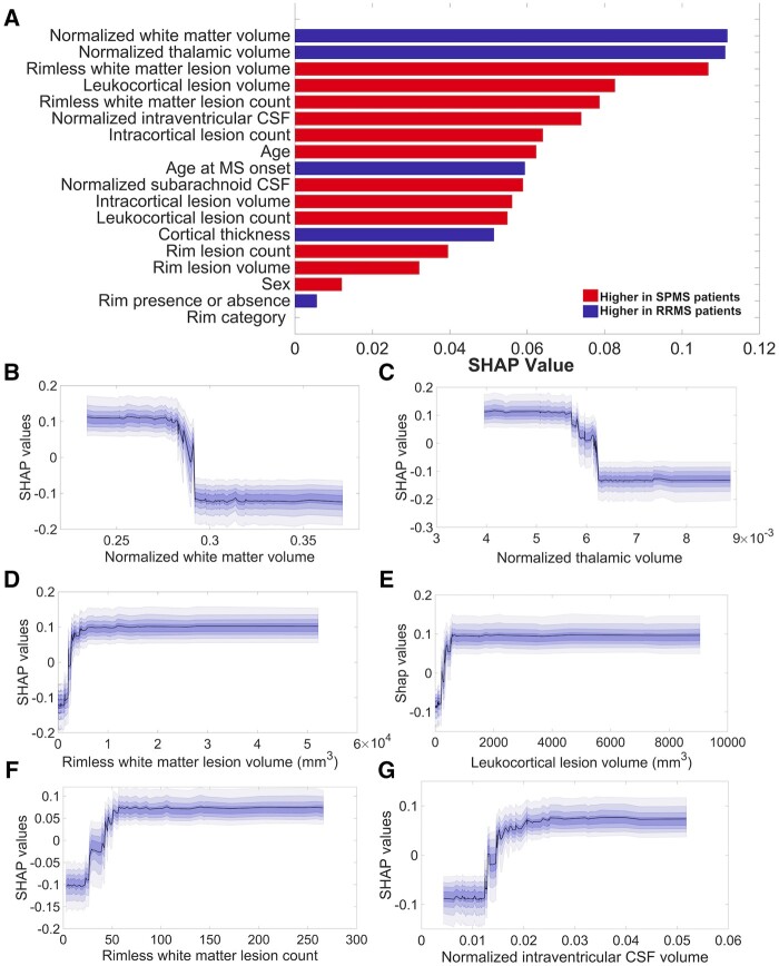
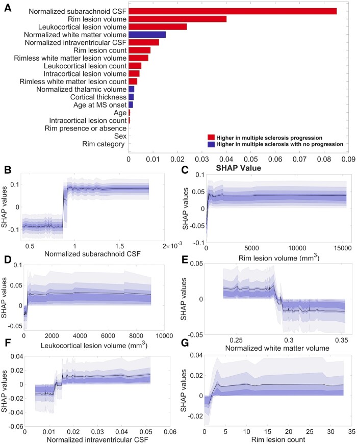
In multiple sclerosis, individual lesion-type patterns on magnetic resonance imaging might be valuable for predicting clinical outcome and monitoring treatment effects. Neuropathological and imaging studies consistently show that cortical lesions contribute to disease progression. The presence of chronic active white matter lesions harbouring a paramagnetic rim on susceptibility-weighted magnetic resonance imaging has also been associated with an aggressive form of multiple sclerosis. It is, however, still uncertain how these two types of lesions relate to each other, or which one plays a greater role in disability progression. In this prospective, longitudinal study in 100 multiple sclerosis patients (74 relapsing-remitting, 26 secondary progressive), we used ultra-high field 7-T susceptibility imaging to characterize cortical and rim lesion presence and evolution. Clinical evaluations were obtained over a mean period of 3.2 years in 71 patients, 46 of which had a follow-up magnetic resonance imaging. At baseline, cortical and rim lesions were identified in 96% and 63% of patients, respectively. Rim lesion prevalence was similar across disease stages. Patients with rim lesions had higher cortical and overall white matter lesion load than subjects without rim lesions (P = 0.018-0.05). Altogether, cortical lesions increased by both count and volume (P = 0.004) over time, while rim lesions expanded their volume (P = 0.023) whilst lacking new rim lesions; rimless white matter lesions increased their count but decreased their volume (P = 0.016). We used a modern machine learning algorithm based on extreme gradient boosting techniques to assess the cumulative power as well as the individual importance of cortical and rim lesion types in predicting disease stage and disability progression, alongside with more traditional imaging markers. The most influential imaging features that discriminated between multiple sclerosis stages (area under the curve±standard deviation = 0.82 ± 0.08) included, as expected, the normalized white matter and thalamic volume, white matter lesion volume, but also leukocortical lesion volume. Subarachnoid cerebrospinal fluid and leukocortical lesion volumes, along with rim lesion volume were the most important predictors of Expanded Disability Status Scale progression (area under the curve±standard deviation = 0.69 ± 0.12). Taken together, these results indicate that while cortical lesions are extremely frequent in multiple sclerosis, rim lesion development occurs only in a subset of patients. Both, however, persist over time and relate to disease progression. Their combined assessment is needed to improve the ability of identifying multiple sclerosis patients at risk of progressing disease.
Keywords: MRI; cortical lesions; machine learning; multiple sclerosis; phase rim lesions.
© The Author(s) (2021). Published by Oxford University Press on behalf of the Guarantors of Brain. All rights reserved. For permissions, please email: journals.permissions@oup.com.
Figures