. 2021 Oct 31;9(2):e0047021.
doi: 10.1128/Spectrum.00470-21. Epub 2021 Oct 27.

Deciphering the Role of Mucosal Immune Responses and the Cervicovaginal Microbiome in Resistance to HIV Infection in HIV-Exposed Seronegative (HESN) Women

Affiliations

Deciphering the Role of Mucosal Immune Responses and the Cervicovaginal Microbiome in Resistance to HIV Infection in HIV-Exposed Seronegative (HESN) Women

Sivasankaran Munusamy Ponnan et al. Microbiol Spectr. .

Abstract

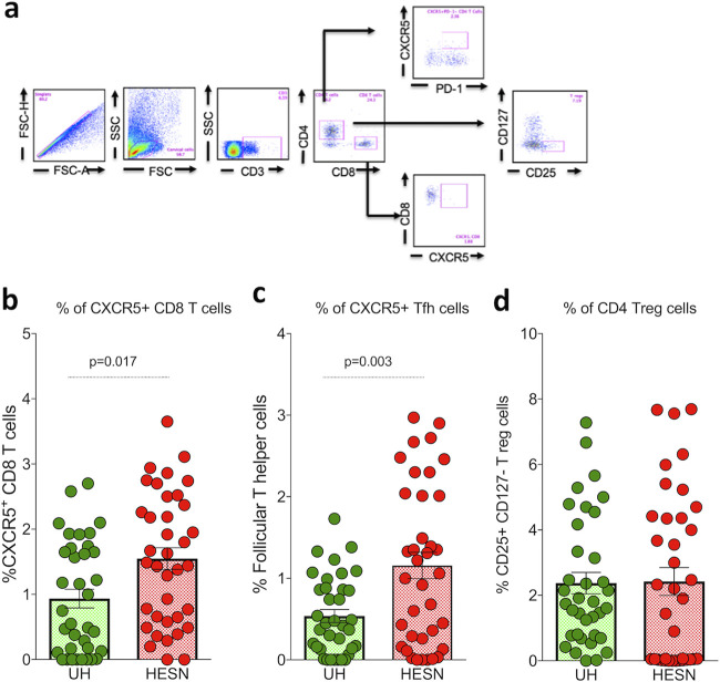
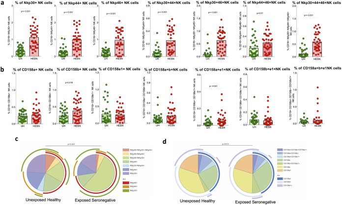
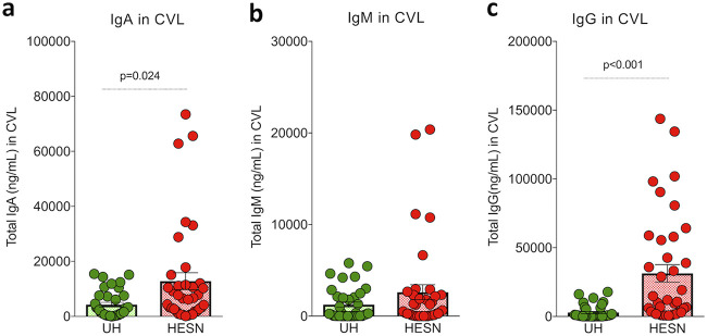
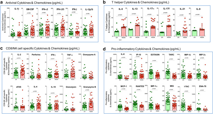
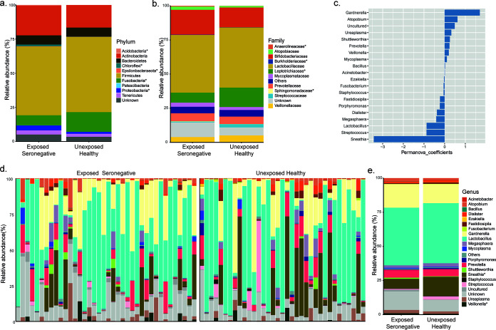
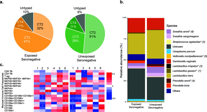
The female genital tract (FGT) is an important site of human immunodeficiency virus (HIV) infection. Discerning the nature of HIV-specific local immune responses is crucial for identifying correlates of protection in HIV-exposed seronegative (HESN) individuals. The present study involved a comprehensive analysis of soluble immune mediators, secretory immunoglobulins (sIg), natural killer (NK) cells, CXCR5+ CD8+ T cells, T follicular helper (Tfh) cells, and T regulatory cells (Tregs) in the vaginal mucosa as well as the nature and composition of the cervicovaginal microbiome in HESN women. We found significantly elevated antiviral cytokines, soluble immunoglobulins, and increased frequencies of activated NK cells, CXCR5+ CD8+ T cells, and Tfh cells in HESN females compared to HIV-unexposed healthy (UH) women. Analysis of the genital microbiome of HESN women revealed a greater bacterial diversity and increased abundance of Gardnerella spp. in the mucosa. The findings suggest that the female genital tract of HESN females represents a microenvironment equipped with innate immune factors, antiviral mediators, and critical T cell subsets that protect against HIV infection. IMPORTANCE The vast majority of human immunodeficiency virus (HIV) infections across the world occur via the sexual route. The genital tract mucosa is thus the primary site of HIV replication, and discerning the nature of HIV-specific immune responses in this compartment is crucial. The role of the innate immune system at the mucosal level in exposed seronegative individuals and other HIV controllers remains largely unexplored. This understanding can provide valuable insights to improve vaccine design. We investigated mucosal T follicular helper (Tfh) cells, CXCR5+ CD8+ T cells, natural killer (NK) cells subsets, soluble immune markers, and microbiome diversity in HIV-exposed seronegative (HESN) women. We found a significantly higher level of mucosal CXCR5+ CD8+ T cells, CD4+ Tfh cells, activated NK cell subsets, and antiviral immune cell mediators in HESN women. We also found a higher abundance of Gardnerella spp., microbiome dysbiosis, and decreased levels of inflammatory markers to be associated with reduced susceptibility to HIV infection. Our findings indicate that increased distribution of mucosal NK cells, CXCR5+ CD8+ T cells, Tfh cells, and soluble markers in HIV controllers with a highly diverse cervicovaginal microbiome could contribute effectively to protection against HIV infection. Overall, our findings imply that future vaccine design should emphasize inducing these highly functional cell types at the mucosal sites.

Keywords: B cells; CBA; CXCR5+ CD8+ cells; HESN; NK cells; TSCM cells; Tfh cells; cervicovaginal lavage; cervicovaginal microbiota; cytobrush; memory B cells.


Figures

FIG 1
FIG 1
Mucosal CXCR5+ CD8+ T cells, T follicular helper cells and Tregs. (a) Representative flow plots showing the percent frequency of CXCR5+ T cells and Tregs in the study groups (HESN, HIV-exposed seronegative [N = 37]; UH, HIV-unexposed seronegative [N = 35]). (b) Figure showing the cumulative frequency of CXCR5+ CD8+ T cells in the HESN and UH groups. (c) Figure showing the cumulative frequency of T follicular helper cells in the HESN and UH groups. (d) Figure showing the cumulative frequency of CD127 CD25+ T regs in the HESN and UH groups. The scatter dot plots summarize the percent frequency of total CD8+ CXCR5+ T cells, Tfh cells, and Tregs (median, 1st, and 3rd quartiles). P values were determined using the Mann-Whitney test.
FIG 2
FIG 2
Distribution of natural cytotoxicity receptor (NCR)- and killer cell immunoglobulin-like inhibitory receptor (KIR)-coexpressing mucosal NK cells. (a) Proportion of NK cells expressing each activating receptor (Nkp30, Nkp44, Nkp46). (b) Proportion of NK cells expressing each inhibitory receptor (CD158a, CD158b, CD158e1). The scatter dot plots summarize the percent frequency of total NCR- and KIR-expressing NK cells from cytobrush specimens (HESN, HIV-exposed seronegative [N = 37]; UH, HIV-unexposed seronegative [N = 35]). The graphical plots show the median, 25th, and 75th percentiles and interquartile range (IQR). P values were determined using the Mann-Whitney test. (c) SPICE analysis of different combinations of NCR-expressing NK cells indicated statistically significant differences in the permutation of different combinations. (d) SPICE analysis of the different combination of KIR-expressing NK cells indicated statistically significant differences in the permutation of different combinations.
FIG 3
FIG 3
Levels of mucosal immunoglobulins (IgG, IgA, and IgM) in cervical vaginal lavage. Mucosal immunoglobulin (IgG, IgA, and IgM) levels in CVL specimens measured by cytometric bead array and reported in ng/ml. (a) Total IgA level in cervicovaginal lavage. (b) Total IgM level in cervicovaginal lavage. (c) Total IgG level in cervicovaginal lavage. The box-and-whisker plots represent median, 25th, and 75th percentiles and range (IQR). P values were determined using the Mann-Whitney test. (HESN, HIV-exposed seronegative [N = 37]; UH, HIV-unexposed seronegative [N = 35]).
FIG 4
FIG 4
Levels of antiviral mediators, T helper cell-specific and CD8+ T cell/NK cell-specific effector molecules, cytokines and chemokines in the cervicovaginal lavage. Levels of (a) antiviral molecules (IL-1, GM-CSF, IFN-2, IFN-λ2/3, IFN-, IL-12p70), (b) T helper cell cytokines (IL-5, IL-13, IL-17α, IL-17f, IL-21, IL-9), (c) CD8/NK cell specific cytokines (IL-2, perforin, IFN-γ, TNF-α, granzyme A, sFAS, IL-4, IL-10, granulysin, granzyme B) (d) Pro-inflammatory chemokines (IL-8, IP-10, EOTAXIN, TARC, MIP-1β, MIP-3α MCP-1, RANTES, MIP-1, MIG, I-TAC, ENA-78), in the CVL of HESN (N = 37) and UH (N = 35) groups. The data shown is the median level of cytokines and chemokines (pg/ml). The scatter dot plots show the median, 25th, and 75th percentiles and range (IQR). P values were determined using the Mann-Whitney test. HESN, HIV exposed seronegative; UH, HIV unexposed seronegative.
FIG 5
FIG 5
Composition of the cervicovaginal microbiome in the study population. (a) Mean relative abundance in the phylum. (b) Mean relative abundance in family. (c) Top 20 taxa at the genus level that have a significant effect on separating the groups as per PERMANOVA analysis. (d) Visualization of sample-wise relative abundance (%) in the two experimental groups. (e) Mean relative abundance in genus. The asterisk (*) denotes significance in the Mann-Whitney test.
FIG 6
FIG 6
Distribution of bacterial species and their association with inflammation. (a) Distribution of CT2, CT3, and CT4 bacteria in HESN and UH women. (b) Bar graph showing average relative abundance (%) of various species. The asterisk (*) denotes significance in the Mann-Whitney test. (c) Heat map showing Spearman’s correlation values between significant species (from Mann-Whitney test) and mucosal immune cell frequencies. The asterisk (*) denotes a significant correlation.

Similar articles

Cited by

References

    1. Cole AM. 2006. Innate host defense of human vaginal and cervical mucosae. Curr Top Microbiol Immunol 306:199–230. doi:10.1097/01.lgt.0000265775.52044.2b. - DOI - PubMed
    1. Mestecky J, Fultz PN. 1999. Mucosal immune system of the human genital tract. J Infect Dis 179 Suppl 3:S470–S474. doi:10.1086/314806. - DOI - PubMed
    1. Mselle TF, Meadows SK, Eriksson M, Smith JM, Shen L, Wira CR, Sentman CL. 2007. Unique characteristics of NK cells throughout the human female reproductive tract. Clin Immunol 124:69–76. doi:10.1016/j.clim.2007.04.008. - DOI - PubMed
    1. Wang H, Zheng X, Wei H, Tian Z, Sun R. 2012. Important role for NKp30 in synapse formation and activation of NK cells. Immunol Invest 41:367–381. doi:10.3109/08820139.2011.632799. - DOI - PubMed
    1. Kulkarni AG, Paranjape RS, Thakar MR. 2014. Higher expression of activating receptors on cytotoxic NK cells is associated with early control on HIV-1C multiplication. Front Immunol 5:222. doi:10.3389/fimmu.2014.00222. - DOI - PMC - PubMed

Publication types

MeSH terms