Review
. 2021 Dec;47(12):1393-1414.
doi: 10.1007/s00134-021-06548-2. Epub 2021 Oct 27.

Brain injury after cardiac arrest: pathophysiology, treatment, and prognosis

Affiliations
Review

Brain injury after cardiac arrest: pathophysiology, treatment, and prognosis

Claudio Sandroni et al. Intensive Care Med. 2021 Dec.

Abstract

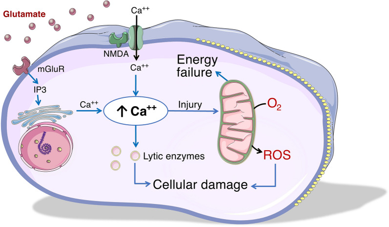
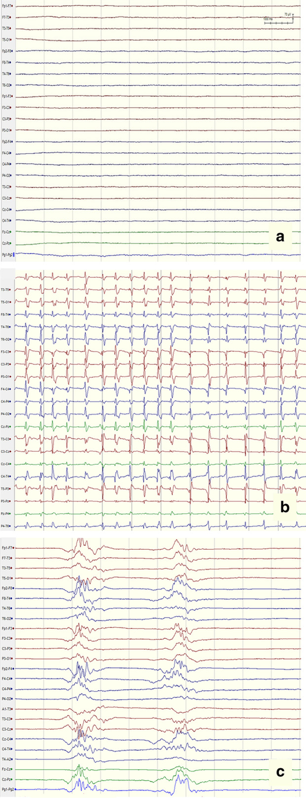
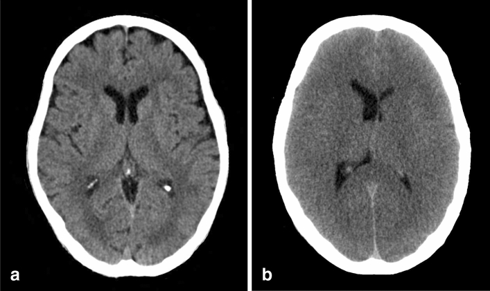
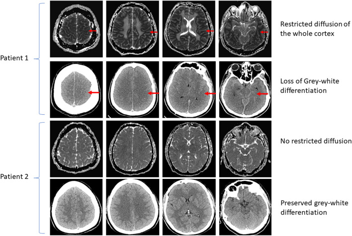
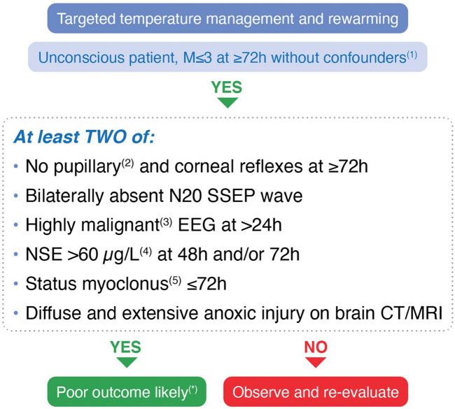
Post-cardiac arrest brain injury (PCABI) is caused by initial ischaemia and subsequent reperfusion of the brain following resuscitation. In those who are admitted to intensive care unit after cardiac arrest, PCABI manifests as coma, and is the main cause of mortality and long-term disability. This review describes the mechanisms of PCABI, its treatment options, its outcomes, and the suggested strategies for outcome prediction.

Keywords: Brain; Cardiac arrest; Coma; EEG; Hypoxia-Ischemia; Prognostication.


Conflict of interest statement

CS is member of the Editorial Board of Intensive Care Medicine and co-author of studies included in the review. TC is co-author of studies included in the review. MS is a co-author of studies included in the review. He is funded by the Michael Smith Foundation of Health Research Health Professional Investigator Award, Vancouver Coastal Health Research Institute’s Clinician Scientist Award and the Canadian Institute of Health Research (grant #437644).

Figures

Fig. 1
Fig. 1
Role of calcium in reperfusion injury. The neurotransmitter glutamate is released by cells following ischaemic injury and binds to two main receptors on the cell membrane: the mGlu receptor (left), which via an intracellular mediator called IP3 releases calcium stores from the endoplasmic reticulum, and the N-methyl-d-aspartic acid (NMDA; top), which opens a channel on the cell membrane letting calcium in. The resulting excess in intracellular calcium levels activates calcium-dependent lytic enzymes, such as caspase, proteases, and phospholipases, which cause damage to the cell structure; in addition, calcium enters the mitochondria and disrupts the electron transport chain. The result is production of reactive oxygen species (ROS) from oxygen, which further aggravate intracellular damage, and energy failure, inducing a vicious cycle leading to cell injury and death
Fig. 2
Fig. 2
The role of the innate immune system’s inflammatory response in ischaemia–reperfusion injury. Upon reperfusion of the cerebrovascular bed following tissue ischaemia, the innate immune system incites an inflammatory response characterized by astroglial activation by brain hypoxia/ischaemia. Principally, resident macrophages, termed microglia, are activated and secrete pro-inflammatory cytokines (interleukin 6, interleukin 1-beta) and chemokines which attract circulating mononuclear cells from the bloodstream. The endothelium upregulates leukocyte adhesion molecules which enable tissue infiltration of monocytes from the bloodstream and in turn, the secretion of pro-inflammatory cytokines is exacerbated with resulting injury to the cells of the neurovascular unit. Complement cascade activation also ensues which further propagates the inflammatory injury and is pro-thrombotic in the cerebral microvasculature
Fig. 3
Fig. 3
Time course of the outcome during the first 3 weeks after ROSC in 939 comatose patients included in the TTM trial. The stacked area chart shows the cumulative percentage of patients who regained consciousness or died. The causes of death are also displayed. Based on original data from [61, 145]. MOF multiorgan failure
Fig. 4
Fig. 4
Health-related quality of life, cognition, and return to work at 6–12 months after PCABI, with gender differences.
Fig. 5
Fig. 5
Normal short-latency somatosensory evoked potentials (SSEPs) pattern after stimulation of the right median nerve at the wrist in a patient with good outcome after cardiac arrest. The N20 wave (top tracing) is recorded on the contralateral scalp area corresponding to the somatosensory cortex. From the ProNeCA study database [82]
Fig. 6
Fig. 6
Examples of EEG-patterns after cardiac arrest that are classified as highly malignant: a suppression without discharges, b suppression with continuous discharges, c burst-suppression
Fig. 7
Fig. 7
CT brain images showing; a normal CT brain from a 70-year-old man. b CT brain 40 h post-arrest in a 48-year-old man. Note generalized oedema with sulcal effacement, ventricular narrowing and reduction of the grey-white matter differentiation
Fig. 8
Fig. 8
Neuroimaging of hypoxic ischemic brain injury after cardiac arrest. Two patients are presented with magnetic resonance imaging sequences pertaining to diffusion weighted imaging (apparent diffusion coefficient mapping). In patient 1, restricted diffusion is demonstrated throughout the entire cortex, depicted by hypoattenuation (red arrows). Conversely, patient 2 demonstrates no restricted diffusion, depicted by normal attenuation on diffusion weighted imaging. The corresponding head computed tomography scans are also shown. For patient 1, diffuse loss of grey-white differentiation is demonstrated and for patient 2, preserved grey-white differentiation is shown
Fig. 9
Fig. 9
ERC-ESICM 2021 algorithm for prognostication in PCABI. EEG electroencephalography, NSE neuron specific enolase, SSEP short-latency somatosensory evoked potentials, ROSC return of spontaneous circulation. 1Major confounders may include sedation, neuromuscular blockade, hypothermia, severe hypotension, hypoglycaemia, sepsis, and metabolic and respiratory derangements. 2Use an automated pupillometer, when available, to assess pupillary light reflex. 3Suppressed background ± periodic discharges or burst suppression, according to ACNS. 4Increasing NSE values between 24 and 48 h or 24/48 h and 72 h further confirm a likely poor outcome. 5Defined as a continuous and generalised myoclonus persisting for 30 min or more. *Caution in case of discordant signs indicating a potentially good outcome (see text for details)

References

    1. Lemiale V, Dumas F, Mongardon N, Giovanetti O, Charpentier J, Chiche JD, Carli P, Mira JP, Nolan J, Cariou A. Intensive care unit mortality after cardiac arrest: the relative contribution of shock and brain injury in a large cohort. Intensive Care Med. 2013;39:1972–1980. - PubMed
    1. Geocadin RG, Callaway CW, Fink EL, Golan E, Greer DM, Ko NU, Lang E, Licht DJ, Marino BS, McNair ND, Peberdy MA, Perman SM, Sims DB, Soar J, Sandroni C, American Heart Association Emergency Cardiovascular Care C Standards for studies of neurological prognostication in comatose survivors of cardiac arrest: a scientific statement from the American Heart Association. Circulation. 2019;140:e517–e542. - PubMed
    1. Sekhon MS, Ainslie PN, Griesdale DE. Clinical pathophysiology of hypoxic ischemic brain injury after cardiac arrest: a "two-hit" model. Crit Care. 2017;21:90. - PMC - PubMed
    1. Buunk G, van der Hoeven JG, Meinders AE. Cerebral blood flow after cardiac arrest. Neth J Med. 2000;57:106–112. - PubMed
    1. Rossen R, Kabat H, Anderson JP. Acute arrest of cerebral circulation in man. Arch Neurol Psychiatr. 1943;50:510–528.