Review
. 2021 Oct 27;11(1):41.
doi: 10.1186/s12348-021-00269-9.

Choroidal involvement in non-infectious posterior scleritis

Affiliations
Review

Choroidal involvement in non-infectious posterior scleritis

Sandra Vermeirsch et al. J Ophthalmic Inflamm Infect. .

Abstract

Purpose: To provide a comprehensive overview of choroidal involvement in non-infectious posterior scleritis; including different imaging modalities and their clinical usefulness.

Methods: Narrative review.

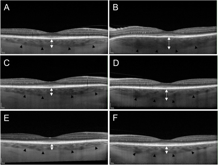
Results: Posterior scleritis is an uncommon yet potentially sight-threatening inflammation of the sclera. During the disease process, inflammation can spread to the adjacent choroid, causing different manifestations of choroidal involvement: (1) increased choroidal thickness, (2) choroidal vasculitis, (3) presentation as a choroidal or subretinal mass in nodular posterior scleritis, and (4) choroidal folds, choroidal effusion and exudative retinal detachment.

Conclusions: Clinical characteristics and multimodal imaging can aid in diagnosing and monitoring disease progression and response to treatment in non-infectious posterior scleritis with choroidal involvement.

Keywords: Choroid; Choroidal folds; Choroidal involvement; Choroidal mass; Choroidal thickness; Choroidal vasculitis; Exudative retinal detachment; Posterior scleritis.


Conflict of interest statement

The authors have no relevant affiliations or financial involvement with any organization or entity with a financial interest in or financial conflict with the subject matter or materials discussed in the manuscript. This includes employment, consultancies, honoraria, stock ownership or options, expert testimony, grants or patents received or pending, or royalties.

Figures

Fig. 1
Fig. 1
Enhanced-depth imaging optical coherence tomography (EDI-OCT) in a case of left posterior scleritis. The black arrow heads delineate the choroidal-scleral junction. (B) Spectralis EDI-OCT demonstrates left subfoveal choroidal thickening on presentation (442μm). The B-scan ultrasound showed thickening of posterior coats (2.5mm) and fluid in Tenon's capsule and optic nerve sheath (not shown). (D) Subfoveal choroidal thickness in the left eye decreased 1 week (406μm) and (F) 4 weeks (292μm) after initiation of oral prednisolone. (A,C,E) Normal subfoveal choroidal thickness in the clinically unaffected right eye of the same patient at corresponding points in time
Fig. 2
Fig. 2
Selected imaging findings in nodular posterior scleritis. (A) Pseudocolour fundus photograph of nodular posterior scleritis. (B) Corresponding B-scan ultrasound findings, showing gross thickening of the posterior coats (6.6mm) with increased echogenicity. An ab externo biopsy was negative for malignancy. (C) The nodular lesion resolved with topical and systemic steroids, and Rituximab-infusions
Fig. 3
Fig. 3
Serous macular detachment secondary to posterior scleritis. (A) Colour fundus photograph and Topcon optical coherence tomography (OCT) at presentation. Delineation of the exudative detachment can be seen on the colour fundus photograph (black arrowheads). (B) Resolution of the serous macular detachment 3 weeks after initiation of oral prednisolone

References

    1. Watson PG, Hayreh SS. Scleritis and episcleritis. Br J Ophthalmol. 1976;60(3):163–191. doi: 10.1136/bjo.60.3.163. - DOI - PMC - PubMed
    1. Berchicci L, Miserocchi E, Di Nicola M, La Spina C, Bandello F, Modorati G. Clinical features of patients with episcleritis and scleritis in an Italian tertiary care referral center. Eur J Ophthalmol. 2014;24(3):293–298. doi: 10.5301/ejo.5000394. - DOI - PubMed
    1. Bin Ismail MA, Lim RHF, Fang HM, Wong EPY, Ling HS, Lim WK, Teoh SC, Agrawal R. Ocular autoimmune systemic inflammatory infectious study (OASIS)-report 4: analysis and outcome of scleritis in an east Asian population. J Ophthalmic Inflamm Infect. 2017;7(1):6. doi: 10.1186/s12348-017-0124-5. - DOI - PMC - PubMed
    1. Erkanli L, Akova YA, Guney-Tefekli E, Tugal-Tutkun I. Clinical features, prognosis, and treatment results of patients with scleritis from 2 tertiary eye care centers in Turkey. Cornea. 2010;29(1):26–33. doi: 10.1097/ICO.0b013e3181ac9fad. - DOI - PubMed
    1. Keino H, Watanabe T, Taki W, Nakashima C, Okada AA. Clinical features and visual outcomes of Japanese patients with scleritis. Br J Ophthalmol. 2010;94(11):1459–1463. doi: 10.1136/bjo.2009.171744. - DOI - PubMed

LinkOut - more resources