. 2021 Oct 29;7(44):eabh0050.
doi: 10.1126/sciadv.abh0050. Epub 2021 Oct 27.

Brain inflammation triggers macrophage invasion across the blood-brain barrier in Drosophila during pupal stages

Affiliations

Brain inflammation triggers macrophage invasion across the blood-brain barrier in Drosophila during pupal stages

Bente Winkler et al. Sci Adv. .

Abstract

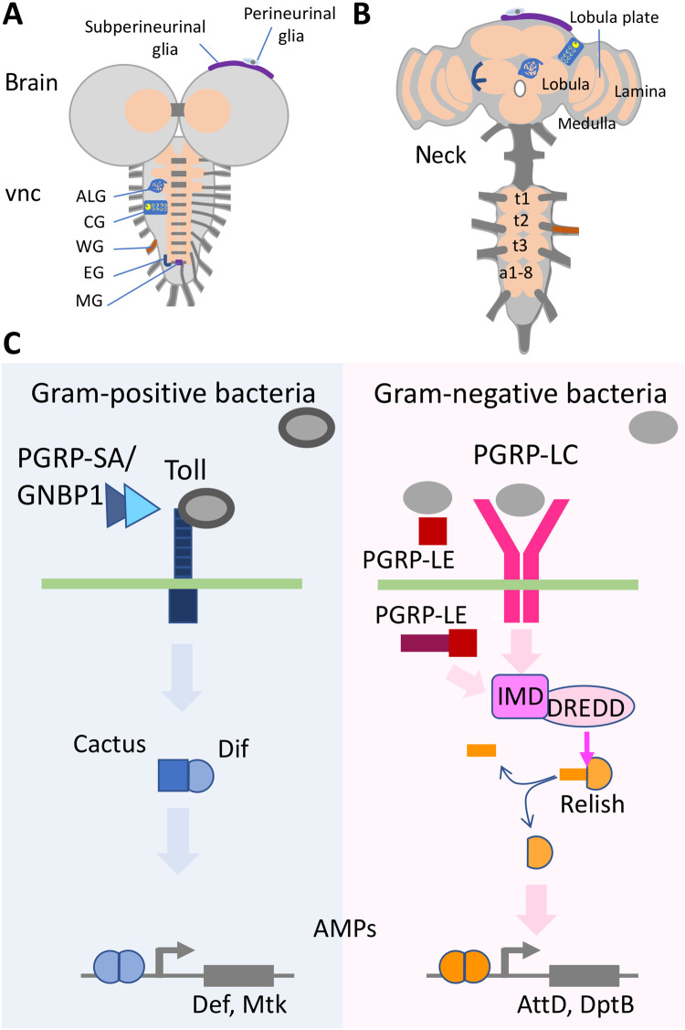
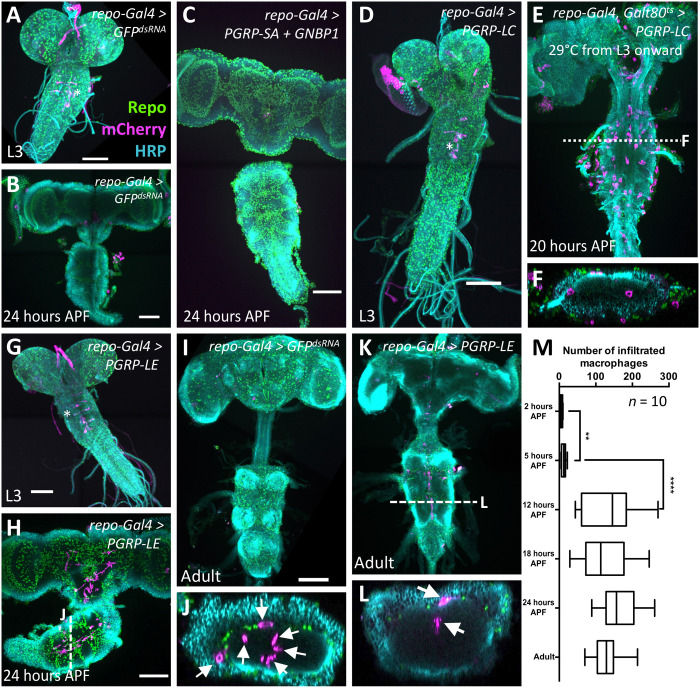
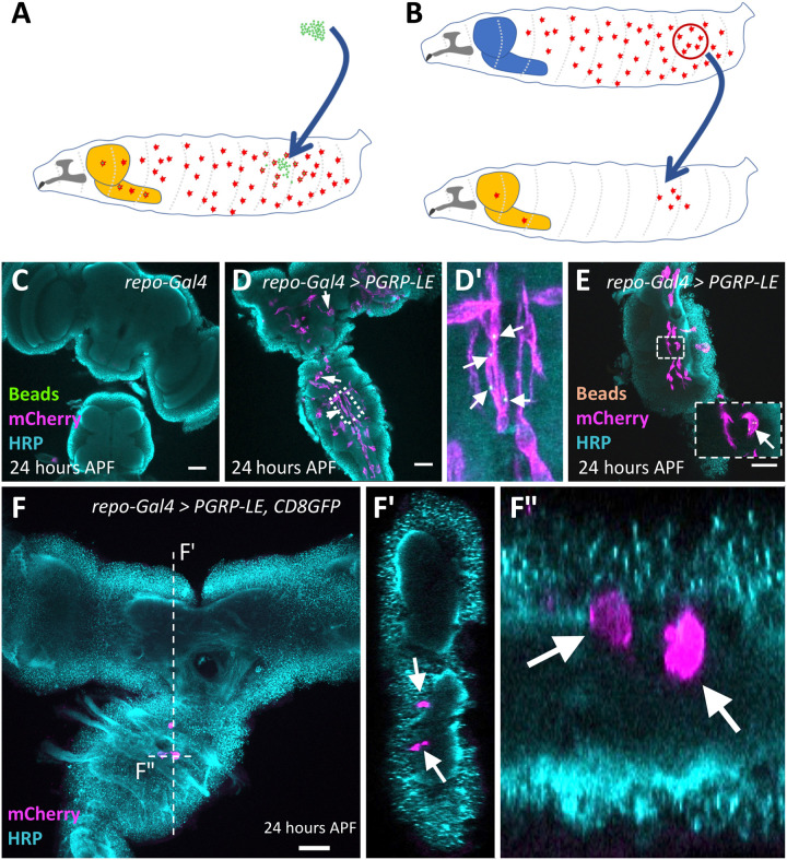
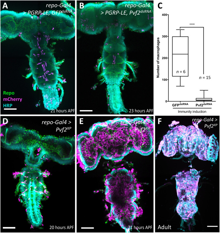
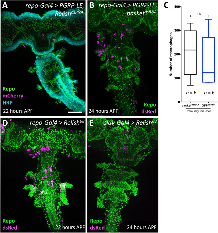
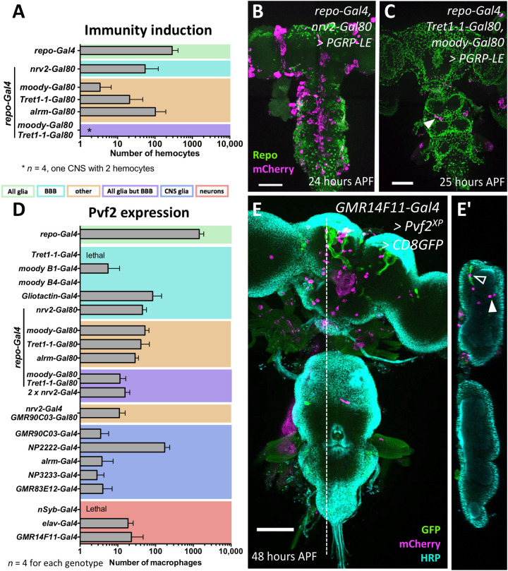
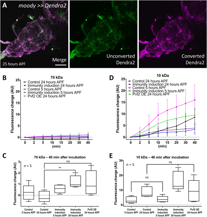
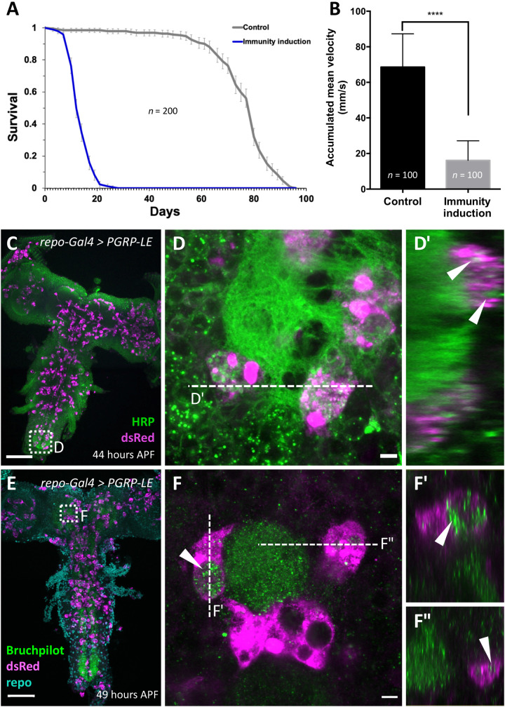
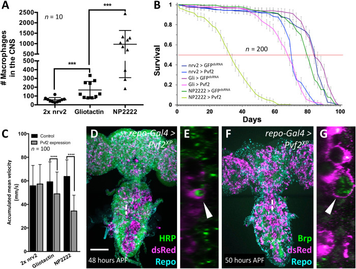
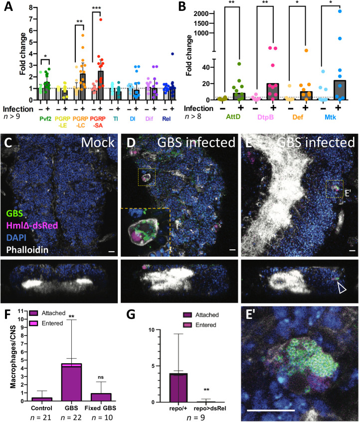
The nervous system is shielded from circulating immune cells by the blood-brain barrier (BBB). During infections and autoimmune diseases, macrophages can enter the brain where they participate in pathogen elimination but can also cause tissue damage. Here, we establish a Drosophila model to study macrophage invasion into the inflamed brain. We show that the immune deficiency (Imd) pathway, but not the Toll pathway, is responsible for attraction and invasion of hemolymph-borne macrophages across the BBB during pupal stages. Macrophage recruitment is mediated by glial, but not neuronal, induction of the Imd pathway through expression of Pvf2. Within the brain, macrophages can phagocytose synaptic material and reduce locomotor abilities and longevity. Similarly, we show that central nervous system infection by group B Streptococcus elicits macrophage recruitment in an Imd-dependent manner. This suggests that evolutionarily conserved inflammatory responses require a delicate balance between beneficial and detrimental activities.


Figures

Fig. 1.
Fig. 1.. Drosophila brains and immunity induction.
(A and B) Schematic view of (A) a third instar larval CNS and (B) an adult brain each composed of the ventral nerve cord (vnc) and the brain lobes. The subperineurial and the perineurial glia establish the BBB. The remaining glial cell types are astrocyte-like glia (ALG), cortex glia (CG), wrapping glia (WG), ensheathing glia (EG), and midline glia (MG). The thoracic neuromeres (t1 to t3) expand during pupal development, while the abdominal neuromers a1-8 condense. Some neuropil areas of the adult are indicated. (C) Signaling pathways directing the innate immune response. The Toll pathway is preferentially activated by Gram-positive bacteria, and the peptidoglycan recognition proteins (PGRP-LC and PGRP-LE) detect mostly Gram-negative bacteria. All relevant components are indicated. For further details, see main text.
Fig. 2.
Fig. 2.. Macrophages enter the brain during pupal stages upon immunity induction.
Dissected brains of indicated age stained for Repo (green) to label glial nuclei, HRP (cyan) to label neuronal membranes, and mCherry expression (magenta) directed by the macrophage marker srpHemo-moe::3xmCherry. wL3, wandering third instar larva. Adults were 7 days old. Scale bars, 100 μm. (A) Control larva expressing double-stranded RNA directed against green fluorescent protein (GFP). The asterisk indicates six neurons expressing the macrophage marker srpHemo-moe::3xmCherry. (B) Control pupa expressing double-stranded RNA directed against GFP. (C) Pupal brain expressing PGRP-SA and GNBP1 in glial cells. Note the absence of mCherry-expressing cells in the brain. (D) Larval brain expressing PGRP-LC in glial cells, no macrophages are found in the brain. The asterisk denotes neurons weakly expressing the marker srpHemo-moe::3xmCherry. (E) PGRP-LC expression is restricted to late larval and pupal stages. Macrophages invade the brain. The dashed line indicates the orthogonal section shown in (F). (G) Larva with PGRP-LE induction in glial cells. Note the elongated ventral nerve cord. The asterisk indicates neurons weakly expressing the macrophage marker. (H and J) Pupal brain expressing PGRP-LE. Note the presence of srpHemo-moe::3xmCherry–expressing cells in the brain. The white dashed line indicates the position of the orthogonal section shown in (J) (arrows indicate macrophages). (I) Adult control brain expressing GFPdsRNA. (K and L) Adult brain with immunity induction in glia. Cherry-expressing cells are found in the brain. The white dashed line indicates the orthogonal section shown in (L) (arrows indicate macrophages). (M) Quantification of the infiltration rate. To count the number of Cherry-positive cells, we used the srpHemo-H2A::3xmCherry marker. Pupae of increasing age were dissected, and the number of Cherry-expressing cells was determined using Imaris. P values are **P2–5 hours APF = 0.0034 and ****P5–12 hours APF < 0.0001 (t test); n = 10 for every time point.
Fig. 3.
Fig. 3.. Cherry-expressing cells originate from the hemolymph.
(A and B) Schematic view on the experimental strategies. The light orange–colored structure corresponds to the inflamed brain. The blue-colored brain reflects a control animal without infection. (A) Injection of fluorescent latex beads into the hemolymph of wandering third instar larvae. (B) Transplantation of macrophages expressing the srpHemo-moe::3xmCherry into the hemolymph of wandering third instar larvae. (C to F) Twenty-four hours APF pupal brains were stained for horseradish peroxidase (HRP) (cyan) and mCherry (magenta). (C) Control brain with no immunity induction. (D and E) Following pan-glial immunity induction, macrophages containing 1-μm green latex beads (D and D′) (arrows indicate macrophages containing latex beads) or 0.5-μm orange latex beads (E) (see arrows) are found in the CNS. (F and F″) Pupal brain after immunity induction and transplantation of genetically labeled macrophages (arrows). Several labeled macrophages are found in the ventral nerve cord. The position of the orthogonal sections shown in (F’) and (F″) is indicated by a white dashed line. Scale bars, 100 μm.
Fig. 4.
Fig. 4.. Pvf2 acts downstream of PGRP-LE to direct macrophage invasion.
(A) Pupal brain with pan-glial immunity induction. Note the presence of macrophages in the CNS. (B) Pupal brain with pan-glial immunity induction and concomitant silencing of Pvf2. Note that fewer macrophages are found in the CNS. (C) Quantification of the number of invading macrophages using srpHemo-H2A::3xmCherry marker in brains with immunity induction without [see (A)] and with RNA interference–mediated Pvf2 knockdown [see (B)] (****P < 0.0001, Mann-Whitney). (D) Pan-glial expression of Pvf2 triggers the invasion of srpHemo-moe::3xmCherry–expressing macrophages into the nervous system 20 hours APF. The macrophages appear to follow axon tracts toward the neuropil. (E) Upon pan-glial Pvf2 expression, the number of macrophages invading the brain increases further during later pupal stages (71 hours APF). (F) Upon pan-glial Pvf2 expression, macrophages are still present in the adult brain (20-day-old female brain). Scale bars, 100 μm.
Fig. 5.
Fig. 5.. Relish is required for macrophage invasion.
(A, B, D, and E) Twenty-two to 24-hour-old pupal brains stained as indicated. Glial nuclei (anti-Repo, green), neuronal membranes (anti-HRP, cyan), and invading macrophages (mCherry or dsRed, magenta). Scale bar, 100 μm. (A) Upon concomitant expression of PGRP-LE and silencing of relish expression using RNA interference, no macrophages enter the brain. (B and C) No suppression of macrophage invasion induced by PGRP-LE expression is observed following concomitant suppression of basket expression. For quantification, see (C) (n = 6; P = 0.6753, Mann-Whitney). ns, not significant. (D) Pan-glial expression of activated Relish causes an invasion of macrophages into the brain. (E) Neuronal expression of activated Relish is not sufficient to trigger invasion of macrophages into the CNS.
Fig. 6.
Fig. 6.. Cell type–specific induction of immunity induces invasion of macrophages.
(A) Pan-glial activation of immunity response triggers invasion of many macrophages into the CNS (n = 10 for repo>>PGRP-LE; n = 4 for all other genotypes). When pan-glial PGRP-LE expression was blocked in various glial subtypes, the number of invading macrophages is reduced. The color coding of the different glial subtypes is indicated below. For further information about the different Gal4 drivers, see Materials and Methods, Table 3. (B) Concomitant silencing in cortex and ensheathing glia using nrv2-Gal80 causes a similar reduction in the number of invading macrophages as concomitant silencing in astrocyte-like glial cells using alrm-Gal80. (C) Upon PGRP-LE expression in all glial cells but the BBB, only very few macrophages entered the brain (arrowhead) (repo-Gal4, moody-Gal80, and Tret1-1-Gal80 UAS-PGRP-LE). (D) Average number of invading macrophages in different expression regimes (n = 10 for repo>>Pvf2; otherwise, n = 4). Color coding is as in (A). In all expression regimes, macrophages enter the brain, except for moodyB4-Gal4–driven Pvf2 expression. (E) Notably, even expression of Pvf2 in only few neurons (GMR14F11-Gal4 is active in the mushroom bodies only, shown by concomitant expression of UAS-CD8-GFP), is able to recruit macrophages into the brain lobes [open arrowhead indicates macrophage associated with mushroom body, and filled arrowhead indicates macrophage located in some distance (E and E′)]. Scale bars, 100 μm.
Fig. 7.
Fig. 7.. Immunity induction does not affect BBB integrity.
(A) The BBB-forming cells are not replaced during onset of pupal development. Confocal image of a 24- to 25-hour APF pupal brain. Larvae with the genotype (moody-Gal4 UAS-tub-Dendra2) were subjected to photoconversion of the subperineurial glia covering the ventral nerve cord. The resultant red fluorescent Dendra2 protein can be detected in pupal brain 25 hours APF. Scale bar, 100 μm. (B) Quantification of dye uptake experiments using fluorescein-labeled 70-kDa dextran in control (repo-Gal4, UAS-GFPdsRNA) and after pan-glial immunity induction (repo-Gal4, UAS-PGRP-LE). Datasets were obtained 5 and 24 hours APF. In addition, we included 24-hour APF pupal brains expressing Pvf2. AU, arbitrary units. OE, overexpression. (C) Quantification of changes in fluorescence uptake after 40 min. A slight but significant increase in fluorescein-labeled dextran can be detected following immunity induction (**P = 0.0079) but not following Pvf2 expression (P = 0.0556). (D) Quantification of dye uptake experiments using Texas Red–labeled 10-kDa dextran in control and after pan-glial immunity induction. The same genotypes and time points as in (A) were used. A large variability of data points was found resulting in large error bars. In all genotypes analyzed, an increase in Texas-red–labeled dextran in the CNS can be detected. (E) Quantification of changes in Texas-red uptake after 40 min. A significant increase in Texas-red–labeled dextran was found for control brains (P = 0.0317) and those with a pan-glial immunity induction (P = 0.0079). However, the levels of Texas-red–labeled dextran were not significantly different between control and pan-glial immunity induction (P5hAPF = 0.667 and P24hAPF = 0.095). Likewise, no differences in Texas-red–labeled dextran uptake were noted in 24-hour APF brains expressing Pvf2 (P = 0.944). n = 5.
Fig. 8.
Fig. 8.. Pan-glial immunity induction affects longevity and climbing ability.
(A) Longevity of flies with pan-glial immunity induction compared to control flies (repo-Gal4, UAS-CD8Cherry versus repo-Gal4 UAS-PGRP-LE). The viability is markedly reduced (n = 200 females; P = 3.75 × 10−92). (B) Same genotypes as in (A). The climbing ability of 7-day-old flies is markedly reduced upon immunity induction (****P < 0.0001). (C to F) Two-day-old pupae with immunity induction stained for macrophages using [hmldsRed] and subsequent anti-dsRed staining, and either anti-HRP (green) (C and D) to label neuronal membranes or anti-Brp (green) (E and F) to label synapses. Note that macrophages harbor vesicles containing neuronal membrane material [arrowheads in (D′), (F′), and (F″)]. Scale bars, 100 μm (C and E) and 5 μm (D and F).
Fig. 9.
Fig. 9.. CNS resident macrophages affect survival and phagocytose neuronal membranes.
(A) Number of macrophages invading the brain of 1-week-old adult flies expressing Pvf2 with different glial cell–specific Gal4 drivers. The P values are ***Pnrv2 vs. Gli = 0.0001 and ***Pgli vs. NP2222 = 0.0002 (Mann-Whitney). (B) Same genotypes as in (A). Longevity of female flies with glial subtype–specific Pvf2 expression compared to control flies. The corresponding Gal4 driver element is indicated in the figure. For control, we expressed GFPdsRNA. The viability is reduced upon Pvf2 expression in all glial subtypes tested and inversely correlates with the number of invading macrophages (n = 200 females in groups of 20 each; ****Pnrv2 = 4.425 × 10⁻³², ****PGli = 5.2984 × 10−41, and ****PNP2222 = 1.9936 × 10−89). (C) Same genotypes as in (A). The climbing ability of 7-day-old females is reduced upon Pvf2 expression and again inversely correlates with the number of invaded macrophages (Pnrv2 = 0.237, ****PGli < 0.0001, and ****PNP2222 < 0.0001). (D to G) Two-day-old pupae with pan-glial Pvf2 expression stained for macrophages using [hmldsRed] and subsequent anti-dsRed staining, and either anti-HRP (green) (D and E) to label neuronal membranes or anti-Brp (green) (F and G) to label synapses. Note that macrophages harbor vesicles containing neuronal membrane material (arrowheads). Scale bar, 100 μm.
Fig. 10.
Fig. 10.. GBS infection leads to immunity induction and macrophage recruitment in early pupal brains.
(A and B) Wandering third instar Drosophila larvae injected with GBS were dissected 4 to 5 hours after infection, and the brains were subjected to qPCR. Expression of the genes PGRP-LC, PGRP-SA, Pvf2, and all AMPs tested (AttD, DtpB, Def, and Mtk) was up-regulated upon infection. Mann-Whitney test for Pvf2, *P = 0.0227; PGRP-LC, **P = 0.0024; PGRP-SA, ***P = 0.0002; AttD, **P = 0.0055; DtpB, **P = 0.0025; Def, *P = 0.0207; Mtk, *P = 0.0499. Unpaired t test for PGRP-LE, P = 0.5297; Tl, P = 0.0988; Dl, P = 0.6202; Dif, P = 0.6758. (C to E) Macrophage recruitment and infiltration to GBS-infected pupal brains. Confocal images (top and orthogonal views) showing Drosophila CNS of hml-dsRed pupae 4 to 5 hours after injection in the hemolymph of either (C) Mock or (D and E) GBS. Macrophages (hml-dsRed in magenta) containing GBS (anti-GBS in green) are detected attached to the CNS (D, inset) or inside the CNS (E). (E′) is a close-up of the dotted boxes from (E). The arrowhead points to a macrophage within the CNS cortex. Phalloidin is in white, and 4′,6-diamidino-2-phenylindole (DAPI) is in blue. Scale bars, 10 μm. (F) Quantification of macrophage localization in the whole CNS 4 to 5 hours after injection in the hemolymph of mock (control), GBS, or formaldehyde-fixed GBS. Kruskal-Wallis test followed by Dunn’s multiple comparisons test on brain-associated (attached + entered) macrophages was performed generating adjusted P values: **P(control vs. GBS) = 0.0011 and P(control vs. fixed-GBS) = 0.7165. Control, n = 21 CNS; GBS, n = 22 CNS; fixed-GBS, n = 10 CNS. (G) Number of srpHemo-moe::3xmCherry–expressing macrophages that attach to the brain upon GBS infection of repo-Gal4 animals and repo-Gal4>UAS-relishdsRNA animals [n = 9; **P = 0.0078, Mann-Whitney on brain-associated (attached + entered) macrophages].

References

    1. Niederkorn J. Y., See no evil, hear no evil, do no evil: The lessons of immune privilege. Nat. Immunol. 7, 354–359 (2006). - PubMed
    1. Saijo K., Glass C. K., Microglial cell origin and phenotypes in health and disease. Nat. Rev. Immunol. 11, 775–787 (2011). - PubMed
    1. Hilu-Dadia R., Kurant E., Glial phagocytosis in developing and mature Drosophila CNS: Tight regulation for a healthy brain. Curr. Opin. Immunol. 62, 62–68 (2020). - PubMed
    1. Logan M. A., Glial contributions to neuronal health and disease: New insights from Drosophila. Curr. Opin. Neurobiol. 47, 162–167 (2017). - PMC - PubMed
    1. Logan M. A., Hackett R., Doherty J., Sheehan A., Speese S. D., Freeman M. R., Negative regulation of glial engulfment activity by Draper terminates glial responses to axon injury. Nat. Neurosci. 15, 722–730 (2012). - PMC - PubMed

LinkOut - more resources