. 2022 Jan 11;88(1):e0152221.
doi: 10.1128/AEM.01522-21. Epub 2021 Oct 27.

A Novel Actinobacterial Cutinase Containing a Noncatalytic Polymer-Binding Domain

Affiliations

A Novel Actinobacterial Cutinase Containing a Noncatalytic Polymer-Binding Domain

Kofi Abokitse et al. Appl Environ Microbiol. .

Abstract

The single putative cutinase-encoding gene from the genome of Kineococcus radiotolerans SRS30216 was cloned and expressed in Escherichia coli as a secreted fusion protein, designated YebF-KrCUT, where YebF is the extracellular carrier protein. The 294-amino-acid sequence of KrCUT is unique among currently characterized cutinases by having a C-terminal extension that consists of a short (Pro-Thr)-rich linker and a 55-amino-acid region resembling the substrate binding domain of poly(hydroxybutyrate) (PHB) depolymerases. Phylogenetically, KrCUT takes a unique position among known cutinases and cutinase-like proteins of bacterial and fungal origins. A modeled structure of KrCUT, although displaying a typical α/β hydrolase fold, shows some unique loops close to the catalytic site. The 39-kDa YebF-KrCUT fusion protein and a truncated variant thereof were purified to electrophoretic homogeneity and functionally characterized. The melting temperatures (Tm) of KrCUT and its variant KrCUT206 devoid of the putative PHB-binding domain were established to be very similar, at 50 to 51°C. Cutinase activity was confirmed by the appearance of characteristic cutin components, C16 and C18 hydroxyl fatty acids, in the mass chromatograms following incubation of KrCUT with apple cutin as the substrate. KrCUT also efficiently degraded synthetic polyesters such as polycaprolactone and poly(1,3-propylene adipate). Although incapable of PHB depolymerization, KrCUT could efficiently bind PHB, confirming the predicted characteristic of the C-terminal region. KrCUT also potentiated the activity of pectate lyase in the degradation of pectin from hemp fibers. This synergistic effect is relevant to the enzyme retting process of natural fibers. IMPORTANCE To date, only a limited number of cutinases have been isolated and characterized from nature, the majority being sourced from phytopathogenic fungi and thermophilic bacteria. The significance of our research relates to the identification and characterization of a unique member of the microbial cutinases, named KrCUT, that was derived from the genome of the Gram-positive Kineococcus radiotolerans SRS30216, a highly radiation-resistant actinobacterium. Given the wide-ranging importance of cutinases in applications such as the degradation of natural and synthetic polymers, in the textile industry, in laundry detergents, and in biocatalysis (e.g., transesterification reactions), our results could foster new research leading to broader biotechnological impacts. This study also demonstrated that genome mining or prospecting is a viable means to discover novel biocatalysts as environmentally friendly and biotechnological tools.

Keywords: Kineococcus; biopolymer degradation; biotechnology; cutinase; enzyme technology; microbial world; α/β fold hydrolase.


Figures
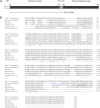
FIG 1
FIG 1
(A) Schematic representation of the domain organization of KrCUT. SP, signal peptide; TP-rich, threonine-proline-rich linker. KrCUT206 is truncated KrCUT without the polymer-binding domain. Cloning of the KrCUT and KrCUT206-encoding genes is facilitated by the primers described in Table 1. (B) Clustal Omega (1.2.4) multiple-sequence alignment of KrCUT from K. radiotolerans (YP_001363838.1) with Cellulomonas bogoriensis (WP_156968314), Microlunatus sagamiharensis (WP_197680654), Klenkia marina (WP_207798241), Fusarium solani (AAA33335.1 and 1CUS), and Aspergillus oryzae RIB40 (3GBS). Asterisks indicate totally conserved amino acids, the catalytic triad residues are highlighted in green and conserved C (cysteine) in red, double-underlined residues in KrCUT indicate the experimentally determined amino acid sequence, the 9-amino-acid sequence in purple is the insertion in KrCUT as described in the text, the threonine-proline-rich sequence is shown in blue, and the boldfaced amino acid residues in the C terminus are conserved residues of the SBD-binding sequence of known PHB depolymerases.
FIG 2
FIG 2
Phylogenetic relationship of KrCUT to other cutinases or cutinase-like sequences. The GenBank accession numbers of the sequences are as indicated preceding the organism names. Those with available crystal structures are accompanied by the Protein Data Bank (PDB) numbers. Numbers in branches indicate bootstrap values per 1,000 replicates.
FIG 3
FIG 3
Secondary-structure plot of KrCUT. Identified in the dashed boxes are the locations of insertions (green) and deletions (red) compared to the F. solani cutinase. Orange text indicates the location of the catalytic triad, while yellow lines show locations of disulfide bonds within the catalytic domain. The substrate binding domain is shown as a cartoon sphere, as there is no structural template available with high similarity for this region.
FIG 4
FIG 4
Purification of recombinant K. radiotolerans cutinase and truncated derivatives. Proteins were analyzed by 10% SDS-PAGE and silver stained. The molecular weight markers are indicated on the sides of the gels. Lane 1, fraction from SP-Sepharose showing YebF-KrCUT fusion protein (39 kDa) and mature KrCUT (31 kDa); lanes 2 and 3, fractions from HiLoad Superdex 75 showing the separated protein bands; lane 4, YebF-KrCUT206 (33 kDa) from HiLoad Superdex 75.
FIG 5
FIG 5
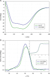
CD spectra of KrCUT and KrCUT206 (A) and thermal denaturation curves (B) at 222 nm. CD derivatives were calculated using Spectra Manager (Jasco). The inset shows the original thermodenaturation curves of the two proteins, measured at 222 nm while increasing the temperature.
FIG 6
FIG 6
Degradation of synthetic polyesters by YebF-KrCUT and KrCUT206. Enzyme concentration is 2.5 μM, unless otherwise stated. (A) Degradation of PCL films (■, YebF-KrCUT; ▴, YebF-KrCUT, 1.25 μM; □, KrCUT206) and PCL pellets (●, YebF-KrCUT; ○, KrCUT206). The curve of PCL film degradation by 5 μM YebF-KrCUT was found to approximate that of 2.5 μM enzyme concentration (data not shown). (B) Degradation of PLA (●, YebF-KrCUT; ○, KrCUT206) and polypropylene adipate (■, YebF-KrCUT; □, KrCUT206).
FIG 7
FIG 7
Adsorption isotherm of cutinase and PHB granules at 25°C. Purified KrCUT at concentrations ranging from 0.16 to 3.2 mg/mL were mixed with 25 mg PHB in 50 mM sodium phosphate (pH 8.0) in a total volume of 1 mL and mixed by gently shaking for 3 h. The PHB polymer was removed by centrifugation, and the concentration of protein in the supernatant was determined by the BCA method. The concentration of bound protein at a particular concentration of cutinase was calculated as the difference between a control without added PHB and free cutinase after incubation with PHB. P value < 0.001.
FIG 8
FIG 8
Amount of pectin released from natural hemp fibers by pectate lyase (PL) or in combination with cutinase (Cut) at different concentrations. The shaded bars indicate the two incubation periods. The amount of unsaturated product released is reported. PLCut40, pectate lyase and 40 units of cutinase; PLCut100, pectate lyase and 100 units of cutinase. P value = 0.024277.
FIG 9
FIG 9
Pocket analysis. Pocket residues were identified by superimposing crystal structures (enabling accent secondary-structure matches refined with Gaussian weight) following alignment in MOE. Residues within 4.5 Å of the covalent ligand in Trichoderma reesei cutinase in complex with a C11Y4 phosphonate inhibitor (PDB code 4PSE) were assigned as pocket residues. Orange, catalytic triad; light gray, pocket residues; magenta, oxyanion hole residues; cyan, gatekeeper residues.

References

    1. Riederer M, Mueller C. 2006. Annual plant reviews, vol 23. Biology of the plant cuticle. Blackwell Publishing Ltd, Oxford, United Kingdom.
    1. Kolattukudy PE. 2001. Polyesters in higher plants. Adv Biochem Eng Biotechnol 71:1–49. 10.1007/3-540-40021-4_1. - DOI - PubMed
    1. Carvalho CM, Aires-Barros MR, Cabral JMS. 1999. Cutinase: from molecular level to bioprocess development. Biotechnol Bioeng 66:17–34. 10.1002/(SICI)1097-0290(1999)66:1<17::AID-BIT2>3.0.CO;2-F. - DOI - PubMed
    1. Soliday CL, Flurkey WH, Okita TW, Kolattukudy PE. 1984. Cloning and structure determination of cDNA for cutinase, an enzyme involved in fungal penetration of plants. Proc Natl Acad Sci USA 81:3939–3943. 10.1073/pnas.81.13.3939. - DOI - PMC - PubMed
    1. Skamnioti P, Gurr SJ. 2008. Cutinase and hydrophobin interplay. Plant Signal Behav 3:248–250. 10.4161/psb.3.4.5181. - DOI - PMC - PubMed

Publication types

LinkOut - more resources